an article by Claire Weston, PhD student at Imperial College London
The Biomicrofluidics System Group at the Dalian Institute of Chemical Physics in China have published an exciting paper in Lab on a Chip where they have used paper as a material to grow and differentiate human pluripotent stem cells.
Recently, there has been much research into generating biocompatible materials for creating microenvironments for the growth of stem cells, with the aim of improving their regenerative potential. Using paper as the material has several advantages over the conventional polymers – it is cheap and readily available, it is biocompatible, and the bundles of cellulose microfibers that make up paper provide a porous 3D structure.
The authors used three different types of paper to identify which were best for stem cell growth – printing paper, filter paper, and nitrocellulose membrane. The paper was pre-coated with the required gels and the stem cells were seeded onto the surface. Initially, the stem cells were differentiated into cardiomyocytes prior to being added to the paper to test if the differentiated cells were able to grow on the different types of paper. The cells aggregated on both printing and filter paper and demonstrated spontaneous beating function, but not on the nitrocellulose membrane. These tissues also maintained their beating function for up to three months. The stem cells were then added to the paper prior to differentiation and the required cardiac differentiation procedures were carried out. The cells differentiated to the cardiomyocytes on all three paper types, however the cardiac-specific marker was only expressed weakly on the nitrocellulose membrane. Within two weeks a strong beating function was observed for the printing paper, but not the other paper types. The authors suggest the printing paper had a better pore size to support the cells than the filter paper, while the nitrocellulose membrane didn’t have a favourable microstructure to support growth of cardiac tissue.
Along with this article, there are some impressive videos showing the cardiac tissue beating that are well worth a watch!
To download the full article for free* click the link below:
Human induced pluripotent stem cell-derived beating cardiac tissues on paper
Li Wang, Cong Xu, Yujuan Zhu, Yue Yu, Ning Sun, Xiaoqing Zhang, Ke Feng and Jianhua Qin
Lab Chip, 2015, 15 , 4283-4290
DOI: 10.1039/C5LC00919G
—————-
About the webwriter
Claire Weston is a PhD student in the Fuchter Group, at Imperial College London. Her work is focused on developing novel photoswitches and photoswitchable inhibitors.
—————-
*Access is free through a registered RSC account until 18/12/2015 – click here to register
The µTAS 2015 Conference was held in October at the Hwabaek International Convention Center in Gyeongju, Korea.
Sarah Ruthven, Executive Editor of Lab on a Chip, was in attendance at the conference to announce the prestigious Lab on a Chip awards which include the Pioneers of Miniaturisation Lectureship (in partnership with Corning Inc), the Widmer Young Researcher Poster Prize, the Art in Science award (sponsored by NIST) and the µTAS video competition (in partnership with Dolomite Microfluidics).
“Pioneers of Miniaturization” Lectureship
Professor Dino Di Carlo was announced as the winner of the 10th “Pioneers of Miniaturization” Lectureship, sponsored by Lab on a Chip and Corning Incorporated and supported by the Chemical and Biological Microsystems Society (CBMS). The “Pioneers of Miniaturization” Lectureship rewards early to mid-career scientists who have made extraordinary or outstanding contributions to the understanding or development of miniaturised systems. Professor Di Carlo received a certificate, a monetary award and delivered a short lecture titled ‘Microfluidic Frontiers’ at the conference. More information can be found on the competition blog.
Art in Science Award
Lab on a Chip and the National Institute of Standards Technology (NIST) presented the Art in Science award to Matteo Cornaglia from the Laboratory of Microsystems, EPFL in Switzerland. The award aims to highlight the aesthetic value in scientific illustrations while still conveying scientific merit. More information on the winning photograph can be found on the competition blog.
µTAS Video Competition
Lab on a Chip and Dolomite Microfluidics announced Dan Kirby and the Ducrée Lab, Dublin City University the winner of the 2015 µTAS Video Competition supported by the Chemical and Biological Microsystems Society (CBMS).
µTAS participants were invited to submit short videos with a scientific or educational focus. The winners, the Ducrée Lab, recreated an 80’s music video titled “Spin me right round” to promote new areas of research in lab-on-a-disc platforms. The full video can be viewed on the competition blog.
Widmer Young Researcher Poster Prize
The Widmer Poster Prize was awarded to Jinho Kim from Inje University, Korea, with a poster titled “Single-cell isolation of circulating tumor cells by microfluidic technology”.
Congratulations to all the winners at the conference! We look forward to seeing you at µTAS 2016 in Dublin, Ireland.
Lab on a Chip and the National Institute of Standards Technology (NIST) were pleased to present the Art in Science award titled “Under the Looking Glass: Art from the World of Small Science” at the µTAS 2015 Conference.
The award highlights the aesthetic value in scientific illustrations while still conveying scientific merit. Many fantastic submissions were received this year with the winner selected by a panel of senior scientists who attended the conference.
And the winner is…
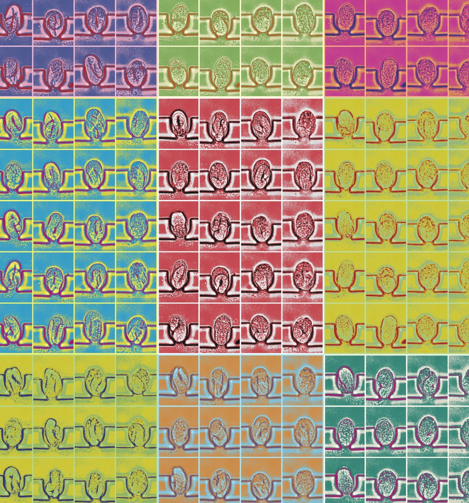
“Through Warhol’s eyepiece” photographed by Matteo Cornaglia, Laboratory of Microsystems, EPFL, Switzerland.
The winning artwork, “Through Warhol’s eyepiece” photographed by Matteo Cornaglia from the Laboratory of Microsystems, EPFL in Switzerland, was created by on-chip multi-dimensional imaging of C.elegans embryogenesis as observed through an Andy Warhol microscope, equipped with a 63x oil immersion objective and a pop art optical filter. For the first time, automated longitudinal studies of C.elegans embryos are made possible by microfluidics. For this artwork, 20 embryos are isolated from an on-chip worm culture upon egg laying and transferred into dedicated micro-incubators for long-term time-lapse imaging of the whole population at single-organism resolution. Each colour corresponds to a different instant of the same population development.
And the runners up are…
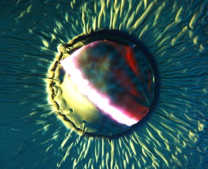
“Reflections of micro-ocean escaping” photographed by Maoxiang Guo, KTH Royal Institute of Technology, Sweden.
A tiny drop of liquid is encapsulated in a polymer microwell, covered with thin gold film. As the device is heated, the liquid is expanding, creating a bulge in the gold film, stress and ripples in the rest of the gold sheet.
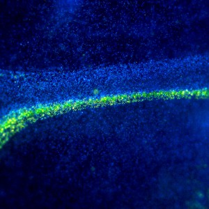
“Microparticle Microgalaxy” photographed by Ghulam Destgeer, Department of Mechanical Engineering (KAIST), South Korea.
Microparticles are manipulated inside a sessile droplet of water placed on top of a vibrating acoustofluidic platform. Surface acoustic waves leaking into the water drive the concentration of the larger diameter (yellow) particles while smaller (blue) particles remain scattered in the background. The particles resembles stars spread in a celestial galaxy.
A big thank you to all the contributors this year.
Lab on a Chip and Dolomite Microfluidics are pleased to announce Dan Kirby and the Ducrée Lab, Dublin City University the winner of the 2015 µTAS Video Competition supported by the Chemical and Biological Microsystems Society (CBMS).
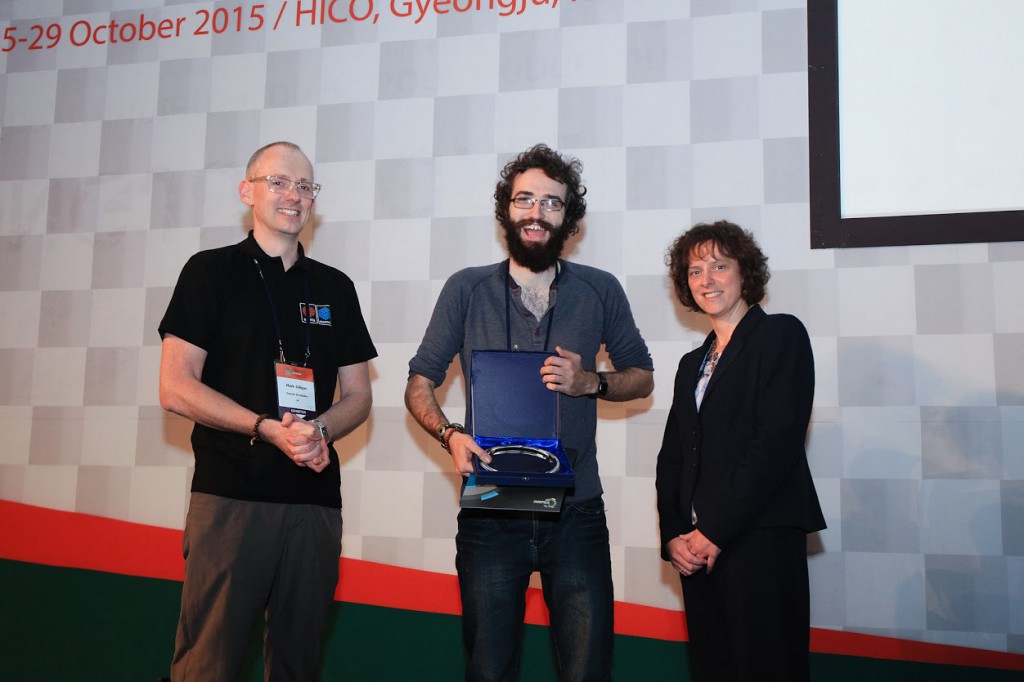
At the µTAS conference in October 2015 Lab on a Chip Executive Editor, Sarah Ruthven (right) and Dolomite Microfluidics Group Chief Executive Officer, Mark Gilligan (left) presented Dan Kirby (centre) with the award and a $2500 gift certificate to spend on Dolomite equipment.
Registered µTAS participants were invited to submit short videos with a scientific or educational focus. Videos could be fun, artistic or just surprising and unusual to be in contention for the prize. The winners, the Ducrée Lab, produced a video titled “Spin me right round” focussing on centrifugal microfluidics. They recreated a classic 80’s music video in the lab to highlight the new areas of research in lab-on-a-disc platforms. They hope that viewers enjoy the “new spin” they have put on biomedical diagnostics!
Thank you very much to all the participants for submitting such high quality entries.
Congratulations to the Ducrée Lab!
A Lab on a Chip article highlighted in Chemistry World by Cesar Palmero
Scientists in China have trapped bacteria in 3D-printed structures and used them to pump materials along customised paths.
Transporting materials in the microscopic world is complex. Conventionally, macroscopic pumps drive motion, but pumps are bulky and not ideal for miniaturisation. Now, Hepeng Zhang and colleagues at Shanghai Jiao Tong University have tackled this problem using native inhabitants of the microscopic world – motile bacteria. Not only are they already present in the media, but their energy conversion efficiency is estimated to be greater than existing man-made micro-motors.
Please visit Chemistry World to read the full article.
Using confined bacteria as building blocks to generate fluid flow
Zhiyong Gao, He Li, Xiao Chen and H. P. Zhang
Lab Chip, 2015, Advance Article
DOI: 10.1039/C5LC01093D