New YouTube Videos


Monitoring CO2 invasion processes at pore scale using Geological Labs on Chip

Transitioning from multi-phase to single-phase microfluidics for long-term culture and treatment of multicellular spheroids

Magnetic force-assisted self-locking metallic bead array for fabrication of diverse concave microwell geometries


Digg This
Reddit This
Stumble Now!
Share on Facebook
Bookmark this on Delicious
Share on LinkedIn
Bookmark this on Technorati
Post on Twitter
Google Buzz (aka. Google Reader)

Recent Advances in 3D Printing

Guest edited by Jennifer Lewis (Harvard University) and Howard Stone (Princeton University) this collection of papers showcases recent advances in the rapidly evolving field of 3D printing, with an emphasis on themes that impact lab-on-a-chip applications.

Free* Access: The upcoming 3D-printing revolution in microfluidics
Critical Review
Nirveek Bhattacharjee, Arturo Urrios, Shawn Kang and Albert Folch
Lab Chip, 2016,16, 1720-1742 DOI: 10.1039/C6LC00163G

Free* Access: High density 3D printed microfluidic valves, pumps and multiplexers
HOT Article
Hua Gong, Adam Trticle. Woolley and Gregory P. Nordin
Lab Chip, 2016,16, 2450-2458 DOI: 10.1039/C6LC00565A

Free* Access: Bioprinted Thrombosis-on-a-Chip
HOT Article
Rahmi Oklu et al.
Lab Chip, 2016, Accepted Manuscript, C6LC00380J

Open Access: 3D- printed microfluidic devices: enablers and barriers
Michael C. Breadmore., et al
Lab Chip, 2016,16, 1993-2013
DOI: 10.1039/C6LC00284F

This collection also features a video demonstration:

3D printing of liquid metals as fugitive inks for fabrication of 3D microfluidic channels
Dishit P. Parekh, Collin Ladd, Lazar Panich, Khalil Moussa and Michael D. Dickey
Lab Chip, 2016,16, 1812-1820 DOI: 10.1039/C6LC00198J

Browse our 3D Printing collection – we hope you enjoy the articles

*Access is free until 10th October via a registered RSC account.

Digg This
Reddit This
Stumble Now!
Share on Facebook
Bookmark this on Delicious
Share on LinkedIn
Bookmark this on Technorati
Post on Twitter
Google Buzz (aka. Google Reader)

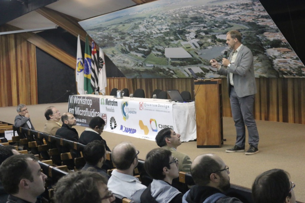
6th Workshop on Microfluidics, State University of Campinas, Brazil

Invited keynote talk “Lab, Cells and Organ on a Chip” by Dr. Albert van den Berg (University of Twente, The Netherlands)

The 6th Workshop on Microfluidics was held at the Convention Center of UNICAMP (State University of Campinas) in Campinas, Sao Paulo (Brazil), from 20-22 July, 2016. Since 2011, the workshop has brought together young students, researchers, and companies from different states of Brazil to discuss topics related to fundamentals, fabrication technologies, innovations, and applications in microfluidic science. This field has been spread out around different regions of Brazil presenting outstanding contributions for microfabrication and microfluidic technologies.

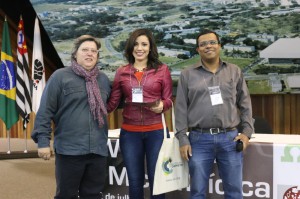
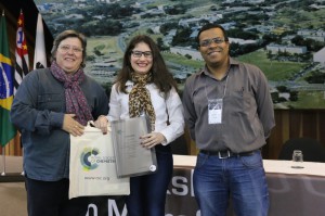
The event was supported by the Royal Society of Chemistry for the fourth time. Prof Dosil P. de Jesus (UNICAMP), member of the 6th Workshop on Microfluidics scientific board, presented the poster competition awards, sponsored by the Royal Society of Chemistry’s Lab on a Chip and Analytical Methods journals.


The winner of the Analytical Methods Poster Competition was Gabriela B. Almeida, from State University of Campinas, for her work “Microfluidic Devices Combining Dielectrophoresis Trapping and Surface Enhanced Raman Spectroscopy”.

Gabriela F. Giordano, from The Brazilian Nanotechnology National Laboratory (LNNano), Campinas, was the winner of the Lab on a Chip Poster Competition for her work “Gravity-Assisted Distillation on a Chip: a Novel Concept for Sample Preparation in Microfluidics”.

Gabriela Brito Almeida (centre), winner of the Analytical Methods Competition, with Prof. Dosil P. de Jesus (UNICAMP) (right) and Dr Elizabeth Magalhaes (RSC Manager, Brazil) (left)

Gabriela F. Giordano (centre), winner of the Lab on a Chip Competition, with Prof. Dosil P. de Jesus (UNICAMP) (right) and Dr Elizabeth Magalhaes (RSC Manager, Brazil) (left)

Digg This
Reddit This
Stumble Now!
Share on Facebook
Bookmark this on Delicious
Share on LinkedIn
Bookmark this on Technorati
Post on Twitter
Google Buzz (aka. Google Reader)

Chlorophyll lasers

a blog article by by Burcu Gumuscu, PhD researcher at University of Twente

From space shuttles to military equipment, or even the kitchenware that we use on daily basis, lasers have found use in more places than we often realise. Interestingly, the solid-state lasers were believed to be a solution to an unknown problem after their invention in 1950s. In that time nobody—including their developer Charles Townes—noticed that the lasers were one of the game changer inventions in the world’s history. After tens of years, in 1964, the laser technology was awarded with a Nobel prize when its potential for diverse applications were realized.

So far we have only discussed solid-state lasers, but there is definitely capacity in the laser world to improve the performance and versatility with the help of little twists, such as optofluidic lasers. “Optofluidics” is a synergic combination of optical systems and microfluidics. In other words, optical systems are built or synthesized from liquids, aiming to serve as good as their solid-state equivalents. For example, two immiscible liquids form a smooth surface in their interface, leading to a laser cavity or an optical resonator with a very high Q-factor that allows for operating at low energy levels. This emerging field gains importance when considering most of the biochemical reactions that occur in aqueous environments. Optofluidic laser systems are flexible to change their optical properties by just replacing the liquid media; and with this twist, lasers have new application areas including diagnosis of genetic disorders at the cellular level and in vivo biosensing.

What is more exciting about the optofluidic lasers is that they can be biodegradable and easily tunable in microenvironments. Researchers in University of Michigan recently showed that one of the most abundant pigments on earth, chlorophylls, can maintain both biodegradability and tunability in optical systems owing to their fluorescence capabilities. Chlorophylls have a high Q-factor, dual-absorption bands in the visible spectrum, and a large shift between absorption and emission bands, suggesting that chlorophylls can be used as donors in fluorescence resonance energy transfer (FRET) laser (Figure 1). In this study, chlorophyll a was isolated from spinach leaf and used as the gain medium and the donor to develop a novel optofluidic laser. Two lasing bands of chlorophyll a was investigated by both theoretical and experimental means. Concentration-dependent studies enabled more insight for the mechanism determining when, where, and why the laser emission band appears. This new technique seems to gain increasing attention for applications in in vivo and in vitro biosensing, solar lighting and energy harvesting.
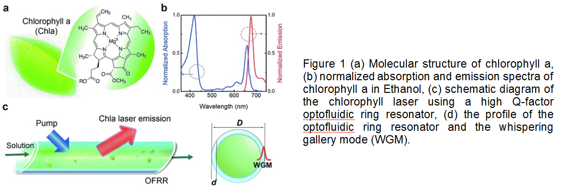
This article, published on 12th May 2016, is included in the Lab on a Chip Recent HOT Articles themed collection.

To download the full article for free* click the link below:

Optofluidic chlorophyll lasers
Yu-Cheng Chen, Qiushu Chena, Xudong Fan
Lab Chip, 2016, 16, 2228-2235
DOI: 10.1039/C6LC00512H

About the Webwriter

Burcu Gumuscu is a PhD researcher in BIOS Lab on a Chip Group at University of Twente in The Netherlands. Her research interests include development of microfluidic devices for second generation sequencing, organ-on-chip development, and desalination of water on the micron-scale.

*Access is free until 23/09/2016 through a registered RSC account.

Digg This
Reddit This
Stumble Now!
Share on Facebook
Bookmark this on Delicious
Share on LinkedIn
Bookmark this on Technorati
Post on Twitter
Google Buzz (aka. Google Reader)

Pioneers of Miniaturization Lectureship 2016

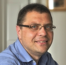
We are delighted to announce that Dr. Daniel Irimia is the winner of the 2016 “Pioneers of Miniaturization” Lectureship.

The 11th “Pioneers of Miniaturization” Lectureship, sponsored by Lab on a Chip and Corning Incorporated, and supported by the Chemical and Biological Microsystems Society (CBMS), is for early to mid-career scientists who have made extraordinary or outstanding contributions to the understanding or development of miniaturised systems.

This “Pioneers of Miniaturization” Lectureship will be presented to Daniel at the µTAS 2016 Conference in Dublin, Ireland, 9-13 October 2016. Daniel will receive a certificate, a monetary award and will give a short lecture during the conference.

Many congratulations to Dr. Daniel Irimia on this achievement from the Lab on a Chip team

About the winner

Dr. Daniel Irimia is a bioengineer trained as a physician and passionate about understanding the clinical consequences of neutrophil activities during disease. He received his Ph.D. in bioengineering from the University of Illinois, Chicago in 2002 before becoming a Research Fellow at Massachusetts General Hospital.

Daniel is currently an Associate Professor in Surgery and Bioengineering and Deputy Director of the BioMEMS Resource Center at the Center for Engineering in Medicine, USA. His research is focused on designing sophisticated tools to measure relevant neutrophil behavior with the highest precision. He leads a team of scientists and doctors that employ microfluidic devices and novel measurements of neutrophil functions to monitor burn patients, optimize treatments, and uncover neutrophil-targeting interventions that could prevent infections and sepsis in burn patients.

As the organizer of the Cell World Races, aimed at encouraging scientists and clinician-researchers to utilize microfluidic tools in their research for higher level of precision and detail, Daniel increases the awareness for the technological changes taking place in the field of cell motility. This has been featured on the front page of Wall Street Journal (March 2014) and in 2012 Daniel was one of the winners of the Wellcome Image Awards for the depiction of “Cancer cells in motion.”

For more details on Dr. Daniel Irimia’s research please visit his homepage.

Digg This
Reddit This
Stumble Now!
Share on Facebook
Bookmark this on Delicious
Share on LinkedIn
Bookmark this on Technorati
Post on Twitter
Google Buzz (aka. Google Reader)

New YouTube Videos

On-demand droplet splitting using surface acoustic waves

A microfluidic cell-trapping device for single-cell tracking of host–microbe interactions

The physical origins of transit time measurements for rapid, single cell mechanotyping

Digg This
Reddit This
Stumble Now!
Share on Facebook
Bookmark this on Delicious
Share on LinkedIn
Bookmark this on Technorati
Post on Twitter
Google Buzz (aka. Google Reader)

New YouTube Videos

Time-lapse lens-free imaging of cell migration in diverse physical microenvironments

On-Chip Micromagnet Frictionometer Based on Magnetically Driven Colloids for Nano-Bio Interface

Real-time assessment of nanoparticle-mediated antigen delivery and cell response

Digg This
Reddit This
Stumble Now!
Share on Facebook
Bookmark this on Delicious
Share on LinkedIn
Bookmark this on Technorati
Post on Twitter
Google Buzz (aka. Google Reader)

New YouTube Videos

Lab-on-a-disc for simultaneous determination of total phenolic content and antioxidant activity of beverage samples

Flow control using audio tones in resonant microfluidic networks: towards cell-phone controlled lab-on-a-chip devices

Microfluidic droplet trapping, splitting and merging with feedback controls and state space modelling

Digg This
Reddit This
Stumble Now!
Share on Facebook
Bookmark this on Delicious
Share on LinkedIn
Bookmark this on Technorati
Post on Twitter
Google Buzz (aka. Google Reader)

New YouTube Videos

Integrative volumetric bar-chart chip for rapid and quantitative point-of-care detection of myocardial infarction biomarkers

Deterministic sequential isolation of floating cancer cells under continuous flow

Thermally robust and biomolecule-friendly room temperature bonding for the fabrication of elastomer-plastic hybrid microdevice

Digg This
Reddit This
Stumble Now!
Share on Facebook
Bookmark this on Delicious
Share on LinkedIn
Bookmark this on Technorati
Post on Twitter
Google Buzz (aka. Google Reader)

What is the best way to study brain?

an article by Burcu Gumuscu, PhD researcher at University of Twente

Before the 1700’s, when dissection techniques were not yet available, the cause of mood changes were thought to be the replacement of liquids and vapors in the body. The biology of the brain has been better understood since the discovery of research and test tools. However, occupying only about 1/50 of the body mass, the brain is perhaps the most complicated organ to study. National Institute of Neurological Disorders and Stroke’s list of over 400 neurological disorders can be seen as a sound proof of this exciting- and frustrating- fact.

Figure 1. The effects of biomechanical forces on the brain

Biomechanical forces on neurons play a fundamental role in neuronal physiology, which, in turn, affect brain development and disorders. During the growth of neurons, the tension created by the biomechanical forces are suggested to influence the cells’ motor activities, gene expression, neurotransmitter release, together with neurite growth and network connections (Figure 1). Research on the biomechanical forces can definitely help us to understand how the brain works, but many questions related to these forces remain unanswered. Quantitative measurements of the cell activity seem to be the only possible path to find satisfying answers to those questions.

A comprehensive list of experimental techniques involving both conventional and alternative micro&nanotechnology approaches have been recently brought to the attention of scientific community by Di Carlo and his coauthors. In their recent critical review, both advantages and disadvantages of conventional toolsnamely motor-driven pressure, patch membrane pressure, osmotic pressure, fluid shear stresses, and deformation of flexible elastomers—, microtechnology toolsincluding atomic force microscopy, micropatterning, and some other potential techniques—, and nanotechnology toolssuch as ferromagnetic and piezoelectric nanoparticles— are discussed.

The literature reports provided in the paper suggest that micro and nanotechnology tools offer better spatiotemporal resolution and throughput when compared to conventional techniques. The cellular functions and the possible technologies for the characterization of those functions are further described (Figure 2). For instance, behind-the-scene biological mechanisms for recovery in traumatic brain injuries can be determined by applying the biomechanical forces at the right place and right time to ultimately mitigate the injuries.

Figure 2. The influence of biomechanical forces on the neuron functions and available technologies for their investigation.

This article, published on 26 April 2016, is included in the Lab on a Chip Recent HOT Articles themed collection.

To download the full article for free* click the link below:

Micro- and nano-technologies to probe the mechano-biology of the brain
Andy Tay, Felix E. Schweizer, and Dino Di Carlo
Lab Chip, 2016,16, 1962-1977
DOI: 10.1039/C6LC00349D, Critical Review

—————-

About the webwriter

Burcu Gumuscu is a PhD researcher in BIOS Lab on a Chip Group at University of Twente in The Netherlands. Her research interests include development of microfluidic devices for second generation sequencing, organ-on-chip development, and desalination of water on the micron-scale.

—————-

*Access is free until 15/08/2016 through a registered RSC account.

Digg This
Reddit This
Stumble Now!
Share on Facebook
Bookmark this on Delicious
Share on LinkedIn
Bookmark this on Technorati
Post on Twitter
Google Buzz (aka. Google Reader)