Art in Science Competition Winner Announced at MicroTAS 2016

Lab on a Chip and the National Institute of Standards Technology (NIST) were pleased to present the Art in Science award at the µTAS 2016 Conference on 13 October 2016. The award highlights the aesthetic value in scientific illustrations while still conveying scientific merit. Many fantastic submissions were received this year with the winner selected by Sam Keltie, Lab on a Chip Executive Editor, Darwin Reyes, NIST and Dino Di Carlo, Lab on a Chip Editorial Board member.

And the winner is…

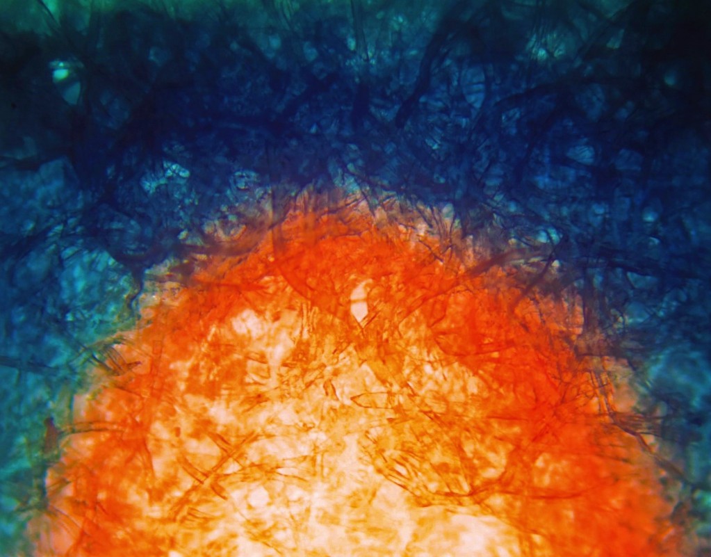
Vaibhav Jain, Purdue University

The Rising Sun

Dark field micrograph of sweat advancing in a paper channel dyed with Cobalt chloride dihydrate (Bluish), changing it to cobalt chloride hexahydrate (Reddish orange)

And the runners up are…

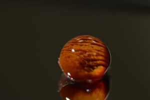
Gokce Ozkazanc, Bilkent University

Composition of Particles in a Droplet”

Visualization of Particle Composition inside a Water Droplet

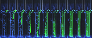
Susanna Lladó Maldonado, TU Braunschweig

Wellness microresort for cells

Mixing performance in a microbubble column-bioreactor

A big thank you to all the contributors this year!

Digg This
Reddit This
Stumble Now!
Share on Facebook
Bookmark this on Delicious
Share on LinkedIn
Bookmark this on Technorati
Post on Twitter
Google Buzz (aka. Google Reader)

Sepsis’ sticking point in for a shock

Written by Alex Pashley for Chemistry World

Simple microfluidic method helps detect life-threatening condition earlier

A new method for monitoring the onset of sepsis touted by US researchers may assist clinicians in helping more of their patients survive this potentially fatal condition.

Sepsis, often referred to as blood poisoning, can be the result of an innocuous infection or injury. Without treatment, the immune system becomes overwhelmed, leading to organ shutdown. In the UK alone, 30,000 people die as a direct result of the condition. Sepsis needs to be caught early to maximise the chances of a full recovery. However, existing diagnostics rely on antibody labelling, which requires samples to be prepared and tested by specialist laboratory staff. This requires time that a patient might not be able to spare. ‘Existing methods are too cumbersome,’ explains Joel Voldman, heading up the new study at Massachusetts Institute of Technology.

detecting sepsis with electrochemical monitoring

The tiny device can process large amounts of blood and detect activated white blood cells that are an indicator for sepsis. Source: © Royal Society of Chemistry

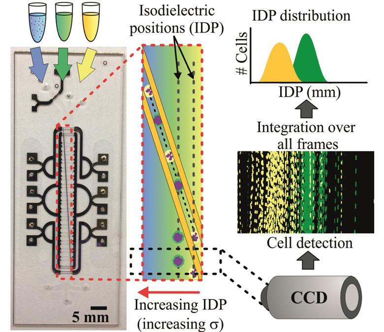
Voldman’s group devised a microfluidic method that takes advantage of the white blood cells’ electrical properties, which makes it possible to detect sepsis quickly in its earliest stages. Sepsis triggers white blood cells to become activated and circulate in the blood. The number of these activated cells indicates the disease’s progression. Applying an electric field then separates activated and non-activated cells based on subtle differences in their electrical properties.

Read the full article in Chemistry World.


Monitoring sepsis using electrical cell profiling

Javier L. Prieto, Hao-Wei Su, Han Wei Hou, Miguel Pinilla Vera, Bruce D. Levy, Rebecca M. Baron, Jongyoon Han and Joel Voldman

Lab Chip, 2016, Advance Article

DOI: 10.1039/C6LC00940A, Paper

Digg This
Reddit This
Stumble Now!
Share on Facebook
Bookmark this on Delicious
Share on LinkedIn
Bookmark this on Technorati
Post on Twitter
Google Buzz (aka. Google Reader)

Blood matters

a blog article by by Burcu Gumuscu, PhD researcher at University of Twente

Iron deficiency anemia (IDA) is not a trivial illness. Every individual in the world’s population has the potential to suffer from this nutritional disease. According to estimations, 900 million people worldwide are already afflicted with it. IDA is known to lower cognitive ability, work capacity, and future productivity of both children and adults. The situation appears to be grave when we consider the economic consequences of these problems.

The human body needs iron to produce red blood cells, and having low iron levels in the body leads to IDA. Diagnosis of IDA requires a complete blood count is performed by a bulky hematology analyzer. IDA has been a common disease for a really long time; however, its associated diagnosis costs are considerably high, and the diagnosis equipment is not available in many places of the world. Given the facts, IDA diagnosis actually deserves cheaper and easily accessible equipment, which has unfortunately remained elusive—up until now.

In last month’s issue of Lab on a Chip, Whitesides research group at Harvard University came up with a sound idea to diagnose IDA in a shorter and inexpensive way. They developed a low-cost and rapid-screening tool to diagnose IDA using aqueous multiphase systems containing layer of polymer-salt mixtures. These mixtures are loaded in a microhematocrit tube (depicted in the figure) together with a drop of blood from a fingerprick. Diagnosis results become available after a 2-minute low-cost centrifuging process.

The reported data suggest that diagnosis of IDA is improved by means of sensitivity and specificity when compared to the bulky hematology analyzer’s results. Several important red blood cell parameters, such as concentration of hemoglobin in a given volume of red blood cells, can be predicted. The technique’s ability to diagnose IDA was further improved using automated digital analysis. They also show that the tool is able to detect a wider range of anemia types including microcytic and hypochromic anemia. The portable and low cost screening tool could possibly find use in rural clinics where large fractions of the population at risk of IDA. Before entering the market, the performance of this technique will still have to be validated to demonstrate feasibility of using and interpreting the assay.

Design of the presented test loaded with blood before and after centrifugation for a representative IDA and Normal sample. Blood is loaded into the top of the tube, from a fingerprick, using capillary action provided by a hole in the side of the tube. Normal blood packs at the bottom of the tube, while less dense blood cells can be seen packing at the interfaces between the phases and inside the tube. Normal and IDA blood can be differentiated by eye after only 2 minutes of centrifugation. It is also possible to read the analysis results in an automated way. A commonly used software is used to convert the image to red intensity graphs.

This article was published in Lab on a Chip on 30th August 2016.

To download the full article for free* click the link below:

Diagnosis of iron deficiency anemia using density-based fractionation of red blood cells

Jonathan W. Hennek, Ashok A. Kumar, Alex B. Wiltschko, Matthew R. Patton, Si Yi Ryan Lee, Carlo Brugnara, Ryan P. Adams and George M. Whitesides
Lab Chip, 2016,16, 3929-3939
DOI: 10.1039/C6LC00875E

—————-

About the Webwriter


Burcu Gumuscu is a postdoctoral fellow in BIOS Lab on a Chip Group at University of Twente in The Netherlands. Her research interests include development of microfluidic devices for next generation sequencing, compartmentalized organ-on-chip studies, and desalination of water on the microscale.

—————-

*Access is free until 7th November 2016 through a registered RSC account – register here

Digg This
Reddit This
Stumble Now!
Share on Facebook
Bookmark this on Delicious
Share on LinkedIn
Bookmark this on Technorati
Post on Twitter
Google Buzz (aka. Google Reader)

New YouTube Videos

Autonomous microfluidic capillaric circuits replicated from 3D-printed molds

The intercell dynamics of T cells and dendritic cells in a lymph node-on-a-chip flow device

A reconfigurable stick-n-play modular microfluidic system using magnetic interconnects
Digg This
Reddit This
Stumble Now!
Share on Facebook
Bookmark this on Delicious
Share on LinkedIn
Bookmark this on Technorati
Post on Twitter
Google Buzz (aka. Google Reader)

Introducing our new Lab on a Chip Advisory Board members

We are delighted to welcome our new Advisory Board members!
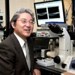
Yoshinobu Baba – Nagoya University

Yoshinobu is a Professor in the Department of Advanced Medical Science Graduate School of Medicine at Nagoya University. His major area of interest is nanobiosicence and nanobiotechnology for omics, systems biology, cancer diagnosis, cancer therapy regenerative medicine, and molecular in vivo imaging.

Jean-Christophe Baret – University of Bordeaux, France

Jean-Christophe is a Professor at the University of Bordeaux. His research group focuses on the fundamental study of interfaces in liquid systems through the dynamics of droplets, bubbles and emulsions.

Anja Boisen – Technical University of Denmark, Denmark

Anja is a Professor in the Department of Micro- and Nanotechnology at the Technical University of Denmark.  Her research group focuses on development and application of micro and nano mechanical sensors and microfabricated solutions for oral drug delivery. The group also explores integration of micro and nano sensors onto centrifugal microfluidic platforms.

Qun Fang – Zhejiang University

Qun is a Qiushi Distinguished Professor in the Department of Chemistry at Zhejiang University, and the Director of Institute of Microanalytical Systems at Zhejiang University. His research interests include microfluidic analysis, capillary electrophoresis, flow injection analysis, and miniaturization of analytical instruments, especially the development of automated and high-throughput droplet-based microfluidic analysis and screening techniques.

Martin A. M Gijs – EPFL, Switzerland

Martin is Professor in the School of Engineering at EPFL and Head of the Laboratory of Microsystems. His present interests are in developing technologies for novel magnetic devices, new microfabrication technologies for microsystems fabrication in general and the development and use of microsystems technologies for microfluidic and biomedical applications in particular.

Noo Li Jeon – Seoul National University, South Korea

Noo Li is a Professor at the School of Mechanical and Aerospace Engineering, Seoul National University. His research group use cell culture and microfluidic techniques to investigate biological processes.

Gwo-Bin Lee – National Tsing Hua University

Gwo-Bin is a Professor at the National Tsing Hua University. His research interests are on nano-biotechnology, micro/nanofluidics and their biomedical applications. He is currently developing integrated micro/nano systems incorporated with nano/biotechnology for a variety of applications, including fast diagnosis of infectious diseases, screening of biomarkers for cancer and cardiovascular diseases, and optofluidics.

Hang Lu – Georgia Institute of Technology, USA

Hang is a Professor at the School of Chemical & Biomolecular Engineering, Georgia Institute of Technology. Her research group work at the interface of engineering and biology. They engineer BioMEMS and microfluidic devices to address questions across a variety of disciplines.

Adrian Neild – Monash University, Australia

Adrian is an Associate Professor and Director of Research in the Department of Mechanical and Aerospace Engineering Department at Monash University. His research interest are focused on non-linear ultrasound including acoustic radiation forces and acoustic streaming as well as viscosity, microfluidic systems, micron-scale particle and biological cell handling, air-coupled ultrasound, transducer design, non-destructive testing, and finite-element analysis.

Nicole Pamme – University of Hull, UK

Nicole is a Professor in Analytical Chemistry at the University of Hull. Following her PhD, where she worked on single particle analysis in microfluidic chips, Nicole spent a year in Japan, working as an independent research fellow in the International Centre for Young Scientists (ICYS) at the National Institute for Materials Science. She has been at the University of Hull since 2005 and is currently Director of Research.

Sámuel Sánchez – Institute of Bioengineering of Catalonia, Spain

Sámuel leads the lab-in-a-tube and nanorobotic biosensors research group at the Institute of Bioengineering of Catalonia. His research focuses on the design of miniaturized devices that bridge multidisciplinary fields from material science, chemistry and biology. His research group studies a broad range of phenomena occurring at the interface between materials and biology, from fundamental studies to applications.
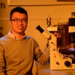
Anderson Shum – University of Hong Kong, China

Anderson is an Associate Professor in the Department of Mechanical Engineering and the University of Hong Kong. His research area covers microscaled fluid dynamics, biomedical applications of microfluidics, eye-on-a-chip, and all-aqueous microfluidics; and his  main area of expertise include droplet microfluidics, emulsion-templated materials and microscaled interfacial phenomena.

Digg This
Reddit This
Stumble Now!
Share on Facebook
Bookmark this on Delicious
Share on LinkedIn
Bookmark this on Technorati
Post on Twitter
Google Buzz (aka. Google Reader)

Introducing our new Lab on a Chip Editorial Board members!

Please join us in welcoming Dino Di Carlo, Piotr Garstecki and Shoji Takeuchi to the Lab on  a Chip Editorial Board!

Dino Di Carlo, University of California at Los Angeles

Dino is Professor and Vice Chair of the Department of Bioengineering and a Professor of Mechanical Engineering at the University of California at Los Angeles. He also Directs the Cancer Nanotechnology Program of the Jonsson Comprehensive Cancer Center. His research group are currently focusing on exploiting unique physics, microenvironment control, and the potential for automation associated with miniaturized systems for applications in basic biology, medical diagnostics, and cellular engineering.

Piotr Garstecki, Polish Academy of Sciences

Piotr is a Professor at the Institute of Physical Chemistry, Polish Academy of Sciences. He currently leads the Research Group of Microfluidics and Complex Fluids. The group conducts research on fundamental aspects of the physics of soft matter systems and develops microfluidic tools for chemistry and biology.


Shoji Takeuchi, University of Tokyo

Shoji is a Professor in the Center for International Research on Integrative Biomedical Systems (CIBiS), Institute of Industrial Science (IIS), University of Tokyo. Since 2008, he has been a director of Collaborative Research Center for Bio/Nano Hybrid Process at IIS. His current research interests include 3D tissue fabrication, implantable devices, artificial cells/lipid bilayer systems, and biohybrid MEMS.

Digg This
Reddit This
Stumble Now!
Share on Facebook
Bookmark this on Delicious
Share on LinkedIn
Bookmark this on Technorati
Post on Twitter
Google Buzz (aka. Google Reader)

New YouTube Videos

A conductive liquid-based surface acoustic wave device

Highly efficient sample stacking by enhanced field amplification on a simple paper device

A lab-on-a-disc with reversible and thermally stable diaphragm valves


Digg This
Reddit This
Stumble Now!
Share on Facebook
Bookmark this on Delicious
Share on LinkedIn
Bookmark this on Technorati
Post on Twitter
Google Buzz (aka. Google Reader)

New Lab on a Chip Associate Editor: Petra S. Dittrich

At Lab on a Chip we are very pleased to announce our newest Associate Editor – Petra Dittrich!

Petra is Associate Professor for Bioanalytics at the Department of Biosystems Science and Engineering. Her research in the field of lab-on-chip-technologies focuses on the miniaturization of high-sensitivity devices for chemical and biological analyses, and microfluidic-aided organization of materials.

Petra’s research group develop miniaturized devices (lab-on-chip technology or microfluidics) for applications in the life sciences. Their interdisciplinary approach combines chemical, physical, biological and engineering aspects of microfluidics-based technology. You can find out more about her research on her homepage: http://www.dittrich.ethz.ch/

Petra is now handling papers so submit your paper to her today!

Digg This
Reddit This
Stumble Now!
Share on Facebook
Bookmark this on Delicious
Share on LinkedIn
Bookmark this on Technorati
Post on Twitter
Google Buzz (aka. Google Reader)

New YouTube Videos

An In-line Spectrophotometer on Centrifugal Microfluidics Platform for Real-time Protein Determination

and Calibration

Hydrodynamic trapping for rapid assembly and in situ electrical characterization of droplet interface bilayer arrays

A fully automated microfluidic micellar electrokinetic chromatography analyzer for organic compound detection

Digg This
Reddit This
Stumble Now!
Share on Facebook
Bookmark this on Delicious
Share on LinkedIn
Bookmark this on Technorati
Post on Twitter
Google Buzz (aka. Google Reader)

Top 10 Reviewers for Lab on a Chip

In celebration of Peer Review Week, with the theme of Recognition for Review – we would like to highlight the top 10 reviewers for Lab on a Chip in 2016, as selected by the editor for their significant contribution to the journal.

Name Institution
Dr David Erickson Cornell University
Dr Edmond Young University of Toronto
Dr David Sinton University of Toronto
Dr Daniel Irimia Harvard University
Dr Kevin Dorfman University of Minnesota
Dr Piotr Garstecki Polish Academy of Sciences
Professor Jaap Den Toonder Delft University
Dr Petra Dittrich ETH Zürich
Dr Michael C Breadmore University of Tasmania
Dr David Juncker McGill University

We would like to say a massive thank you to these reviewers as well as the Lab on a Chip board and all of our community for their continued support of the journal, as authors, reviewers and readers.

Review to win!
As a little added bonus to celebrate Peer Review Week, for the next four weeks our reviewers will be in with a chance of winning a fantastic prize! Simply submit a review for any of our journals between 19 September and 16 October 2016 and you will be automatically eligible for a chance to win one of our fantastic prizes.

Digg This
Reddit This
Stumble Now!
Share on Facebook
Bookmark this on Delicious
Share on LinkedIn
Bookmark this on Technorati
Post on Twitter
Google Buzz (aka. Google Reader)