Archive for the ‘Uncategorized’ Category

Look Mum, no pumps!

GA

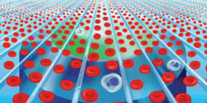
Richard Crooks and colleagues, researchers at the University of Texas, Austin developed a way to locally concentrate and move analytes using internal bipolar electrodes (bypassing the need for an outside driver of fluid flow). Electrodes printed on the bottom of the microfluidic channels create controllable gates which balance the convective and electrokinetic forces acting on charged sample molecules. A single DC power supply and controller box is needed to open/close these gates to deliver analytes to different regions of the chip.­­

Crooks and his group have extensively investigated bipolar electrochemistry theory and in this paper demonstrate the use of bipolar electrodes to separate, enrich, and transport ­­­­­bands of analytes in microfluidic channels. Electric potentials applied across a channel induce an electric field within the buffer whilst conductive substrates present on the floor of the microchannel also adopt a potential between their two poles. H+ ions within the buffer are partially neutralized by electrogenerated OH and thus regions of ion depletion appear. These depletion zones attract charged analytes from the solution to maintain the charge gradient induced by the electric field (the speed of electromigration of analytes to the depletion zone is proportional to the electric field). Bipolar electrodes on the channel bottom contain their own local field and so analytes concentrate in these areas, leading to enrichment near the electrodes.

In this work, Crooks and his team demonstrate separation and enrichment of two common fluorescent dyes:  BODIPY2- and MPTS3-. The two dye bands are then directed to two separate reservoirs. In previous papers, the group focused on optimizing enrichment and achieved enrichment rates of up to 0.57 BODIPY2 1, . The current extension and integration of online separation and enrichment achieves comparable rates of enrichment, 0.11 and 0.31 fold/second for BODIPY and MPTS, respectively, while also enabling control over separating analytes of different electromobility (μep) and transporting these bands to designated areas of the device.

To create the devices presented, the group used conventional photolithography techniques to pattern gold bipolar passive electrodes (BPEs) on glass and bonded PDMS channels on top of the regions. This method can be easily multiplexed as additional BPEs can be activated to guide separated and enriched analytes to different areas of the chip.

 References:

1 R. K. Anand, E. Sheridan, D. Hlushkou, U. Tallarek and R. M. Crooks, Lab on a Chip, 2011, 11, 518-527.

Electrochemically-gated delivery of analyte bands in microfluidic devices using bipolar electrodes
Karen Scida, Eoin Sheridan and Richard M. Crooks, Lab Chip, 2013, 13, 2292-2299.
DOI: 10.1039/c3lc50321f

Digg This
Reddit This
Stumble Now!
Share on Facebook
Bookmark this on Delicious
Share on LinkedIn
Bookmark this on Technorati
Post on Twitter
Google Buzz (aka. Google Reader)

New YouTube videos

View the new videos on the Lab on a Chip YouTube site using the links below:


Aqueous two-phase microdroplets with reversible phase transitions


Passive Droplet Sorting using Viscoelastic Flow Focusing 


Formation of polymersomes with double bilayers templated by quadruple emulsions


A doubly cross-linked nano-adhesive for the reliable sealing of flexible microfluidic devices


Ultrafast cell switching for recording cell surface transitions: new insights into epidermal growth factor receptor signalling

Flow-switching allows independently programmable, extremely stable, high-throughput diffusion-based gradients

Highly reproducible chronoamperometric analysis in microdroplets

Disaggregation of microparticle clusters by induced magnetic dipole–dipole repulsion near a surface

Exploring a direct injection method for microfluidic generation of polymer microgels

Droplet morphometry and velocimetry (DMV): a video processing software for time-resolved, label-free tracking of droplet parameters

Digg This
Reddit This
Stumble Now!
Share on Facebook
Bookmark this on Delicious
Share on LinkedIn
Bookmark this on Technorati
Post on Twitter
Google Buzz (aka. Google Reader)

Circulating cancer cells spiral towards separation

Repeated biopsies of tumours can be a painful and distressing procedure for cancer patients. A new biochip developed by researchers in Singapore can isolate tumour cells from blood samples, and may one day be an alternative to more invasive methods for tracking later stage cancers. 

Operating principle of circulating tumour cell enrichment by a spiral channel with trapezoid cross-section

Operating principle of circulating tumour cell enrichment by a spiral channel with trapezoid cross-section

Deaths from cancer generally occur after the cancer has spread. Cells detach from the primary tumour and travel through the blood, subsequently forming new tumours. Being able to isolate and characterise these circulating tumour cells (CTCs) can provide information about the original tumour. However, CTCs exist in very low numbers in the blood stream and hence require enrichment and separation before analysis. 

Read the full article in Chemistry World 

Slanted spiral microfluidics for the ultra-fast, label-free isolation of circulating tumor cells
E W Majid et al, Lab Chip, 2013, Accepted manuscript, Paper
DOI: 10.1039/C3LC50617G

Digg This
Reddit This
Stumble Now!
Share on Facebook
Bookmark this on Delicious
Share on LinkedIn
Bookmark this on Technorati
Post on Twitter
Google Buzz (aka. Google Reader)

New Youtube videos

View the new videos on the Lab on a Chip YouTube site using the links below:


Microfluidic Synthesis of Atto-liter Scale Double Emulsions toward Ultrafine Hollow Silica Spheres with Hierarchical Pore Networks


Droplet sorting based on the number of encapsulated particles using a solenoid valve


Temperature-driven self-actuated microchamber sealing system for highly integrated microfluidic devices


Vector separation of particles and cells using an array of slanted open cavities

A simple route to functionalize polyacrylamide hydrogels for the independent tuning of mechanotransduction cues

Microfluidic chemostat for measuring single cell dynamics in bacteria

Electrostatic charging and control of droplets in microfluidic devices 

Dynamic pH mapping in microfluidic devices by integrating adaptive coatings based on polyaniline with colorimetric imaging techniques

 An integrated microfluidic cell culture system for high-throughput perfusion three-dimensional cell culture-based assays: effect of cell culture model on the results of chemosensitivity assays

Rapid generation and manipulation of microfluidic vortex flows induced by AC electrokinetics with optical illumination

Digg This
Reddit This
Stumble Now!
Share on Facebook
Bookmark this on Delicious
Share on LinkedIn
Bookmark this on Technorati
Post on Twitter
Google Buzz (aka. Google Reader)

A new function for your cell-phone: analysing blood at point of care

Today, calling is not the only function of the cell-phone, but in some cases just a nice side function. A new function developed by Aydogan Ozcan and co-workers is the ability to perform a rapid blood analysis using your cell phone.

In a previous article the group at University of California, Los Angeles, USA, showed that a cell-phone with some add-on components can be used to test for the presence of peanuts in cookies1. In this new article, a module is demonstrated which can be used to measure characteristics of blood. Three variables which can be tested with their system are the haemoglobin content and white and red blood cell concentrations.

After connecting a base attachment to the cell phone (in this case an Android phone), three different add-on components can then be attached. Each component consists of a lens, light source and chamber for the sample. For the white blood cell count, the cells are first fluorescently labelled and placed in a chamber with known volume. Subsequently the sample is excited and the fluorescence is measured in the perpendicular direction. In case of the red blood cell count, unlabelled cells in a specific volume are optically detected using bright field illumination. For the last application, the measurement of the haemoglobin content, the absorbance of the lysed blood sample is determined, which is directly related to the concentration of haemoglobin. The user-friendly phone app allows you to choose one of the three analyses and input parameters, such as the sample dilution factor. It subsequently processes the captured images to generate the test results, which can be uploaded to a database or sent on to clinicians

Although some sample pre-processing is necessary, the blood analysis will take about 10 seconds for each image taken. The results of the cell phone module are in good agreement with a standard test, thereby making it applicable for blood analysis at point of care.

References

1. Ahmet F. Coskun, Justin Wong, Delaram Khodadadi et alA personalized food allergen testing platform on a cellphone. Lab Chip, 2013, 13, 636–640

Cost-effective and rapid blood analysis on a cell-phone
Hongying Zhu, Ikbal Sencan, Justin Wong, Stoyan Dimitrov, Derek Tseng, Keita Nagashima and Aydogan Ozcan  
DOI: 10.1039/C3LC41408F

Loes Segerink is a Post-Doctoral researcher in the BIOS Lab on a Chip group, University of Twente, The Netherlands

Digg This
Reddit This
Stumble Now!
Share on Facebook
Bookmark this on Delicious
Share on LinkedIn
Bookmark this on Technorati
Post on Twitter
Google Buzz (aka. Google Reader)

SERS-active nanostructures synthesized within a microfluidic channel

A new method for the direct, bottom-up growth of a SERS substrate within a microfluidic channel, developed at the University of Connecticut, will enable the inexpensive fabrication of SERS-integrated devices.

Surface enhanced Raman scattering (SERS) spectroscopy is a powerful method for analyte detection and identification.  SERS relies on the electric field enhancement provided by nanostructured metal surfaces to amplify the Raman scattering “fingerprint” of adsorbed molecules, enabling specific detection down to the single molecule level1.  Previously, SERS integration into practical devices has been limited, partly due to the cost and difficultly of fabricating SERS substrates and microfluidic channels separately, then aligning and bonding them together.  In this HOT article, researchers at the University of Connecticut led by Prof. Yu Lei have devised a method to fabricate a novel nanostructured SERS substrate directly within existing microfluidic channels, greatly simplifying the construction of such devices2

Previous SERS-integrated microfluidic devices have utilized silver plates fabricated via laser writing3 and silver nanoparticles created using physical vapor deposition4 as SERS substrates. These devices provide high detection sensitivity, but are costly to produce due to the top-down fabrication methods used.  The advantage of the new method by Parisi et al. is the ability to synthesize a SERS substrate bottom-up and in situ, reducing the complexity and cost of device fabrication.  SERS experiments can then be carried out immediately.

In situ electrodeposition and galvanic displacement are used to fabricate a nanostructured SERS substrate (right) within a microfluidic device (left)

In the new technique, copper nanowalls coated with carbon are synthesized inside a microfluidic channel via electrodeposition.  This is accomplished by applying a voltage across part of the channel while copper acetate flows through.  Next, silver nitrate flows through the channel, and a galvanic replacement reaction results in silver nanoparticles coating the nanowalls.  Then, an analyte solution flows through the channel and adsorbs onto the substrate, allowing the user to measure SERS.  The authors used crystal violet as an example analyte to demonstrate the sensitive in-channel detection capability, measuring the SERS of analyte concentrations down to 50 pM.

The researchers hope that their fabrication technique will ultimately allow SERS to be integrated into various microfluidics-based biological and chemical sensing platforms, increasing the power and flexibility of these devices. 

References

1. J. Kneipp et al., Chem. Soc. Rev., 37, 1052–1060, 2008. 
2. J. Parisi et al., Lab on a Chip, 13, 1501–1508, 2013.
3. B.-B. Xu et al., Lab on a Chip, 11, 3347–3351, 2011.
4. Z. Geng et al., Sensors and Actuators A, 169 (1), 37–42, 2011.

Read this HOT article in Lab on a Chip today:

In situ synthesis of silver nanoparticle decorated vertical nanowalls in a microfluidic device for ultrasensitive in-channel SERS sensing
Joseph Parisi, Liang Su and Yu Lei
DOI: 10.1039/C3LC41249K

 This article is featured in the web collection Lab on a Chip Top 10%


Katie Mayer is a post-doctoral researcher in the Walt Laboratory at Tufts University, USA

Digg This
Reddit This
Stumble Now!
Share on Facebook
Bookmark this on Delicious
Share on LinkedIn
Bookmark this on Technorati
Post on Twitter
Google Buzz (aka. Google Reader)

Taming shape-shifters: analyzing dynamic protein structures on-chip

Researchers at York University in Toronto have developed an integrated microfluidic device to facilitate analysis of modified protein structures after biochemical reactions.  

The platform guides a protein right through from a controlled reaction, functional labeling, and fragmentation zones to a coupled mass spectrometer for determination of the protein’s secondary structure. Using this device, the group conducts the first dynamic structural analysis of the β-lactamase enzyme TEM-1 to identify key regions of the enzyme that affect catalytic activity.  TEM-1 is the enzyme that provides bacterial resistance to β-lactam antibiotics.

Protein binding events crucial to protein function can change protein shape directly at the binding site or through another site (an allosteric mechanism). One example of allostery is when the binding of oxygen to one heme site in hemoglobin increases oxygen affinity at other heme sites due to structural changes. For hemoglobin, functional labeling during binding events and mass spectrometry (MS) protein structure analysis was used to find the 3 sets of residues responsible for the allosteric mechanism at each heme site.1 The integrated microfluidic platform described here by Derek J. Wilson’s group enables similar experiments with faster sample transfer to electrospray ionization for MS analysis.

This integrated microfluidic platform first incubates the protein of interest with a reactant for a controlled time. Then, the amide protons of the backbone are functionally labeled with deuterium and the labeled protein is digested in a pepsin-functionalized region. Finally, the fragments are ionized and transferred into a time-of-flight mass spectrometer (Q-TOF MS). Labeled peptides are quickly delivered to the spectrometer resulting in high retention of the proton label.

Evaluating TEM-1 after incubation with its inhibitor clavulanate, Wilson’s group notes that regions of the protein which undergo slow changes in deuterium uptake correspond to allosteric transitions and occur on the protein periphery. Rapid changes occur to reposition specific residues, “loosening” some structures and “locking up” others near the active site.

These measurements are the first to analyze TEM-1 inhibition dynamics and demonstrate the great utility of this microfluidic platform to facilitate exploration of allosteric mechanisms and other dynamic structural changes affecting protein function.

1. I. A. Kaltashov and S. J. Eyles, in Mass Spectrometry on the Frontiers of Molecular Biophysics and Structural Biology: Perspectives and Challenges, Wiley Online Library, 2012, pp.186-208.

An electrospray MS-coupled microfluidic device for sub-second hydrogen/deuterium exchange pulse-labelling reveals allosteric effects in enzyme inhibition
Tamanna Rob, Preet Kamal Gill, Dasantila Golemi-Kotra, and Derek J. Wilson
DOI: 10.1039/C3LC00007A

Sasha is a PhD student at Stanford University working with Professor Beth Pruitt’s Microsystems Lab. Her research interests focus on designing microscale devices for studying cell mechanobiology and the biophysical underpinnings of cell-cell and cell-substrate interactions.

Digg This
Reddit This
Stumble Now!
Share on Facebook
Bookmark this on Delicious
Share on LinkedIn
Bookmark this on Technorati
Post on Twitter
Google Buzz (aka. Google Reader)

New YouTube videos

View the new videos on the Lab on a Chip YouTube site using the links below:


A phaseguided passive batch microfluidic mixing chamber for isothermal amplification


Vacuum-assisted cell loading enables shear-free mammalian microfluidic culture


Configuration change of liquid crystal microdroplets coated with a novel polyacrylic acid block liquid crystalline polymer by protein adsorption


Formation and optogenetic control of engineered 3D skeletal muscle bioactuators


Self-priming compartmentalization digital LAMP for point-of-care

 Reactive deposition of nano-films in deep polymeric microcavities

RhoA mediates flow-induced endothelial sprouting in a 3-D tissue analogue of angiogenesis

Optical microplates for high-throughput screening of photosynthesis in lipid-producing algae

Optoelectronic reconfigurable microchannels

Blood plasma separation in a long two-phase plug flowing through disposable tubing

DropletMicroarray: facile formation of arrays of microdroplets and hydrogel micropads for cell screening applications

Digg This
Reddit This
Stumble Now!
Share on Facebook
Bookmark this on Delicious
Share on LinkedIn
Bookmark this on Technorati
Post on Twitter
Google Buzz (aka. Google Reader)

Label-free biomolecules detection with smartphone app

Researchers at the University of Illinois led by Brian Cunningham use an iPhone camera as a spectrometer to detect biomolecules.

A custom-designed cradle holds the smartphone in place so that all of the optical components are perfectly aligned for detection in the path of a photonic crystal biosensor made on a plastic substrate.

The smartphone’s computational capabilities and simple user interface enable it to guide a user through the steps of the assay using touchscreen commands via an iPhone App. The phone processes the image converting a sequence of photos into a spectrum and then converts this spectrum into a value for the resonant wavelength of the photonic crystal.

The team are now working to expand the range of assays possible. They envision that this device could be made even more practical by incorporating microfluidic channels for wet samples.

Label-free biodetection using a smartphone
Dustin Gallegos, Kennet D. Long, Hojeong Yu, Peter P. Clark, Yixiao Lin, Sherine George, Pabitra Natha and Brian T. Cunningham
DOI: 10.1039/C3LC40991K

Digg This
Reddit This
Stumble Now!
Share on Facebook
Bookmark this on Delicious
Share on LinkedIn
Bookmark this on Technorati
Post on Twitter
Google Buzz (aka. Google Reader)

Microfluidic sugar paper

Researchers in the US have demonstrated that the speed of fluid in a paper microfluidic device can be controlled by sugar solutions dried onto the paper.

Many chemical tests require washing steps and the addition of reagents in a precisely timed sequence. These steps can be controlled manually, but if they are automated there is less room for human error.

Conceptual illustration (left) and folding card format (right) of paper with dissolvable delays for automated multi-step assay

Conceptual illustration (left) and folding card format (right) of paper with dissolvable delays for automated multi-step assay

 Read the original Chemistry World article here.

Dissolvable fluidic time delays for programming multi-step assays in instrument-free paper diagnostics
Barry Lutz, Tinny Liang, Elain Fu, Sujatha Ramachandran, Peter Kauffmana and Paul Yagera
Lab Chip, 2013, Advance Article
DOI: 10.1039/C3LC50178G

Digg This
Reddit This
Stumble Now!
Share on Facebook
Bookmark this on Delicious
Share on LinkedIn
Bookmark this on Technorati
Post on Twitter
Google Buzz (aka. Google Reader)