Archive for the ‘Uncategorized’ Category

What we know about cancer tumors

Cancer tumors are a lot more complex than we think: besides cancer cells, supportive tissue cells, fat, and even immune cells can be found in a tumor. Combined crosstalk in between these cell groups influences the way the tumor develops or responses to drug treatment. On the other hand, the majority of what we know about cancer tumors has been acquired by studying cell ensembles. Recent strides to improve our understanding of cancer revealed that we have long been missing the stochastic interactions and rare events due to ensemble-average measurements. We can unveil how these cell groups work together and how the rare events change the fate of a tumor thanks to single-cell analysis techniques.

Single cells can be identified by extrinsic and intrinsic markers. Extrinsic markers are definitive of genetic and proteomic states of a cell. Flow cytometry and mass spectrometry have been the workhorse of extrinsic marker analysis, where genetic or proteomic materials are often fluorescently labeled for detection. With these techniques, multiplexed analysis of thousands of cells can be employed simultaneously. Intrinsic markers include size, shape, density, optical, mechanical, and electrical properties which do not require labelling. Microfluidic techniques provide with a plethora of different functionalities to sort the cells based on intrinsic markers. Combination of both extrinsic and intrinsic data advances our understanding of how cell heterogeneity is reflected in cell-to-cell variations in tumor development and drug-response. Although many powerful methods are available for determining extrinsic markers, not many techniques can gather information about a panel of different intrinsic markers.

A recent study from Biological Microtechnology and BioMEMS group at MIT represents an important microfluidic approach for the development of multiparameter intrinsic cytometry tool. The approach includes several different microfluidic modules combined with microscope imaging and image processing by machine learning. Separate modules measuring cell size, deformability, and polarization can be combined and organized within the tool (Figure 1). (i) Size module detects the cell size optically in a flow through system. Cell size module is necessary to separate different cell types that can give important cues about disease state. (ii) In deformability module, cells pass through narrow channels, and their transit time defines the deformability. Cell deformability gives cues about cytoskeletal and nuclear changes associated with cancer progression. (iii) In the polarization module, dielectrophoretic force at a fixed frequency is applied on cells driven by opposing hydrodynamic forces. Cells approach coplanar electrodes with different equilibrium positions depending on their polarizability. Cell polarizability allows for distinguishing subtle changes in biological phenotypes. As a proof-of-concept work, drug-induced structural changes in cells were detected for the first time using five different intrinsic markers, including size, deformability, and polarizability at three frequencies. The authors indicate that this powerful tool can further be equipped with visual readout capabilities, such as deterministic lateral displacement array, inertial microfluidics, acoustophoresis, optical techniques.

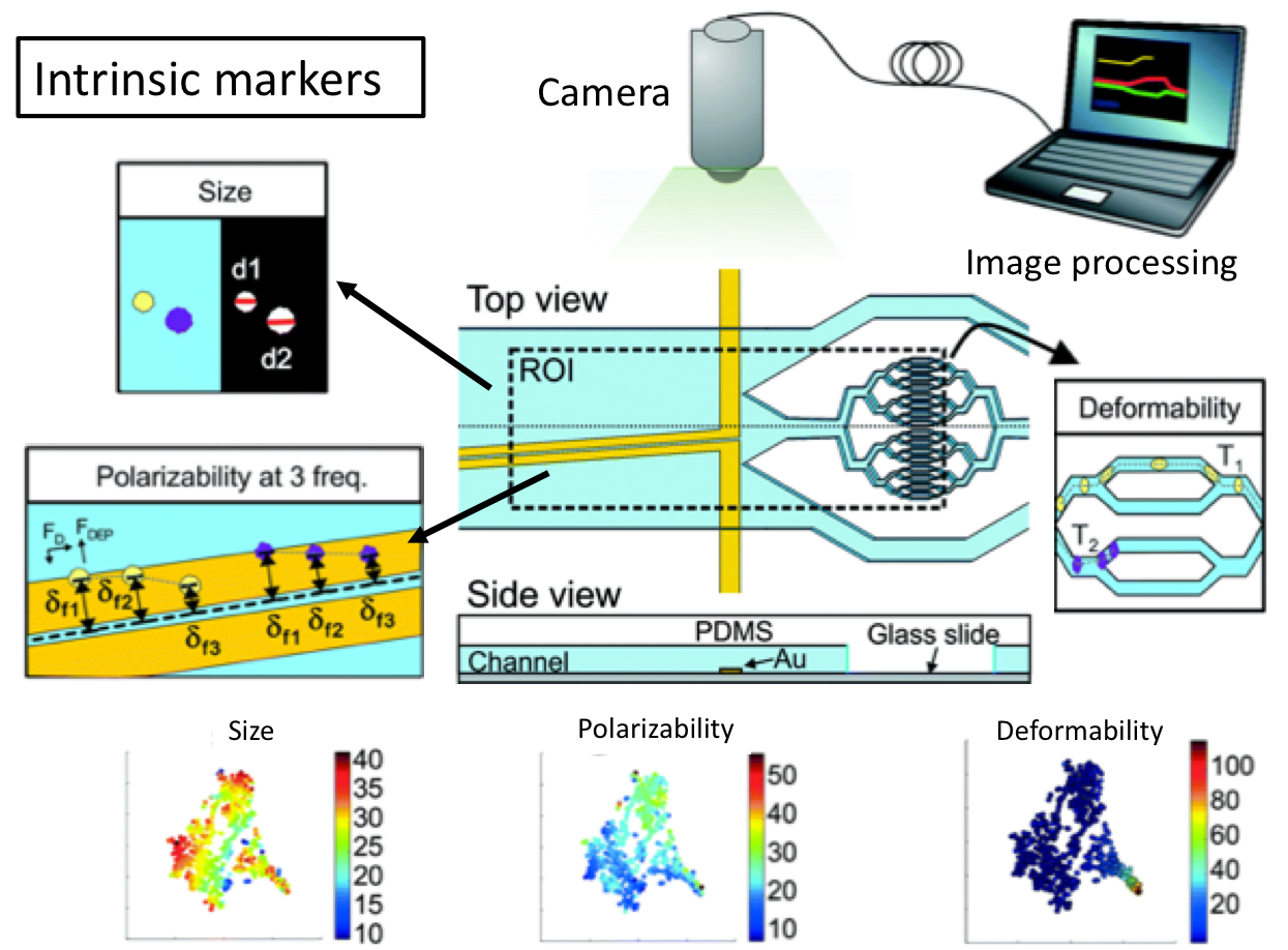
Figure 1. Multiparameter intrinsic cytometry combines different microfluidic modules on one substrate along with cell tracking to correlate per-cell information across modules for different intrinsic properties including size, polarizability, and deformability.


To download the full article for free* click the link below:

Multiparameter cell-tracking intrinsic cytometry for single-cell characterization
Apichitsopa, A. Jaffe, and J. Voldman
Lab Chip, 2018, Lab on a Chip Recent Hot Articles
DOI: 10.1039/C8LC00240A

*Article free to read until 31st August 2018

About the Webwriter

Burcu Gumuscu is a postdoctoral fellow in Herr Lab at UC Berkeley in the United States. Her research interests include development of microfluidic devices for quantitative analysis of proteins from single-cells, next generation sequencing,compartmentalized organ-on-chip studies, and desalination of water on the microscale.


Digg This
Reddit This
Stumble Now!
Share on Facebook
Bookmark this on Delicious
Share on LinkedIn
Bookmark this on Technorati
Post on Twitter
Google Buzz (aka. Google Reader)

Magnetic separation of circulating tumor cells…but it’s not what you think

Cancers typically originate in one organ yet can spread to distant regions of the body forming secondary tumours called metastases. This happens as cells from the primary tumour migrate into the circulatory system and then travel to other organs. These cells, which are a very rare population within the circulatory system, are termed circulating tumor cells (CTCs). Because of their role in cancer biology, they have garnered a lot of interest lately. Their detection and isolation present several analytical challenges. For one, they are the proverbial “needle in a haystack”, with counts on the order of one CTC for every billion blood cells. This has traditionally led to a paradox: these rare cells are best handled in microscale systems but the world-to-chip mismatch limits microfluidic devices from rapidly processing the large (> 5 mL) samples necessary. Second, recent studies have revealed CTCs to be very heterogeneous populations, limiting the use of surface markers for labelling and capturing a broad range of CTCs. Because there is much still to learn about CTCs, there’s also an interest in recovering viable CTCs for further analysis. In their recent report, Zhao et al. demonstrate a microfluidic device capable of enriching CTCs using magnetic separation. But it’s not that typical magnetophoretic separations you may be familiar with!

Magnetic separation of circulating tumor cells - nanoparticles

Rather than using magnetic particles to bind to surface antigens and eventually separate out CTCs, they capitalize on a phenomenon known as “negative magnetophoresis”. Cells are suspended within a uniformly magnetic medium and application of a non-uniform magnetic field results in a magnetic buoyant force. (This is akin to how negative dielectrophoresis exerts a force on particles in a non-uniform electric field.) The advantage of this method is that the “working principle applies to every non-magnetic material,” according to Prof. Leidong Mao. “Naturally,” he thought “it could apply to CTC enrichment.” However, despite previous work separating different cell populations with negative magnetophoresis, moving to CTC enrichment is not so straightforward. CTC enrichment is the most challenging separation. “All previous applications in our group are with cells at high concentrations,” mentioned Prof. Mao. The main challenge in developing the chip was trying to preserve the characteristics of an “ideal” CTC enrichment device; one that could process a significant amount of blood quickly, have a high recovery rate of CTCs, give reasonable purity of isolated CTCs, and retain cell integrity and viability for further analysis.

With this method, heterogeneous populations of CTCs can be enriched as selection is size dependent rather than based on expression of certain surface markers. This also avoids the costs associated with traditional magnetic labelling – typically used to label and deplete the millions of white blood cells. The device is capable of working at flow rates of 5-7 mL/hr, which is what is necessary to process an entire blood sample and can achieve high recovery rates (>90%). While the authors report purities that appear low (10-12%), they are working on improving purity. One strategy they suggest in their report is to follow the route of the iChip and combine size based separations with magnetic WBC depletion.

To read the full paper for free*, click the link below:

Label-free ferrohydrodynamic cell separation of circulating tumor cells
DOI: 10.1039/C7LC00680B (Paper) Lab Chip, 2017, 17, 3097-3111

—————-

About the Webwriter

Darius Rackus is a postdoctoral researcher at the University of Toronto working in the Wheeler Lab. His research interests are in combining sensors with digital microfluidics for healthcare applications.

*free to access until 14th December 2017


Digg This
Reddit This
Stumble Now!
Share on Facebook
Bookmark this on Delicious
Share on LinkedIn
Bookmark this on Technorati
Post on Twitter
Google Buzz (aka. Google Reader)

ICAS 2017 – International Congress on Analytical Sciences

ICAS 2017 is the 5 yearly international congress organised by the Chinese Chemical Society (CCS) and the International Union of Pure and Applied Chemistry (IUPAC). The event takes place at the Hainan International Convention and Exhibition Centre in Hainan, China between 5th and 8th May 2017. The theme of this year’s congress is “Analytical Chemistry – From Tool to Science”, which will contain sessions on advanced instrumental analysis, nanoscience and nanotechnology, biological and bioanalysis, environmental sciences, food safety, micro-analysis and microfluidic, sensors systems, mass spectrometry, separation and chromatography, spectrometry/spectroscopy, and electrochemical analysis. The Royal Society of Chemistry Journals Lab on a Chip, Analyst and Analytical Methods are very pleased to be supporting this event.

Visit the conference website for further details on themes and speakers and to submit your abstract.

Important Dates:
Abstract Submission Deadline: 28th February 2017

Early Bird Registration Deadline: 31st March 2017

 Register now to attend and present your work!

Digg This
Reddit This
Stumble Now!
Share on Facebook
Bookmark this on Delicious
Share on LinkedIn
Bookmark this on Technorati
Post on Twitter
Google Buzz (aka. Google Reader)

A new ‘on-chip’ immunoassay device

Professor Yarmash‘s lab at Rutgers University have developed a proof of concept microfluidic device, capable of running multiple immunoassays in parallel. The device allows 32 samples to be assayed simultaneously and multiple analytes can be tested in each sample.

As shown in the diagram below, each sample inlet has a bead trap that contains antibody-conjugated microbeads. These are commercially available, allowing virtually any analyte to be tested. The sample flows over the beads at an optimised rate, allowing the analytes to bind to their specific antibodies. A secondary antibody is added that binds to antibodies complexed to analytes, followed by a fluorescent tag that binds to the secondary antibody. The microbeads are then collected, placed in a 96 well plate, and analysed.

a) diagram and b) photo of the device; c) diagram of valve configuration and flow pathways during the assay; d) key steps in assay.

Device layout and assay principle

The authors assayed several proteins from an in vitro supernatant and their results corroborated well with a standard benchtop immunoassay. Compared to the benchtop standard, the device has significantly reduced sample consumption as well as large reductions in microbead and detection antibody consumption. It has comparable sensitivity to the benchtop standard and has a large working range, meaning that analytes present at different concentrations in the sample can be measured simultaneously. In addition to this, it is compatible with commercial reagents and analyte concentration can be quantified. Although previously published devices have addressed some of these characteristics, this the first example where they are combined into one device.

Moving on from their proof-of-concept study, the Yarmash group hopes to develop a device capable of in vivo measurements. One example they give is analysis of cerebrospinal fluid in rats, an important animal model in Alzheimer’s research, where immunoassays are currently limited by the small volumes available.


To download the full article for free* click the link below:

Development and validation of a microfluidic immunoassay capable of multiplexing parallel samples in microliter volumes
Mehdi Ghodbane, Elizabeth C. Stucky, Tim J. Maguire, Rene S. Schloss, David I. Shreiber, Jeffrey D. Zahn and Martin L. Yarmush
Lab Chip
, 2015,15, 3211-3221
DOI:
10.1039/C5LC00398A

—————-

About the webwriter

Claire Weston is a PhD student in the Fuchter Group, at Imperial College London. Her work is focused on developing novel photoswitches and photoswitchable inhibitors.

—————-

*Access is free until 19/11/2015 through a registered RSC account – click here to register

Digg This
Reddit This
Stumble Now!
Share on Facebook
Bookmark this on Delicious
Share on LinkedIn
Bookmark this on Technorati
Post on Twitter
Google Buzz (aka. Google Reader)

New YouTube Videos

Digg This
Reddit This
Stumble Now!
Share on Facebook
Bookmark this on Delicious
Share on LinkedIn
Bookmark this on Technorati
Post on Twitter
Google Buzz (aka. Google Reader)

Top 10 most accessed Lab on a Chip articles in June 2015

In June 2015, our most downloaded Lab on a Chip articles were:

Shia-Yen Teh, Robert Lin, Lung-Hsin Hung and Abraham P. Lee
DOI: 10.1039/B715524G

Ali Kemal Yetisen, Muhammad Safwan Akram and Christopher R. Lowe
DOI: 10.1039/C3LC50169H

Russell H. Cole, Niek de Lange, Zev J. Gartner and Adam R. Abate
DOI: 10.1039/C5LC00333D

Ching-Hui Lin, Yi-Hsing Hsiao, Hao-Chen Chang, Chuan-Feng Yeh, Cheng-Kun He, Eric M. Salm, Chihchen Chen, Ing-Ming Chiu and Chia-Hsien Hsu
DOI: 10.1039/C5LC00541H

Friedrich Schuler, Frank Schwemmer, Martin Trotter, Simon Wadle, Roland Zengerle, Felix von Stetten and Nils Paust
DOI: 10.1039/C5LC00291E

Mei He, Jennifer Crow, Marc Roth, Yong Zeng and Andrew K. Godwin
DOI: 10.1039/C4LC00662C

Joost F. Swennenhuis, Arjan G. J. Tibbe, Michiel Stevens, Madhumohan R. Katika, Joost van Dalum, Hien Duy Tong, Cees J. M. van Rijn and Leon W. M. M. Terstappen
DOI: 10.1039/C5LC00304K

A. Liga, A. D. B. Vliegenthart, W. Oosthuyzen, J. W. Dear and M. Kersaudy-Kerhoas
DOI: 10.1039/C5LC00240K

A. Wasay and D. Sameoto
DOI: 10.1039/C5LC00342C

Ivo Leibacher, Peter Reichert and Jürg Dual
DOI: 10.1039/C5LC00083A

Interesting read? Let us know your thoughts below.

And remember, you can submit directly to Lab on a Chip!

Digg This
Reddit This
Stumble Now!
Share on Facebook
Bookmark this on Delicious
Share on LinkedIn
Bookmark this on Technorati
Post on Twitter
Google Buzz (aka. Google Reader)

New YouTube Videos

Multiplexed Paper Analytical Device for Measuring Airborne Metal Particulates with Distance-Based Detection 


Transportation, Dispersion and Ordering of Dense Colloidal Assemblies by Magnetic Interfacial Rotaphoresis 


Gecko Gaskets for Self-Sealing and High Strength Reversible Bonding of Microfluidics 

Digg This
Reddit This
Stumble Now!
Share on Facebook
Bookmark this on Delicious
Share on LinkedIn
Bookmark this on Technorati
Post on Twitter
Google Buzz (aka. Google Reader)

New You Tube Videos

Generation of stable orthogonal gradients of chemical concentration and substrate stiffness in a microfluidic device 


Continuous Transfer of Liquid Metal Droplets Across a Fluid-Fluid Interface Within an Integrated Microfluidic Chip 


A flow-free droplet-based device for high throughput polymorphic crystallization 

Digg This
Reddit This
Stumble Now!
Share on Facebook
Bookmark this on Delicious
Share on LinkedIn
Bookmark this on Technorati
Post on Twitter
Google Buzz (aka. Google Reader)

Microfluidic Applications

Lab on a Chip Industry Workshop

Microfluidic Applications

Join our event on Facebook and find out who else is attending!

August 2-3 2014 in Dalian, China

This workshop focuses on the innovative developments in Lab-on-a-Chip technology and the applications of microfluidics in diagnostics, biological, material, pharmaceutical, and environmental sciences. For more information, please visit the official webpage.

Register now – deadline is July 15th 2014

Digg This
Reddit This
Stumble Now!
Share on Facebook
Bookmark this on Delicious
Share on LinkedIn
Bookmark this on Technorati
Post on Twitter
Google Buzz (aka. Google Reader)

Reprogrammable microfluidic chips

A microfluidic chip with channels that can be programmed then reset and reconfigured has been developed by scientists from France and Japan.

Water is dispensed into chip reservoirs. By selectively switching on electrodes, water is manipulated to carve out the channels

Water is dispensed into chip reservoirs. By selectively switching on electrodes, water is manipulated to carve out the channels

In recent years, scientists from across of the globe have developed a plethora of microfluidic chips to perform a variety of tasks, from PCR to cell sorting. However, a serious drawback of microfluidic technologies is that each application requires a unique arrangement of inlets, outlets and microchannels, so microfluidic chips are usually specific to one particular purpose. This, combined with the time-consuming and costly manufacturing processes required to construct microfluidic devices, makes the idea of a reprogrammable chip very attractive.

Read the full article here at Chemistry World.

Programmable and reconfigurable microfluidic chip
Raphaël Renaudot, et al.
Lab Chip, 2013, Accepted Manuscript
DOI: 10.1039/C3LC50850A, Paper

Digg This
Reddit This
Stumble Now!
Share on Facebook
Bookmark this on Delicious
Share on LinkedIn
Bookmark this on Technorati
Post on Twitter
Google Buzz (aka. Google Reader)