Archive for the ‘Uncategorized’ Category

Announcing our new Editor-in-Chief – Aaron Wheeler

We are delighted to announce that in January 2021, Professor Aaron Wheeler (University of Toronto, Canada) will take over as the new Editor-in-Chief for Lab on a Chip.

Professor Wheeler is Canada Research Chair in Microfluidic Bioanalysis at the University of Toronto. He has been recognized with a number of honors including the E.W.R. Steacie Memorial Fellowship from the Canadian National Sciences and Engineering Research Council, the Arthur F. Findeis Award from the American Chemical Society, and the Joseph Black Award from the Royal Society of Chemistry. Professor Wheeler’s research group develops microfluidic tools to solve problems in chemistry, biology, and medicine.

Aaron has been an Associate Editor for Lab on a Chip since 2013, and we are delighted that he will continue to handle submissions for Lab on a Chip. Aaron’s experience and knowledge of the journal and its community, combined with his academic experience, mean he will be a fantastic Editor-in-Chief for the journal.

“I’m honored to be taking up the duties of Editor-in-Chief for Lab on a Chip, “Perhaps uniquely relative to other journals, Lab on a Chip played a critical role in the birth and growth of a new field of research. Since the year 2000, the journal has been the ‘home’ for the best that the community has to offer, and I look forward to continuing that tradition going forward” says Aaron, “I am in awe of the diversity of topics that the Lab on a Chip community has made an impact in, be that cells or gels, soil or oil, or trees or bees, there is a Lab on a Chip for that. I can’t imagine a community that is more fun to be a part of.”

This news does of course mean that Professor Abe Lee will be standing down as Editor-in-Chief of Lab on a Chip. Professor Lee has served on the Editorial Board of Lab on a Chip for eleven years, first as an Editorial Board member, then an Associate Editor and finally as our Editor-in-Chief, and we are extremely grateful to Abe for his creativity and leadership throughout this period. We wish him all the best, and look forward to continuing to work with him as he moves to hold a position on our Advisory Board.

Read some of Aaron’s most recent publications below, free to access until 17th December 2020.

Direct loading of blood for plasma separation and diagnostic assays on a digital microfluidic device
Christopher Dixon, Julian Lamanna and Aaron R. Wheeler
Paper
Lab Chip, 2020, 20, 1845-1855

When robotics met fluidics
Junjie Zhong, Jason Riordon, Tony C. Wu, Harrison Edwards, Aaron R. Wheeler, Keith Pardee, Alán Aspuru-Guzik and David Sinton
Perspective
Lab Chip, 2020, 20, 709-716

Digital microfluidics and nuclear magnetic resonance spectroscopy for in situ diffusion measurements and reaction monitoring
Ian Swyer, Sebastian von der Ecken, Bing Wu, Amy Jenne, Ronald Soong, Franck Vincent, Daniel Schmidig, Thomas Frei, Falko Busse, Henry J. Stronks, André J. Simpson and Aaron R. Wheeler
Paper
Lab Chip, 2019, 19, 641-653

Towards a personalized approach to aromatase inhibitor therapy: a digital microfluidic platform for rapid analysis of estradiol in core-needle-biopsies
Sara Abdulwahab, Alphonsus H. C. Ng, M. Dean Chamberlain, Hend Ahmado, Lucy-Ann Behan, Hala Gomaa, Robert F. Casper and Aaron R. Wheeler
Paper
Lab Chip, 2017, 17, 1594-1602

Pre-concentration by liquid intake by paper (P-CLIP): a new technique for large volumes and digital microfluidics
Darius G. Rackus, Richard P. S. de Campos, Calvin Chan, Maria M. Karcz, Brendon Seale, Tanya Narahari, Christopher Dixon, M. Dean Chamberlain and Aaron R. Wheeler
Paper
Lab Chip, 2017, 17, 2272-2280

Digg This
Reddit This
Stumble Now!
Share on Facebook
Bookmark this on Delicious
Share on LinkedIn
Bookmark this on Technorati
Post on Twitter
Google Buzz (aka. Google Reader)

Emerging Investigator Series – Katherine Elvira

Lab on a Chip is delighted to introduce our most recent Emerging Investigator, Katherine Elvira!

Katherine received her undergraduate Master’s degree in Chemistry from Imperial College London in 2007. She started working in the field of microfluidics during her PhD (2012, Imperial College London), by building digital microfluidic platforms to perform automated chemical reactions. Katherine then moved to ETH Zürich (Switzerland) working firstly as a Postdoctoral Researcher and then as a Senior Scientist in the Institute for Chemical and Bioengineering. Since 2017, Katherine is the Canada Research Chair in New Materials and Techniques for Health Applications and an Assistant Professor in the Department of Chemistry at the University of Victoria, Canada. Katherine’s group currently develops microfluidic technologies to build bespoke artificial cells for the quantification of pharmacokinetic parameters in vitro. Katherine has recently presented this work at the Gordon Research Conference on Drug Metabolism (2019), is Co-Chair for the Gordon Research Conference on the Physics and Chemistry of Microfluidics (2023) and is a Scientific Mentor for the Creative Destruction Lab.

Read Dr Elvira’s Emerging Investigator paper* “A bespoke microfluidic pharmacokinetic compartment model for drug absorption using artificial cell membranes” and find out more about her and her research in the interview below.
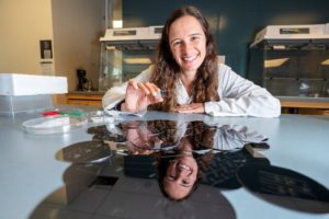
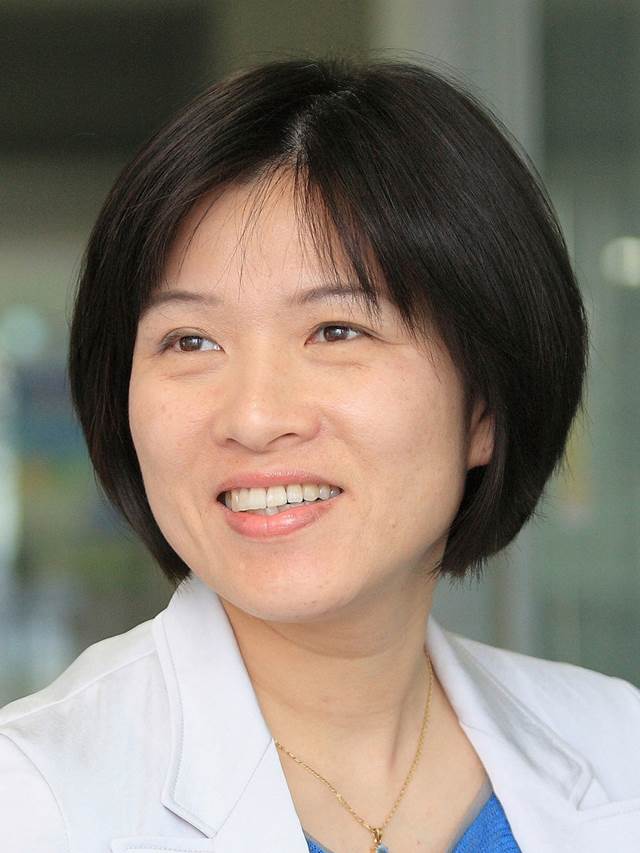
Katherine Elvira

Image credit: UVic Photo Services

Your recent Emerging Investigator Series paper focuses on a new type of pharmacokinetic compartment model for the prediction of drug absorption. How has your research evolved from your first article to this most recent article?

Funnily enough, my first ever article was a very early precursor to this work. We built a microfluidic platform for the formation of droplet interface bilayers (DIBs) in high-throughput. I didn’t work with DIBs again until I started as a Canada Research Chair at the University of Victoria, but they are the basis for the new type of pharmacokinetic compartment model that we show in the Emerging Investigator article. We can now make them mimic human cell membranes and hence they form the building blocks for the compartments in the pharmacokinetic compartment model. In between, my research involved making microfluidic devices for application in many different fields, such as drug discovery, organic chemistry and food science. I also spent some time investigating why microfluidic droplets rarely behave perfectly, and how we can mitigate this. I like that paper because we included a poster in the ESI which my group still uses in the lab to determine what is going wrong with their chips.

What aspect of your work are you most excited about at the moment?

I am really excited about some cool new artificial cell and tissue models that we are building, and how they can be used to model disease and drug behaviour in humans. My group is full of outstanding researchers, which makes it really easy to be excited about the work they are doing!

In your opinion, what is the biggest advantage of using your microfluidic platform over other methods?

It’s two things, really. Firstly, the fact that we are able to build networks of different compartments and artificial cell membranes on a chip. This allows us to build an in vitro model of the pathway that a drug would take in a human, from the intestine to the blood. And secondly, the fact that we can make these artificial cell membranes using phospholipids that are found in human cells. This makes our in vitro model quite biomimetic. In fact, we are able to predict molecular transport into cells three times better than the state-of-the-art in vitro commercial technique.

What do you find most challenging about your research?

Let’s face it, PDMS is awesome in some ways, but awful in others. I would love to find another material that has the advantages of PDMS, such as being cheap, transparent, and good for prototyping, but that has really stable and modifiable surface chemistry, and that we can mass produce for commercial applications.

In which upcoming conferences or events may our readers meet you?

I am part of the Technical Program Committee for MicroTAS 2020, so I will be at the online conference in October. And next year I am co-Vice Chair of the Gordon Research Conference on the Physics and Chemistry of Microfluidics, which will hopefully be taking place in Italy.

How do you spend your spare time?

I am really lucky to live in beautiful British Columbia on the West coast of Canada. During the winter months I get to go backcountry snowboarding in some of the most outstanding mountains in the world. In the summer, I switch my snowboard for a stand-up paddle board along the beaches in Victoria. I also love travelling, which I do a lot, both for work and for fun. I need my yoga classes to keep me relaxed, and have a slight obsession with cooking all the European foods that I miss from home.

Which profession would you choose if you were not a scientist?

I wanted to be an astronaut when I was younger. It still sounds cool but I am also happy staying on earth and being an academic, it’s a pretty great life.

Can you share one piece of career-related advice or wisdom with other early career scientists?

I don’t always find it easy being a woman in science, so I would encourage early career female scientists to persevere, we need diversity in academia. I would tell their male colleagues to be good allies.


*Dr Elvira’s paper is free to access with an RSC account for the next month.

Digg This
Reddit This
Stumble Now!
Share on Facebook
Bookmark this on Delicious
Share on LinkedIn
Bookmark this on Technorati
Post on Twitter
Google Buzz (aka. Google Reader)

Pioneers of Miniaturization Lectureship 2020: Open for Nominations


Lab on a Chip and Dolomite are proud to sponsor the fifteenth Pioneers of Miniaturization Lectureship, to honour and support the up and coming, next generation of scientists who have significantly contributed to the understanding or development of miniaturised systems.

This year’s Lectureship will be presented at the online µTAS 2020 event with the recipient receiving a prize of US$2,000. The µTAS 2020 organisers have made the decision that the meeting will be held as an online event, 4-9th October 2020, and as a result the deadline for nominations for the Pioneers of Miniaturization Lectureship is 31st July 2020.

The Lectureship consists of the following elements:

  • A prize of US$2,000. No other financial contribution will be offered
  • A certificate recognising the winner of the lectureship
  • The awardee is required to give a short lecture at the online µTAS 2020 event

Eligibility Criteria

To be eligible for the lectureship, candidates must:

  • Have completed their PhD
  • Be actively pursuing an independent research career on miniaturised systems.
  • Be at an early-mid career stage of their independent career (typically this will be within 15 years of completing their PhD, but appropriate consideration will be given to those who have taken a career break or followed a different study path).

Nomination process

To be considered for the 2020 lectureship, the following must be sent to the Editorial Office

  • A letter of recommendation with the candidate’s accomplishments and why the lectureship is deserved.
  • The nominee must be aware that he/she has been nominated for this lectureship.
  • A complete nomination form (includes list of the candidate’s relevant publications or recent work, candidate’s scientific CV, and full contact details)
  • Nominations from students and self-nominations are not permissible.

Selection criteria and judging process

  • Nominations must be made via email to loc-rsc@rsc.org using the Dolomite/Lab on a Chip Pioneers of Miniaturization Lectureship nomination form and a letter of recommendation.
  • The decision on the winner of the lectureship will be made by a panel of judges comprising a representative from Dolomite and members from the Lab on a Chip Editorial Board, coordinated by the Executive Editor of Lab on a Chip.
  • The award is for outstanding contributions to the understanding or development of miniaturised systems. This will be judged mainly through their top 1-3 papers and/or an invention documented by patents/or a commercial product. Awards and honorary memberships may also be considered.


Nomination Deadline: 31 July, 2020 


Download Nomination form here 

Dolomite Microfluidics a leading provider of microfluidics-based solutions for a range of applications including drug encapsulation, droplet manufacture and particle generation. They manufacture complete systems as well as individual modular components to balance ease of use with flexibility.

Digg This
Reddit This
Stumble Now!
Share on Facebook
Bookmark this on Delicious
Share on LinkedIn
Bookmark this on Technorati
Post on Twitter
Google Buzz (aka. Google Reader)

Emerging Investigators Series – Ye Ai


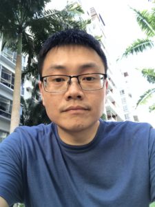
Dr. Ye Ai is currently an Associate Professor at Singapore University of Technology and Design (SUTD). He obtained his B.S. in Mechanical Engineering from Huazhong University of Science and Technology (China) in 2005 and his Ph.D. in Mechanical and Aerospace Engineering from Old Dominion University (USA) in 2011. Prior to joining SUTD as an assistant professor in 2013, he worked as a postdoctoral researcher at the Bioscience Division of Los Alamos National Laboratory from June 2011 to January 2013. He was a visiting scholar at Massachusetts Institute of Technology (MIT) from August 2014 to July 2015. He was promoted to associate professor with tenure in September 2019. Dr. Ai’s research interest focuses on developing novel microfluidic technologies for particle/cell manipulation and single cell analysis. His research team is also striving to translate their innovative microfluidic technologies to commercial market through collaborations with industry.

Read Dr Ai’s recent Emerging Investigator Series paper: Microfluidic impedance cytometry device with N-shaped electrodes for lateral position measurement of single cells/particles in the most recent issue of Lab on a Chip, and find out more about him and his work below.


  1. Your recent Emerging Investigator Series paper focuses on measuring the lateral position of single cells or particles. How has your research evolved from your first article to this most recent article?

My first research article when I was a PhD student was to develop a finite element model with dynamically deformed mesh that can simulate the transient motion of finite-size particles in microscale fluid flows. My PhD research mainly focused on electrokinetics for manipulating particles, cells and ions in micro/nanoscale. My postdoctoral training at the Bioscience Division of Los Alamos National Laboratory exposed me to a lot of biological problems, in particular the need in high-throughput cellular analysis at the single cell level. My previous research experience has somehow shaped my current research focus into single cell manipulation and analysis using novel microfluidic technologies when I become an independent principal investigator in Singapore.

  1. What aspect of your work are you most excited about at the moment?

I am most excited to apply our developed microfluidic technologies for solving real biomedical problems and enabling new biological studies. As an example, in 2017 my team published our single cell sorting technology using a highly focused acoustic beam in Lab Chip (DOI: 10.1039/c7lc00678k). Later, I was approached by quite a number of research teams worldwide who wanted to try our sorting technology. These email communications have encouraged me to apply our developed prototype for real biomedical problems. Right now, I have established collaboration with a few biomedical research institutes in Singapore and we have found that our sorting technology is not causing any cell damage, which is however challenging for conventional FACS machine. We currently have the idea to commercialize this single cell sorting technology. Let us see what is going to happen in the next few years.

  1. In your opinion, what are the key considerations when designing a microfluidic platform for real-time measurements?

My research team is currently developing both hydrodynamic and acoustic cell sorting platforms. The conventional way to quantify the sorting performance (e.g. purity and recovery) is to run additional cell analysis of collected samples, typically using a flow cytometer. In this work (DOI: 10.1039/c9lc00819e), we designed and validated a new impedance cytometry device that enables the measurement of the lateral positions and physical properties of individual particles and cells. The integration of this new device with any cell sorting platform will allow the evaluation of the sorting performance to be implemented in the same system.

The key consideration of integrating these real-time measurements really depends on whether there is a critical need. But I do see a lot of sorting applications need these real-time, in-line measurements for the purposes of quality control and workflow simplification. And any integration will somehow complicate the system and increase the cost, so the other key consideration is the ease of integration. Integration of electronics is generally easier compared to optics, and we are measuring intrinsic biophysical properties rather than labelling approaches; therefore, I do see great opportunities to integrate our new impedance cytometry device with a variety of cell sorting platforms.

  1. What do you find most challenging about your research?

The microfluidics and Lab on a chip research area is interdisciplinary in nature. My challenge is always to find the right people (e.g. students, postdoctoral fellows, collaborators) and secure sufficient resources to work on real impactful research problems.

  1. How do you spend your spare time?

I am trying to make a balance between work and personal life, so I mainly spend my spare time with my family members, especially my second kid is only 8 months old. I also spend some of my spare time to do physical exercise, which can help relax and leave some time for free thinking.

  1. Which profession would you choose if you were not a scientist?

I rarely thought about this before. Perhaps I would choose to be a doctor.

  1. Can you share one piece of career-related advice or wisdom with other early career scientists?

Based on my own experience, it is important to define a unique research domain based on your own expertise and the surrounding research ecosystem when early career scientists start their independent research. It is also wise to have a clear vision about what you want to achieve in the next five years.

Digg This
Reddit This
Stumble Now!
Share on Facebook
Bookmark this on Delicious
Share on LinkedIn
Bookmark this on Technorati
Post on Twitter
Google Buzz (aka. Google Reader)

What are the challenges to sample-in-answer-out technologies in clinical settings?

The boom in microfluidic total analysis systems is spurred by the use of microscale fabrication techniques. In 1999, Agilent Technologies introduced a coin-size device called “LabChip” to the market for the analysis of DNA. The timing of the market release of this device was remarkable as it benefited from the hype of the Human Genome Project. The LabChip has drawn considerable attention from both academia and industry. As a result, the shrink of chemistry labs into coin-sized microfluidic devices has evolved more from technological push than from market pull. Technology-driven development pathway brought challenges, especially for the clinical integration of microfluidic devices due to the lack of standards, focus, and communication between academia and clinics.

A recent Lab on a Chip paper from Martyn Boutelle’s Lab addresses this issue. The authors identify the problem from a well-addressed microdialysis perspective. They state that “taking a microfluidic system into clinical environment brings lots of challenges, not least that during setups and developments the very low the flow rates used in combination with microdialysis means that leaks and misdirection of flows are very hard to see.” The authors define the most significant challenge as the development of a device that’s robust enough to be used by and provide enough information to, the clinical team without micromanagement by experts.

The authors attack the problem by creating a sensor-based online system associated with electrochemical measurement, which would be able to analyze the sample in a miniaturized platform continuously. They developed the microfluidic sensors and chip, but they wanted to increase the use of their technology in real-world scenarios by non-experts, so they looked into ways to introduce more precision and control to the platform. The authors combined their technology with LabSmith microfluidic components and constructed breadboard like layouts for typical lab protocols. The authors add that “the main surprise was the ability to bring the rigor of an analytical laboratory into unusual places such as abattoirs, surgical theatres and public transport!”

In this work and the previous work of the authors, the performance of 3-D printed chips was compared to the PDMS ones. The authors found that PDMS material is much more vulnerable than the 3D print outs when frequently handled. The sensor attached to the chip is programmed to calibrate in regular intervals (e.g., every three hours) in single or multiphase flow conditions. Authors describe the advantage of the system that ”remote access to the scripts allows interaction with the system without the requirement of a highly skilled person being right next to it, which in the context of surgical theatres and hospital wards is a distinct advantage. If the codes and scripts are available to less skilled personnel, they are still able to interact and use the system and by making the system more user-friendly a wider audience and more enthusiasm is generated for the product, increasing interest, uptake, and use.”

The authors would like to improve the platform further by making it wearable since it already has grounds for such an operation with wireless sensors. The next thing to be improved in the system is the feed. At this moment, the syringes have to be regularly refilled. This might not be a problem in the laboratory; however, monitoring can last for days in a clinical setting, and periodically refilling the syringes may lead to noise artifacts. Another improvement could be the ease of operation and troubleshooting when, e.g., a tubing becomes blocked in the middle of the measurement when the user is short on space and time.

Lastly, the authors think that this pioneering platform can help shape the future in the market by giving more people access to an area of science that was previously highly skilled, whilst maintaining analytical robustness. This will be the start to break the barrier between academia-made devices and clinical settings.


To download the full article for free* click the link below:

Clinical translation of microfluidic sensor devices: focus on calibration and analytical robustness

Sally A. N. Gowers, Michelle L. Rogers, Marsilea A. Booth, Chi L. Leong, Isabelle C. Samper, Tonghathai Phairatana, Sharon L. Jewell, Clemens Pahl, Anthony J. Strong and Martyn G. Boutelle, Lab Chip, 2019, Lab on a Chip Hot Articles

DOI: 10.1039/C9LC00400A

*access is free with an RSC account (free to register)


About the Webwriter

Burcu Gumuscu is a researcher in Mesoscale Chemical Systems Group at the University of Twente in the Netherlands. Her research interests include the development of microfluidic devices for quantitative analysis of proteins from single-cells, next-generation sequencing, compartmentalized organ-on-chip studies, and desalination of water on the microscale.

Digg This
Reddit This
Stumble Now!
Share on Facebook
Bookmark this on Delicious
Share on LinkedIn
Bookmark this on Technorati
Post on Twitter
Google Buzz (aka. Google Reader)

We are delighted to announce that Hang Lu is the 2019 winner of the “Pioneers of Miniaturization” Lectureship!

The 14th “Pioneers of Miniaturization” Lectureship, sponsored by Dolomite and Lab on a Chip , is for early to mid-career scientists who have made extraordinary or outstanding contributions to the understanding or development of miniaturised systems.

The 2019 “Pioneers of Miniaturization” Lectureship will be presented to Professor Lu at the µTAS 2019 Conference in Basel, Switzerland, being held on 27-31 October 2019. Professor Lu will receive a certificate, a monetary award and will give a short lecture during the conference.

Many congratulations to Professor Hang Lu on this achievement from the Lab on a Chip Team!

About the Winner

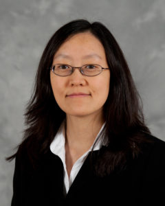
Professor Hang Lu is the Love Family Professor, School of Chemical and Biomolecular Engineering, Georgia Institute of Technology, USA.

Professor Lu earned her PhD in Chemical Engineering from Massachusetts Institute of Technology, USA in 2003. After a postdoctoral fellowship with Professor Cornelia I. Bargmann, at University of California San Francisco and the Rockefeller University, she was appointed as an Assistant Professor at School of Chemical and Biomolecular Engineering, Georgia Institute of Technology.

In recognition of her outstanding achievements, Professor Lu has received numerous awards and international recognition, including being invited to join Board of Directors, Chemical and Biological Microsystems Society, invited to present at the Nobel Symposium on Microfluidics (2017) and the National Academy of Sciences’ Kavli Frontiers of Science Symposia (2014, 2012, 2009), awarded the ACS Analytical Chemistry Young Innovator Award, Chemical and Biological Microsystems Society (2013), Council of Systems Biology in Boston (CSB2) Prize in Systems Biology (2011), a National Science Foundation CAREER award (2010), an Alfred P. Sloan Foundation Research Fellowship (2009), a DARPA Young Faculty Award (2007), a DuPont Young Professor Award (2006), the Saville Lectureship of Princeton University (2013), the H. C. Van Ness Award Lectures of Rensselaer Polytechnic Institute (2011), and is a fellow of the American Institute for Medical and Biological Engineering (AIMBE) and  a fellow of the American Association for the Advancement of Science (AAAS). She has authored more than 140 peer-reviewed publications and has served on the Editorial Board of Lab on a Chip as Associate Editor since 2017. She is currently the director of the Interdisciplinary Bioengineering Program, and the associate director of the NSF-Simons Foundation supported Southeast Center for Mathematics and Biology, Georgia Institute of Technology.

Professor Lu has pioneered the use of microfluidic systems for imaging and performing genetic studies with small organisms, primarily the nematode C. elegans. In a series of studies published since 2008 she established a set of technologies to streamline imaging, phenotyping, and sorting of C. elegans based on features that are difficult to distinguish and discern by human eyes. The throughput of these technologies were often 1,000 times that of conventional approaches. Professor Lu’s technology has enable faster and more accurate experiments and revolutionized how genetic screens and high-content imaging experiments are done currently in other scientists’ labs. In parallel, her lab has also engineered micro systems for high-content experiments with cells, aggregates, organoids, and embryos to extract high-dimensional information for systems biology studies.

The Lu group performs research at the interface of engineering and biology. They engineer automated microfluidic systems, microscopy tools, and image imformatic technologies to address questions in neuroscience, cell biology, and biotechnology that are difficult to answer using conventional techniques. Applied to the study of fundamental biological questions, these new techniques allow the Lu group to gather large-scale quantitative data about complex systems.

Learn about the Lu group online

Digg This
Reddit This
Stumble Now!
Share on Facebook
Bookmark this on Delicious
Share on LinkedIn
Bookmark this on Technorati
Post on Twitter
Google Buzz (aka. Google Reader)

Lab on a Chip thematic collection on droplet-based single cell sequencing

Lab on a Chip is delighted to share with you our Thematic Collection on droplet-based single-cell sequencing.

The droplet-based single-cell sequencing field is advancing very rapidly. Large numbers of studies are underway to collect and explore the new information that is now accessible with single-cell RNA-seq. Improvements to the microfluidics are also advancing rapidly. This collection of papers and reviews focusses on the between the technological advancements and high impact applications of droplet-based single-cell sequencing.

This topical and exciting collection is collated by Thought leader Dave Weitz and the Lab on a Chip Editorial Board. The collection is introduced in a perspective on single cell sequencing by the Thought leader Dave Weitz, and in two editorials, one on “InDrops and Drop-seq” by Allon Klein and Evan Macosko and one on “an engineer and business person’s perspective” by businessman and engineer Mark Gilligan.

Read the full collection at: http://rsc.li/drop-sc-seq

Below is a selection of content highlights featured in the collection. In addition, all papers are free to read until 31st May*

Perspective

Droplet-based single cell RNAseq tools: a practical guide

Robert Salomon, David Gallego-Ortega, et al.

Critical Review

Finding a helix in a haystack: nucleic acid cytometry with droplet microfluidics

Iain C. Clark and Adam R. Abate

Paper

High throughput gene expression profiling of yeast colonies with microgel-culture Drop-seq

Leqian Liu, Adam R. Abate, et al.

Paper

Simplified Drop-seq workflow with minimized bead loss using a bead capture and processing microfluidic chip

Marjan Biočanin, Bart Deplancke, et al.

Lab on a Chip is the leading journal publishing significant and original work related to miniaturisation, at the micro- and nano-scale, of interest to a multidisciplinary readership with an Journal Impact Factor of 5.995**. The journal is guided by Editor-in-Chief Abraham (Abe) Lee (University of California, Irvine) who is supported by our team of Associate Editors (Yoon-Kyoung Cho, Petra Dittrich, Hang Lu, Jianhua Qin, Manabu Tokeshi, Joel Voldman and Aaron Wheeler).

We hope you enjoy reading the papers within this Thematic Collection and we welcome future submissions on droplet-based single-cell sequencing.

Dolomite/Lab on a Chip Pioneers of Miniaturization Lectureshipdeadline approaching-nominate a colleague now!

Organ-on a-chip systems- translating concept into practice thematic collectionSubmit now

Organ-,body- and disease-on-a-chip thematic collectionRead now

Personalised medicine:liquid biopsyRead now

Lab on a Chip Emerging Investigator Series Apply now

Digg This
Reddit This
Stumble Now!
Share on Facebook
Bookmark this on Delicious
Share on LinkedIn
Bookmark this on Technorati
Post on Twitter
Google Buzz (aka. Google Reader)

Lab on a Chip thematic collection on personalised medicine-liquid biopsy

Lab on a Chip is delighted to share with you our Thematic Collection on personalised medicine-liquid biopsy.

This collection of papers and reviews focusses on the interface between the technological advancements and high impact applications of liquid biopsy technologies. This collection is collated by Thought Leaders Mehmet Toner and Stefanie Jeffrey and the Lab on a Chip Editorial Board and is introduced in an Editorial by the Thought Leaders on Liquid biopsy: a perspective for probing blood for cancer

Read the full collection at: http://rsc.li/liquid-biopsy

Below is a selection of content highlights featured in the collection. In addition, all papers are free to read until 31st May*

Tutorial Review

Cancer diagnosis: from tumor to liquid biopsy and beyond

Ramanathan Vaidyanathan, Chwee Teck Lim, et al.

Critical Review

Circulating tumor DNA and liquid biopsy: opportunities, challenges, and recent advances in detection technologies

Lena Gorgannezhad, Nam-Trung Nguyen, Muhammad J. A. Shiddiky et al.

Paper

Dynamic CTC phenotypes in metastatic prostate cancer models visualized using magnetic ranking cytometry

Leyla Kermanshah, Shana O. Kelley et al.

Paper

An ultrasensitive test for profiling circulating tumor DNA using integrated comprehensive droplet digital detection

Chen-Yin Ou, Timothy J. Abram, Weian Zhao et al.

Paper

Cancer marker-free enrichment and direct mutation detection in rare cancer cells by combining multi-property isolation and microfluidic concentration

Soo Hyeon Kim, Teruo Fujii et al.

Paper

Urine-based liquid biopsy: non-invasive and sensitive AR-V7 detection in urinary EVs from patients with prostate cancer

Hyun-Kyung Woo, Hong Koo Ha, Yoon-Kyoung Cho et al.


Lab on a Chip is the leading journal publishing significant and original work related to miniaturisation, at the micro- and nano-scale, of interest to a multidisciplinary readership with an Journal Impact Factor of 5.995**. The journal is guided by Editor-in-Chief Abraham (Abe) Lee (University of California, Irvine) who is supported by our team of Associate Editors (Yoon-Kyoung Cho, Petra Dittrich, Hang Lu, Jianhua Qin, Manabu Tokeshi, Joel Voldman and Aaron Wheeler).

We hope you enjoy reading the papers within this Thematic Collection!

Keep up to date with Lab on a Chip throughout the year by signing up for free table of contents alerts and monthly e-newsletters.

Dolomite/Lab on a Chip Pioneers of Miniaturization Lectureshipdeadline approaching-nominate a colleague now!

Organ-on a-chip systems- translating concept into practice thematic collectionSubmit now

Organ-,body- and disease-on-a-chip thematic collectionRead now

Droplet-based single-cell sequencingRead now

Lab on a Chip Emerging Investigator Series Apply now

Digg This
Reddit This
Stumble Now!
Share on Facebook
Bookmark this on Delicious
Share on LinkedIn
Bookmark this on Technorati
Post on Twitter
Google Buzz (aka. Google Reader)

What we know about cancer tumors

Cancer tumors are a lot more complex than we think: besides cancer cells, supportive tissue cells, fat, and even immune cells can be found in a tumor. Combined crosstalk in between these cell groups influences the way the tumor develops or responses to drug treatment. On the other hand, the majority of what we know about cancer tumors has been acquired by studying cell ensembles. Recent strides to improve our understanding of cancer revealed that we have long been missing the stochastic interactions and rare events due to ensemble-average measurements. We can unveil how these cell groups work together and how the rare events change the fate of a tumor thanks to single-cell analysis techniques.

Single cells can be identified by extrinsic and intrinsic markers. Extrinsic markers are definitive of genetic and proteomic states of a cell. Flow cytometry and mass spectrometry have been the workhorse of extrinsic marker analysis, where genetic or proteomic materials are often fluorescently labeled for detection. With these techniques, multiplexed analysis of thousands of cells can be employed simultaneously. Intrinsic markers include size, shape, density, optical, mechanical, and electrical properties which do not require labelling. Microfluidic techniques provide with a plethora of different functionalities to sort the cells based on intrinsic markers. Combination of both extrinsic and intrinsic data advances our understanding of how cell heterogeneity is reflected in cell-to-cell variations in tumor development and drug-response. Although many powerful methods are available for determining extrinsic markers, not many techniques can gather information about a panel of different intrinsic markers.

A recent study from Biological Microtechnology and BioMEMS group at MIT represents an important microfluidic approach for the development of multiparameter intrinsic cytometry tool. The approach includes several different microfluidic modules combined with microscope imaging and image processing by machine learning. Separate modules measuring cell size, deformability, and polarization can be combined and organized within the tool (Figure 1). (i) Size module detects the cell size optically in a flow through system. Cell size module is necessary to separate different cell types that can give important cues about disease state. (ii) In deformability module, cells pass through narrow channels, and their transit time defines the deformability. Cell deformability gives cues about cytoskeletal and nuclear changes associated with cancer progression. (iii) In the polarization module, dielectrophoretic force at a fixed frequency is applied on cells driven by opposing hydrodynamic forces. Cells approach coplanar electrodes with different equilibrium positions depending on their polarizability. Cell polarizability allows for distinguishing subtle changes in biological phenotypes. As a proof-of-concept work, drug-induced structural changes in cells were detected for the first time using five different intrinsic markers, including size, deformability, and polarizability at three frequencies. The authors indicate that this powerful tool can further be equipped with visual readout capabilities, such as deterministic lateral displacement array, inertial microfluidics, acoustophoresis, optical techniques.

Figure 1. Multiparameter intrinsic cytometry combines different microfluidic modules on one substrate along with cell tracking to correlate per-cell information across modules for different intrinsic properties including size, polarizability, and deformability.


To download the full article for free* click the link below:

Multiparameter cell-tracking intrinsic cytometry for single-cell characterization
Apichitsopa, A. Jaffe, and J. Voldman
Lab Chip, 2018, Lab on a Chip Recent Hot Articles
DOI: 10.1039/C8LC00240A

*Article free to read until 31st August 2018

About the Webwriter

Burcu Gumuscu is a postdoctoral fellow in Herr Lab at UC Berkeley in the United States. Her research interests include development of microfluidic devices for quantitative analysis of proteins from single-cells, next generation sequencing,compartmentalized organ-on-chip studies, and desalination of water on the microscale.


Digg This
Reddit This
Stumble Now!
Share on Facebook
Bookmark this on Delicious
Share on LinkedIn
Bookmark this on Technorati
Post on Twitter
Google Buzz (aka. Google Reader)

Magnetic separation of circulating tumor cells…but it’s not what you think

Cancers typically originate in one organ yet can spread to distant regions of the body forming secondary tumours called metastases. This happens as cells from the primary tumour migrate into the circulatory system and then travel to other organs. These cells, which are a very rare population within the circulatory system, are termed circulating tumor cells (CTCs). Because of their role in cancer biology, they have garnered a lot of interest lately. Their detection and isolation present several analytical challenges. For one, they are the proverbial “needle in a haystack”, with counts on the order of one CTC for every billion blood cells. This has traditionally led to a paradox: these rare cells are best handled in microscale systems but the world-to-chip mismatch limits microfluidic devices from rapidly processing the large (> 5 mL) samples necessary. Second, recent studies have revealed CTCs to be very heterogeneous populations, limiting the use of surface markers for labelling and capturing a broad range of CTCs. Because there is much still to learn about CTCs, there’s also an interest in recovering viable CTCs for further analysis. In their recent report, Zhao et al. demonstrate a microfluidic device capable of enriching CTCs using magnetic separation. But it’s not that typical magnetophoretic separations you may be familiar with!

Magnetic separation of circulating tumor cells - nanoparticles

Rather than using magnetic particles to bind to surface antigens and eventually separate out CTCs, they capitalize on a phenomenon known as “negative magnetophoresis”. Cells are suspended within a uniformly magnetic medium and application of a non-uniform magnetic field results in a magnetic buoyant force. (This is akin to how negative dielectrophoresis exerts a force on particles in a non-uniform electric field.) The advantage of this method is that the “working principle applies to every non-magnetic material,” according to Prof. Leidong Mao. “Naturally,” he thought “it could apply to CTC enrichment.” However, despite previous work separating different cell populations with negative magnetophoresis, moving to CTC enrichment is not so straightforward. CTC enrichment is the most challenging separation. “All previous applications in our group are with cells at high concentrations,” mentioned Prof. Mao. The main challenge in developing the chip was trying to preserve the characteristics of an “ideal” CTC enrichment device; one that could process a significant amount of blood quickly, have a high recovery rate of CTCs, give reasonable purity of isolated CTCs, and retain cell integrity and viability for further analysis.

With this method, heterogeneous populations of CTCs can be enriched as selection is size dependent rather than based on expression of certain surface markers. This also avoids the costs associated with traditional magnetic labelling – typically used to label and deplete the millions of white blood cells. The device is capable of working at flow rates of 5-7 mL/hr, which is what is necessary to process an entire blood sample and can achieve high recovery rates (>90%). While the authors report purities that appear low (10-12%), they are working on improving purity. One strategy they suggest in their report is to follow the route of the iChip and combine size based separations with magnetic WBC depletion.

To read the full paper for free*, click the link below:

Label-free ferrohydrodynamic cell separation of circulating tumor cells
DOI: 10.1039/C7LC00680B (Paper) Lab Chip, 2017, 17, 3097-3111

—————-

About the Webwriter

Darius Rackus is a postdoctoral researcher at the University of Toronto working in the Wheeler Lab. His research interests are in combining sensors with digital microfluidics for healthcare applications.

*free to access until 14th December 2017


Digg This
Reddit This
Stumble Now!
Share on Facebook
Bookmark this on Delicious
Share on LinkedIn
Bookmark this on Technorati
Post on Twitter
Google Buzz (aka. Google Reader)