Archive for the ‘Hot Articles’ Category

HOT article: Digital microfluidics for measuring glucose in human blood serum

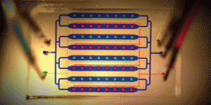
Hywel Morgan and colleagues at Sharp Laboratories of Europe, the University of Southampton and Sharp Corporation, Japan, demonstrate a large area digital microfluidic array in this HOT article.

Using a thin film transistor (TFT) array rather than the traditional patterned electrodes usually used in electrowetting on dielectric (EWOD) devices, the team developed active matrix electrowetting on dielectric (AM-EWOD) devices. The TFT array enables each of the many thousand electrodes to be individually addressable, and the array is ‘fully reconfigurable and can be programmed to support multiple simultaneous operations’.

Read how the device can be used for measuring glucose in human blood serum in the full article (it’s free to access for four weeks*!):

Programmable large area digital microfluidic array with integrated droplet sensing for bioassays
B. Hadwen, G. R. Broder, D. Morganti, A. Jacobs, C. Brown, J. R. Hector, Y. Kubota and H. Morgan
DOI: 10.1039/C2LC40273D

*Following a simple registration.

Digg This
Reddit This
Stumble Now!
Share on Facebook
Bookmark this on Delicious
Share on LinkedIn
Bookmark this on Technorati
Post on Twitter
Google Buzz (aka. Google Reader)

Sharp images of turbid flows in microfluidic devices

Pietro Ferraro and colleagues from three Italian research institutes have used digital holography to image objects in turbid flowing media in microfluidic devices.  Although many current techniques are able to provide in situ images of liquids flowing in microfluidic devices, they are limited to cases of clear liquids  with relatively stable flows.  Due to the Doppler effect of flowing colloidal particles the researchers were able to reconstruct digital holographic images, and demonstrated this with milk:

As with all our HOT articles, this one is free to access for 4 weeks following a simple registration:

Microscopy imaging and quantitative phase contrast mapping in turbid microfluidic channels by digital holography
Melania Paturzo, Andrea Finizio, Pasquale Memmolo, Roberto Puglisi, Donatella Balduzzi, Andrea Galli and Pietro Ferraro
DOI: 10.1039/C2LC40114B

Digg This
Reddit This
Stumble Now!
Share on Facebook
Bookmark this on Delicious
Share on LinkedIn
Bookmark this on Technorati
Post on Twitter
Google Buzz (aka. Google Reader)

Measuring migration of individual cells

Yanyi Huang and colleagues at Peking University have developed a microfluidic-based assay to allow them to quantitatively measure single cell migration speeds.  Existing methods for determining cell migration are abundant, as the process is important for processes such as cancer metastasis, embryonic development and wound healing, but it is very difficult to distinguish between migrating and proliferating cells in current assays.

Using their migration assay, Huang and colleagues were able to show that HUVEC migration is epigenetically regulated:

Live cell imaging analysis of the epigenetic regulation of the human endothelial cell migration at single-cell resolution
Chunhong Zheng, Zhilong Yu, Ying Zhou, Louis Tao, Yuhong Pang, Tao Chen, Xiannian Zhang, Haiwei Qiu, Hongwei Zhou, Zitian Chen and Yanyi Huang
DOI: 10.1039/C2LC40192D

As with all our HOT articles, this one is free to access for 4 weeks following a simple registration.

Also check out the video showing their device in operation:

Digg This
Reddit This
Stumble Now!
Share on Facebook
Bookmark this on Delicious
Share on LinkedIn
Bookmark this on Technorati
Post on Twitter
Google Buzz (aka. Google Reader)

Submicron particle focussing, 3D capillary networks & monitoring cell signalling in Issue 16

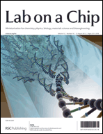
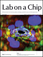
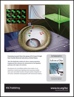
We’ve got some great artwork on the covers of Issue 16,  on topics from particle focusing to creating 3D capillary networks and cell signalling.

Lateral migration and focusing of colloidal particles and DNA molecules under viscoelastic flowOn the outside front cover we have a HOT article from Ju Min Kim et al who have achieved viscoelasticity-driven focusing of particles as small as 200 nm – a technique which had previously only been achieved with micron-sized particles.  The team also showed that DNA focusing is significantly enhanced by medium viscoelasticity and that the focussing of both the colloidal particles and DNA is dependant on length.

Lateral migration and focusing of colloidal particles and DNA molecules under viscoelastic flow
Jae Young Kim, Sung Won Ahn, Sung Sik Lee and Ju Min Kim
DOI: 10.1039/C2LC40147A

In vitro formation and characterization of a perfusable three-dimensional tubular capillary network in microfluidic devicesOn the inside front cover is another HOT article from Noo Li Jeon and colleagues at Seoul University who have designed a 3D array of perfusable capillaries from HUVECs.  The the capillary network can be grown on the microfluidic device in 3-4 days and the authors hope will not only be useful for basic angiogenesis research but also drug screening applications.

In vitro formation and characterization of a perfusable three-dimensional tubular capillary network in microfluidic devices
Ju Hun Yeon, Hyun Ryul Ryu, Minhwan Chung, Qing Ping Hu and Noo Li Jeon
DOI: 10.1039/C2LC40131B

Microfluidically-unified cell culture, sample preparation, imaging and flow cytometry for measurement of cell signaling pathways with single cell resolutionLast but not least, on the back cover is research from Anup Singh and colleagues at Sandia National Laboratory who have developed a chip that enables dynamic monitoring of an entire cell signalling pathway in a single experiment, by combining cell culture, stimulation, and preparation for analysis by multicolor flow cytometry and fluorescence imaging in one device.

Microfluidically-unified cell culture, sample preparation, imaging and flow cytometry for measurement of cell signaling pathways with single cell resolution
Meiye Wu, Thomas D. Perroud, Nimisha Srivastava, Catherine S. Branda, Kenneth L. Sale, Bryan D. Carson, Kamlesh D. Patel, Steven S. Branda and Anup K. Singh
DOI: 10.1039/C2LC40344G

As with all our cover articles these are free to access for 6 weeks (following a simple registration for an RSC Publishing account).

For more exciting miniaturisation research take a look at the rest of the issue

Digg This
Reddit This
Stumble Now!
Share on Facebook
Bookmark this on Delicious
Share on LinkedIn
Bookmark this on Technorati
Post on Twitter
Google Buzz (aka. Google Reader)

HOT article: Ion diode logics for pH control

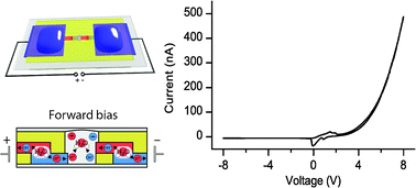
This week’s HOT article is by Magnus Berggren and co-workers at Linköping University, Sweden. The paper presents three different designs of micro-fabricated ion bipolar membrane diodes (IBMDs), which the authors demonstrate overcoming some of the problems of traditional fast switching ion diodes, such as accumulation of ions inside the device structure.

The first two designs are bipolar membranes which are capable of either splitting water or providing high current rectification. The third design incorporates the previous two, connecting the bipolar membranes in series, meaning that suppression of ion accumulation is achieved.

Ion diode logics for pH control
Erik O. Gabrielsson, Klas Tybrandt and Magnus Berggren
DOI: 10.1039/C2LC40093F

This HOT article is free to access for the next four weeks following a simple registration for individual users.

Digg This
Reddit This
Stumble Now!
Share on Facebook
Bookmark this on Delicious
Share on LinkedIn
Bookmark this on Technorati
Post on Twitter
Google Buzz (aka. Google Reader)

HOT article: microfluidic polyplex preparation on a chip

Researchers at Philipps-Universität Marburg, Germany, have developed a new technique for the production of polyplexes that produces more regular sized complexes compared to the standard pipetting methods. Polyplexes are the complexes that form by electrostatic interactions of oppositely charged macromolecules – in this case poly(ethylene imine) (PEI) and plasmid DNA, which are used in gene therapy applications.

The group, led by Thomas Kissel, developed a method that produces the complexes using microfluidics on a chip. Over the course of the study, they investigated the factors that could affect the complex size, and discovered that the most important factor was the ratio of PEI to DNA, whilst other factors were not as important. Using the chip method, regular sized complexes (140 nm and 160 nm) were produced, compared to the more irregular pipetting method. The team also showed that different (targeted) PEI-based vectors could be used for the formation of complexes with pDNA and siRNA.

Read the details of the study here:

Optimized preparation of pDNA/poly(ethylene imine) polyplexes using a microfluidic system
Heiko Debus, Moritz Beck-Broichsitter and Thomas Kissel
DOI: 10.1039/C2LC40176B

This HOT article is free to access for the next four weeks following a simple registration for individual users.

Digg This
Reddit This
Stumble Now!
Share on Facebook
Bookmark this on Delicious
Share on LinkedIn
Bookmark this on Technorati
Post on Twitter
Google Buzz (aka. Google Reader)

Have you seen the best of LOC?

The Editors at Lab on a Chip have been busy picking out the top 10% from all our high quality papers to bring you a collection of recent articles that we think will be of exceptional significance for the miniaturisation community.

Papers in this category will have received excellent reports during peer review, and demonstrate a breakthrough in device technology, methodology or demonstrate important new results for chemistry, physics, biology or bioengineering enabled by miniaturisation.

Here are the papers that have caught our eye so far:

Frontier
Microengineered physiological biomimicry: Organs-on-Chips
Dongeun Huh, Yu-suke Torisawa, Geraldine A. Hamilton, Hyun Jung Kim and Donald E. Ingber
DOI: 10.1039/C2LC40089H

Focus
Education: a microfluidic platform for university-level analytical chemistry laboratories
Jesse Greener, Ethan Tumarkin, Michael Debono, Andrew P. Dicks and Eugenia Kumacheva
DOI: 10.1039/C2LC20951A

Tutorial Review
Brain slice on a chip: opportunities and challenges of applying microfluidic technology to intact tissues
Yu Huang, Justin C. Williams and Stephen M. Johnson
DOI: 10.1039/C2LC21142D

Critical Review
Engineers are from PDMS-land, Biologists are from Polystyrenia
Erwin Berthier, Edmond W. K. Young and David Beebe
DOI: 10.1039/C2LC20982A

Communications
“Fluidic batteries” as low-cost sources of power in paper-based microfluidic devices
Nicole K. Thom, Kimy Yeung, Marley B. Pillion and Scott T. Phillips
DOI: 10.1039/C2LC40126F

Sorting cells by size, shape and deformability
Jason P. Beech, Stefan H. Holm, Karl Adolfsson and Jonas O. Tegenfeldt
DOI: 10.1039/C2LC21083E

Papers
High throughput automated chromatin immunoprecipitation as a platform for drug screening and antibody validation
Angela R. Wu, Tiara L.A. Kawahara, Nicole A. Rapicavoli, Jan van Riggelen, Emelyn H. Shroff, Liwen Xu, Dean W. Felsher, Howard Y. Chang and Stephen R. Quake
DOI: 10.1039/C2LC21290K

A microfluidic device for whole-animal drug screening using electrophysiological measures in the nematode C. elegans
Shawn R. Lockery, S. Elizabeth Hulme, William M. Roberts, Kristin J. Robinson, Anna Laromaine, Theodore H. Lindsay, George M. Whitesides and Janis C. Weeks
DOI: 10.1039/C2LC00001F

Ion diode logics for pH control
Erik O. Gabrielsson, Klas Tybrandt and Magnus Berggren
DOI: 10.1039/C2LC40093F

Human gut-on-a-chip inhabited by microbial flora that experiences intestinal peristalsis-like motions and flow
Hyun Jung Kim, Dongeun Huh, Geraldine Hamilton and Donald E. Ingber
DOI: 10.1039/C2LC40074J

A multifunctional pipette
Alar Ainla, Gavin D. M. Jeffries, Ralf Brune, Owe Orwar and Aldo Jesorka
DOI: 10.1039/C2LC20906C

Visualization of microscale particle focusing in diluted and whole blood using particle trajectory analysis
Eugene J. Lim, Thomas J. Ober, Jon F. Edd, Gareth H. McKinley and Mehmet Toner
DOI: 10.1039/C2LC21100A

DNA electrophoresis in a nanofence array
Sung-Gyu Park, Daniel W. Olson and Kevin D. Dorfman
DOI: 10.1039/C2LC00016D

Bipolar electrochemistry for cargo-lifting in fluid channels
Gabriel Loget and Alexander Kuhn
DOI: 10.1039/C2LC21301J

Rapid screening of antibiotic toxicity in an automated microdroplet system
Krzysztof Churski, Tomasz S. Kaminski, Slawomir Jakiela, Wojciech Kamysz, Wioletta Baranska-Rybak, Douglas B. Weibel and Piotr Garstecki
DOI: 10.1039/C2LC21284F

Dual-electrode microfluidic cell for characterizing electrocatalysts
Ioana Dumitrescu, David F. Yancey and Richard M. Crooks
DOI: 10.1039/C2LC21181E

Rapid, sensitive, and multiplexed on-chip optical sensors for micro-gas chromatography
Karthik Reddy, Yunbo Guo, Jing Liu, Wonsuk Lee, Maung Kyaw Khaing Oo and Xudong Fan
DOI: 10.1039/C2LC20922E

A silicone-based stretchable micropost array membrane for monitoring live-cell subcellular cytoskeletal response
Jennifer M. Mann, Raymond H. W. Lam, Shinuo Weng, Yubing Sun and Jianping Fu
DOI: 10.1039/C2LC20896B

Batch fabrication of disposable screen printed SERS arrays
Lu-Lu Qu, Da-Wei Li, Jin-Qun Xue, Wen-Lei Zhai, John S. Fossey and Yi-Tao Long
DOI: 10.1039/C2LC20926H

Bubbles no more: in-plane trapping and removal of bubbles in microfluidic devices
Conrad Lochovsky, Sanjesh Yasotharan and Axel Günther
DOI: 10.1039/C1LC20817A

A digital microfluidic method for multiplexed cell-based apoptosis assays
Dario Bogojevic, M. Dean Chamberlain, Irena Barbulovic-Nad and Aaron R. Wheeler
DOI: 10.1039/C2LC20893H

Technical Innovation
Three-dimensional microfiber devices that mimic physiological environments to probe cell mechanics and signaling
Warren C. Ruder, Erica D. Pratt, Sasha Bakhru, Metin Sitti, Stefan Zappe, Chao-Min Cheng, James F. Antaki and Philip R. LeDuc
DOI: 10.1039/C2LC21117C

We will be adding to this collection throughout the year so keep checking back for more outstanding articles!

Digg This
Reddit This
Stumble Now!
Share on Facebook
Bookmark this on Delicious
Share on LinkedIn
Bookmark this on Technorati
Post on Twitter
Google Buzz (aka. Google Reader)

Lab on a Chip publishes its 150th issue

This week we’re celebrating our 150th issue at Lab on a Chip. Albert van den Berg, Deputy Chair of our Editorial Board, writes about the journal’s history and its exciting future, in his editorial.

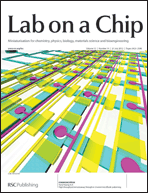
The issue’s front cover features artwork from Kamlesh Patel and colleagues, whose critical review discusses the recent advances in digital microfluidics, focusing on applications for chemistry, biology and medicine. Topics discussed include the use of droplets for chemical synthesis, enzyme assays and the analysis of blood.

Digital microfluidics: a versatile tool for applications in chemistry, biology and medicine
Mais J. Jebrail, Michael S. Bartsch and Kamlesh D. Patel
DOI: 10.1039/C2LC40318H


A communication from Yanyi Huang and co-workers demonstrating an integrated microfluidic immunoassay chip for high-throughput sandwich immunoassay tests is featured on the inside front cover.

The authors state the device can perform ELISA measurements in one hour with just 1 μL of sample for four repeats.

High-throughput immunoassay through in-channel microfluidic patterning
Chunhong Zheng, Jingwen Wang, Yuhong Pang, Jianbin Wang, Wenbin Li, Zigang Ge and Yanyi Huang
DOI: 10.1039/C2LC40145B


The back cover features artwork from Tony Jun Huang and colleagues, whose paper on acoustic-based tunable patterning demonstrates a technique that can arrange microparticles or cells into arrays using pairs of slanted-finger interdigital transducers.

Tunable patterning of microparticles and cells using standing surface acoustic waves
Xiaoyun Ding, Jinjie Shi, Sz-Chin Steven Lin, Shahrzad Yazdi, Brian Kiraly and Tony Jun Huang
DOI: 10.1039/C2LC21021E

Also in this issue is the latest Research highlight from Ali Khademhosseini, and Acoustofluidics 14: Applications of acoustic streaming in microfluidic devices from Martin Wiklund and co-authors.

Read the rest of the issue here.

Digg This
Reddit This
Stumble Now!
Share on Facebook
Bookmark this on Delicious
Share on LinkedIn
Bookmark this on Technorati
Post on Twitter
Google Buzz (aka. Google Reader)

A microfluidic ‘baby-machine’ and nanochannels to allow efficient fluorescent analysis of proteins

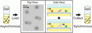
Scott Manalis and coworkers at MIT have developed a microfluidic version of the “baby-machine”, a device used to synchronize eukaryotic cells at a specific point in the cell cycle for study of cellular and molecular processes.

The original method which was developed in the 1960s chemically bound cells to a membrane. As they divide one cell remains attached to the membrane and the ‘newborn’ cell is eluted, producing a synchronised population of cells in the G1 phase of the cell cycle.  However, the method of chemically attaching cells to the membrane is not suitable for all cell types.  Manalis et al have modified this procedure to attach cells by applying pressure differences to capture cells on their microfluidic device, thereby avoiding the use of chemicals that can make cells non-viable.

Read more…
A microfluidic “baby machine” for cell synchronization
Josephine Shaw, Kristofor Payer, Sungmin Son, William H. Grover and Scott R. Manalis
DOI: 10.1039/C2LC40277G


Xing-Hua Xia and colleagues at Nanjing University have recently improved on the fluorescent analysis of proteins by utilising the confines of a nanochannel in a microfluidic device.  The nanochannel allows the efficient concentration of proteins and separates labelled proteins from unreacted fluorescent labels due to size.  This removes the need for purification to remove the unreacted dye on an external chromatographic instrument and allows protein concentration, labelling, and purification in continuous manner in one device.

Read more…
Rapid protein concentration, efficient fluorescence labeling and purification on a micro/nanofluidics chip
Chen Wang, Jun Ouyang, De-Kai Ye, Jing-Juan Xu, Hong-Yuan Chen and Xing-Hua Xia
DOI: 10.1039/C2LC20977B

These latest two HOT articles are free to access for 4 weeks following a simple registration for individual users

Digg This
Reddit This
Stumble Now!
Share on Facebook
Bookmark this on Delicious
Share on LinkedIn
Bookmark this on Technorati
Post on Twitter
Google Buzz (aka. Google Reader)

HOT articles: A new assay to assess drug permeability of cells and new measure for acoustic energy density

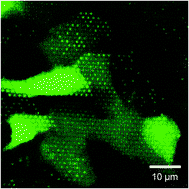
Macroporous silicon chips for laterally resolved, multi-parametric analysis of epithelial barrier functionMeasuring the ability of compounds to cross various tissue barriers is of critical importance during drug development, as most drugs will have to cross at least one tissue barrier to reach their target.  Various assays already exist to assess this process, but have shortcomings such as being prone to artefacts or failing to provide information on permeation pathway. Now Joachim Wegener and colleagues have devised a new silicon chip to visualise permeability, with the ability to see defects within the cell layer and details about the permeation route. Read more…

Macroporous silicon chips for laterally resolved, multi-parametric analysis of epithelial barrier function
Stefanie Michaelis,  Christina E. Rommel,  Jan Endell,  Petra Göring,  Ralf Wehrspohn,  Claudia Steinem,  Andreas Janshoff,  Hans-Joachim Galla and Joachim Wegener
DOI: 10.1039/C2LC00026A

On a completely different topic the next HOT article is on acoustofluidics.  Henrik Bruus et al present a simple method based on measuring light intensity to determine the acoustic energy density in microchannel.  This parameter can be used as a measure of acoustophoretic performance, but is typically tricky to measure.  Read how they achieved it:

Measuring acoustic energy density in microchannel acoustophoresis using a simple and rapid light-intensity method
Rune Barnkob,  Ida Iranmanesh,  Martin Wiklund and Henrik Bruus
DOI: 10.1039/C2LC40120G

Remember – these articles are  free (if you have an RSC Publishing account) for the next four weeks!

Digg This
Reddit This
Stumble Now!
Share on Facebook
Bookmark this on Delicious
Share on LinkedIn
Bookmark this on Technorati
Post on Twitter
Google Buzz (aka. Google Reader)