The Sustainable Nanotechnology Organization (SNO) is a Professional and international non-profit society, which held its 2nd annual conference on November 3 – 5 2013 at the Fess Parker’s Double Tree Hotel in Santa Barbara, California. The conference was chaired by Dr. Arturo Keller of the University of California at Santa Barbara.
SNO focuses on advancing sustainable nanotechnology around the world through education, research, and the promotion of responsible use of nanotechnology (www.susnano.org). The conference brought together scientists and experts from around the world, both from academia, industry and government agencies, to present and discuss current research findings on the subject of nanotechnology and sustainability. The conference was also attended by members of the press and nongovernmental organizations, and had an increase in attendance by 20% over 2012.
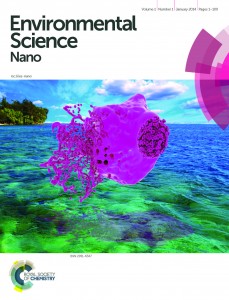
During the meeting it was announced that SNO will partnership with new journal Environmental Science: Nano, published by the Royal Society of Chemistry. Environmental Science: Nano will represent SNO as the Organisation’s official journal and is free to access to all SNO members for two years from launch. It is hoped that members will support this journal with submissions (please see end of post for more details). The journal offers great benefits, including free colour, no page limits and an efficient review process.
As well as this exciting announcement, the SNO conference featured an exciting three days of activities, including outstanding technical programs and cutting-edge research on nanotechnology and sustainability. Presentations included 6 plenary speakers, 154 platform presentations, and 46 poster presentations, which were drawn from early faculty career investigators, postdoctoral fellows, students, and industrial participants. In addition, prior to the Conference, SNO’s first Nanoceria workshop was held, which was led by Robert Yokel in conjunction with the University of Kentucky.
Although SNO is a relatively new organization it has formed an excellent basis to drive it towards a strong and sustainable future. The annual conference provides a place for the formation and advancement of the new community of sustainable nanotechnology. 2013’s conference program was built around providing plenty of time for networking and social interactions; this included the Sunday evening welcome reception and banquet, which allowed for community development.
Attendants at the 2013 Conference represented a vast geographical area, with participants from almost all states of the USA, as well as international participants from Canada, France, Great Britain, India, Korea, Japan and Poland. In addition, approx. 25% of the participants were women, and a sizeable student presence was recorded, indicative of the “recentness” of the field. These young scientists bring fresh appeal to the organization and SNO aims to support and honour them; $500 prizes were awarded to 26 graduate students based on their resumes and the relevance of their research to sustainability and nanotechnology.
If you would like to submit to Environmental Science: Nano, please use the following submission link: http://mc.manuscriptcentral.com/esn
Alternatively, please speak to a member of our Editorial Board at this address esnano-rsc@rsc.org
Associate Editors: Greg Lowry, (Carnegie Mellon University), James Hutchison (University of Oregon), Kristin Schirmer (Eawag, Switzerland)
Chair and Editor-in-Chief: Vicki Grassian (University of Iowa)
Vice-Chair: Christy Haynes (University of Minesota)
Details of our full Editorial Board can be found here: http://rsc.li/19CHl5s


