Archive for the ‘HOT articles’ Category

Carbon nanotubes as a Trojan Horse for heavy metals

Carbon nanotubes (CNTs) are promising nanomaterials because of their many interesting properties: CNTs are flexible yet super strong and practically impossible to break. Depending on their atomic arrangement, CNTs can be semiconductors or very strong conductors of electricity. When it comes to transporting heat, this amazing material can move it very quickly along its length, while behaving as an insulator from side to side. CNTs can also be doped or decorated with different chemical elements, compounds, or nanoparticles and serve as a transport carrier for those materials. For these reasons, along with many others, CNTs are being developed for a myriad of applications, ranging from electronics to drug delivery. Because on this increasing interest in applications of CNTs, we can expect their global production to grow over time, which may lead to an increase in the potential for CNTs to be released into the environment or to be put into contact with people.

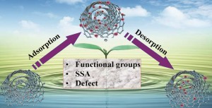
Jie Li and colleagues explored the possibility that CNTs may serve as a “Trojan Horse” by carrying heavy metal ions that were incorporated onto their surface and then releasing those ions when they reach the environment. Imagine the giant Greek wooden horse, arriving the gates of the city of Troy, full of soldiers in its belly. Now, scratch that and imagine a carbon nanotube arriving in your local river, its back teeming with heavy metals. Will these metals dismount the CNTs and impact the wildlife of your local river? Will they go through the local treatment plant and arrive in your home, via your tap water? Or will these metals stay forever stuck onto the CNTs and not cause any harm?

To answer this question, researchers from China studied different types of CNTs with a variety of metal ions: copper, hexavalent chromium (a known carcinogen), and arsenic (a known toxic chemical).

They found that the absorption and desorption of heavy metals from CNTs do not occur at the same pace. In practical terms, the metal ions come off the CNTs slower than the rate at which they are put onto the CNTs. When comparing different types of CNTs, they found that multi-walled CNTs and double-walled CNTs have a higher capacity to carry arsenic and chromium cations than single walled CNTs or oxidized CNTs. For copper (an anion), they observed exactly the opposite.

The sorption of heavy metal ions onto the surface of CNTs is a reversible process.Therefore it is possible that once CNTs enter the environment, they release the metals which they were carrying. The higher the concentration of metals, the more reversible this process might be. The release of metals from CNTs occurs differently for negatively versus positively charged ionic metals and varies greatly among different types of CNTs. This means that we cannot predict what will happen to CNTs (and the metals they carry) in the environment without understanding their structure and knowing exactly what types of chemicals are sorbed onto their surface.

To access the full article, download a copy for free* by clicking the link below.

Jie Li, Changlun Chen, Shouwei Zhang and Xiangke Wang
DOI: 10.1039/C4EN00044G

* Access is free through a registered RSC account – click here to register

About the webwriter

Marina Vance is a research scientist at Virginia Tech and associate director of the Virginia Tech Center for Sustainable Nanotechnology Her work focuses on people’s exposure to nanomaterials.

Follow her on twitter: @marinavance

Digg This
Reddit This
Stumble Now!
Share on Facebook
Bookmark this on Delicious
Share on LinkedIn
Bookmark this on Technorati
Post on Twitter
Google Buzz (aka. Google Reader)

Light Effects the Surface Chemistry of Oxidized Multiwalled Carbon Nanotubes

Environmental transformation of engineered nanomaterials makes it extremely difficult for us to track them under natural conditions because their identity and the related properties are changing constantly. Therefore, much research is being conducted, simulating natural environmental conditions, to understand these nanoparticle transformations and the subsequent behavior. One such parameter which is constantly under investigation is the effect of sun light or more specifically the visible (400-700 nm) – lower energy UVA light (300-400 nm). Since higher energy UV-light (UVB and UVC) is efficiently absorbed by the ozone layer and the atmosphere, not much attention has been given to the transformations caused by them. However, UVC radiation is used heavily in municipal drinking and wastewater treatment plants for water disinfection from microorganisms. Given the increasing usage of carbon nanotubes (CNTs) and their potential release into water systems, Julie L. Bitter and co-workers of John Hopkins University, Baltimore in USA have conducted a thorough investigation on the transformations occurring on oxidized multiwalled CNTs (O-MWCNTs) upon the exposure to UVC (254 nm) irradiation.

In this work, O-MWCNTs suspended in ultrapure water was subjected to UVC irradiation under two configurations;

(1) large batch volumes,

(2) small batch volumes.

Samples treated under large batch volumes were used in characterization studies and mass loss measurements while the latter was used in particle sizing and concentration measurements. Furthermore, the effect of water quality parameters was investigated by varying the solution pH and the ionic strength.

oxidized multiwalled carbon nanotubes

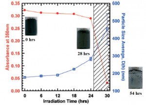
The results indicated that O –MWCNTs surface undergo decarboxylation inducing aggregation causing the particles to settle out of the solution. This process was found to be dominated by one photon, direct excitation mechanism instead of the mechanism mediated by reactive oxygen species. Surface characterization with XPS analysis as well as chemical derivatization showed a significant reduction in the distribution of oxygen-containing functional groups upon irradiation, supporting the above observation. Furthermore, it was interesting to note that aggregation was resisted until a sufficient number of carboxylic acid groups were removed where the electrostatic repulsions between the O-MWCNTs were no longer strong enough to prevent aggregation. This UVC induced aggregation was observed at all the light intensities and under both oxic and anoxic conditions. The resistance towards photo-induced aggregation however, was enhanced under high pH and low ionic strength conditions.

To access the full article, download a copy for free* by clicking the link below.

Transformations of oxidized multiwalled carbon nanotubes exposed to UVC (254 nm) irradiation

Julie L. Bitter,   Jin Yang,   Somayeh Beigzadeh Milani,  Chad T. Jafvert and   D. Howard Fairbrother

DOI: 10.1039/C4EN00073K

*Access is free through a registered RSC account – click here to register

Digg This
Reddit This
Stumble Now!
Share on Facebook
Bookmark this on Delicious
Share on LinkedIn
Bookmark this on Technorati
Post on Twitter
Google Buzz (aka. Google Reader)

Phytotoxicity of Cerium Oxide Nanoparticles

Plants: basic components of the ecosystem and vulnerable to nanoparticle exposure. It is important to understand the interactions between nanoparticles and plants, especially when herbivorous consumers introduce these plants into our food chain.

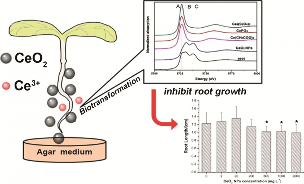
As CeO2 NPs are widely used in many applications, their interactions with the ecosystem are inevitable. It has previously been shown that CeO2 NPs can inhibit root elongation of plants in aqueous suspensions. Dr Zhiyong Zhang et al investigated the toxicity of CeO2 NPs on asparagus lettuce in a plant agar medium, a semisolid, soil-like medium which provides a more realistic environment for plant growth.

A variety of parameters were investigated to understand the plant’s defence and response to abiotic stress caused by CeO2 NPs. Although the agar medium limited the bioavailability of CeO2 NPs, they were still more toxic to asparagus lettuce in the agar medium than in aqueous solution. This could be caused by the production of excess reactive oxygen species causing oxidative stress to the plants.

The increased phytotoxicity of CeO2 NPs in a soil like medium can also be explained by the biotransformation of CeO2 NPs. It has previously been demonstrated that Ce3+ released from CeO2 NPs can cause species-specific toxicity. This study showed that in an agar medium more than 20% of the Ce in the roots was transformed to Ce3+, whereas in aqueous solution only 6% of CeO2 was reduced to Ce3+. It is therefore reasonable to postulate that the phytotoxicity of CeO2 NPs is also attributed to the release of Ce3+.

To read the full paper, download it for free* today!

Effect of Cerium Oxide Nanoparticles on Asparagus Lettuce Cultured in an Agar Medium
Di Cui,   Peng Zhang,   yuhui Ma,   Xiao He,   Yuanyuan Li,  Jing Zhang,   Yuechun Zhao and   Zhiyong Zhang
DOI: 10.1039/C4EN00025K

* Access is free through a registered RSC account – click here to register

Digg This
Reddit This
Stumble Now!
Share on Facebook
Bookmark this on Delicious
Share on LinkedIn
Bookmark this on Technorati
Post on Twitter
Google Buzz (aka. Google Reader)

Nanoparticle detection in a microsecond

Nanoparticles – they are really small; we can’t see them, hear them or feel them and therefore they are pretty hard to detect! Manuel David Montaño, from Colorado School of Mines, and colleagues have made improvements in the detection and characterization of engineered nanoparticles, simply by reducing the time of detection for each nanoparticle – the dwell time.

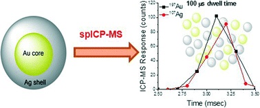
In order to determine the toxicity, fate and transport of nanoparticles in the environment, we first need to determine the size and quantity of nanoparticles in the environment. In order to determine the size and quantity of nanoparticles in the environment we need to be able to accurately detect these nanoparticles. Single particle ICP-MS (spICP-MS) is already a promising technique to detect and characterize low concentrations of engineered nanoparticles in biological and environmental matrices. Initially developed for aerosol particle analysis, spICP-MS uses time resolved analysis with dwell times of approximately 10miliseconds. So how does it work? A discrete pulse of intensity, origination from nanoparticle vaporization and ionization, can be detected – the signal generated by the ions can then be correlated to nanoparticle mass.

The problem – this method of detection only works on low concentrations of nanoparticles. In high concentrations of nanoparticles, two or more nanoparticles can be detected during the same dwell time giving invalid results. It appears that particles are larger in size and lower in concentration that they really would be in the environment. To overcome this problem, often samples have to be diluted considerably, making the results less environmentally relevant. In this study, instead of diluting the samples, researchers simply reduced the dwell time from milliseconds to microseconds. This improved the resolution and working range of spICP-MS, allowing a greater breadth of environmental samples to be analysed.

You can read the full paper for free* by clicking the link below

Improvements in the detection and characterization of engineered nanoparticles using spICP-MS with microsecond dwell times
M. D. Montaño, H. R. Badiei, S. Bazargan and J. F. Ranville
DOI: 10.1039/C4EN00058G

* Access is free through a registered RSC account – click here to register

Digg This
Reddit This
Stumble Now!
Share on Facebook
Bookmark this on Delicious
Share on LinkedIn
Bookmark this on Technorati
Post on Twitter
Google Buzz (aka. Google Reader)

Nanoparticle Crystal Structure Affects Alkali and Acid Digestion

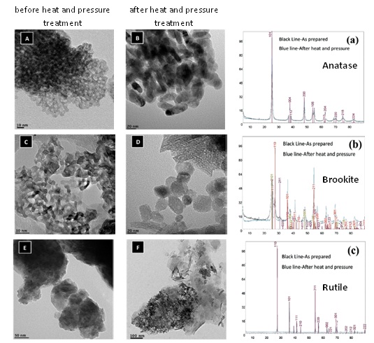
Titanium Oxide nanoparticles (TiO2 NPs) – given their wide applications, exposure scenarios and classification as a class 2B carcinogen (International Agency of Research on Cancer), it is high time for the precise and accurate quantitative analysis of TiO2 NP contamination in environmental samples. Being one of the metal oxides that is extremely hard to solubilize makes accurate and precise measurement of TiO2 a challenge. Recent investigations using mixed acid digestion and alkali potassium hydroxide (KOH) fusion has given improved recoveries of TiO2 when compared to the conventional methods. This is great news for all of us who struggle to quantify TiO2 nanoparticles in complex environmental matrices! However, as with all nanoscale materials there are complications arising from size and polymorph dependent thermodynamic stabilities as well as chemical reactions between Ti and other sample matrices, especially at elevated temperature and pressure. Thus, R. G. Silva and coworkers of United States Environmental Protection Agency (US-EPA) investigate the digestibility of different polymorphs of TiO2 NPs; anatase, rutile and brookite. These samples were used for spiking environmental matrices consisting of river sediment and clay minerals (bentonite and kaolinite). Furthermore, a portion of these were subjected to heat (300OC) and pressure (10.3 bar) treatment to investigate its impact on the Ti recovery from the TiO2 NPs.

Extensive characterization of all three nanoparticle samples with respect to size, shape, crystallinity and surface area before and after the heat and pressure treatments showed significant changes in the physicochemical properties of anatase and brookite. Rutile on the other hand was resistant to changes. In terms of digestion, acid digestion resulted in relatively lower Ti concentration for the pure TiO2 NP samples that underwent heat and pressure treatment. In contrast, alkali fusion resulted in increased levels of Ti. Nevertheless, when the TiO2 NP polymorphs were blended in the environmental matrices, for anatase and brookite the recoveries were similar for both types of digestions. However, for the recovery of rutile the alkali fusion method proved to be superior to that of the mixed acid method. Therefore, this work recommends using the alkali fusion method for the extraction of Ti from TiO2 NP contaminated unknown environmental samples.

To access the full article, download a copy for free* by clicking the link below.

Polymorph-dependent titanium dioxide nanoparticle dissolution in acidic and alkali digestions

R. G. Silva, M. N. Nadagouda, C. L. Patterson, Srinivas Panguluri, T. P. Luxton, E. Sahle-Demessieb and   C. A. Impellitterib
DOI: 10.1039/C3EN00103B

*Access is free through a registered RSC account – click here to register

Digg This
Reddit This
Stumble Now!
Share on Facebook
Bookmark this on Delicious
Share on LinkedIn
Bookmark this on Technorati
Post on Twitter
Google Buzz (aka. Google Reader)

Illuminating the issue of real-time nanomaterial characterization

Fluorescence complexation could hold the key to more detailed monitoring of airborne nanomaterials. Researchers at Texas A&M University, USA describe the successful development of a new method of simultaneous online quantification and characterization.

Fluorescent Complexation

The increased manufacture and use of nanomaterials has led to increased concerns about their associated health risks, particularly in the occupational setting. One of the main barriers to addressing uncertainties in this field is a poor understanding of personal exposure levels. There is currently a lack of sufficient dynamic data regarding the main exposure routes for airborne nanomaterials so appropriate exposure guidelines, profiles and models cannot be established.

It is important, therefore to establish effective means of measuring the levels and chemical/physical properties of nanomaterials in real-time. Preferred traditional means of nanomaterial analysis e.g. mass spectrometry (MS) and electron microscopy (EM) cannot be easily adapted to use in an online capacity. This means exposure analysis is generally slow and can only be carried out for relatively short, unrepresentative time-frames.

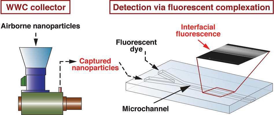
In this study Fanxu Meng and co-workers introduce and demonstrate an integrated methodology that allows continuous online monitoring of the levels and characterization of airborne nanomaterials. This method combines ultra high flow sampling with a sensitive fluorescence-based detection system.

The sampling system comprised a modified wetted wall cyclone (WWC) collector which has an ultra high flow rate (>1000 L min-1) combined with a continuous flow microfluidic network allowing online detection capability. The detection system utilized florescence generated by the combination of a suspension of collected nanoparticles and a tracer dye solution. The resulting dye-nanoparticle complex produces an intense and easily detectible fluorescent signature, dependent on the quantity and the physical/chemical properties of the collected nanomaterials.

The integrated system was tested using prepared suspensions of Al2O3. A scanning mobility particle sizer (SMPS) was used to quantify the concentration and size distribution of nanoparticles inside the test chamber. It was shown that florescence displays a characteristic pattern, dependent on the concentration but also the size and particle surface area of the nanomaterials. A linear correlation between florescence intensity and airborne concentration of Al2O3 at concentrations up to 1.0-1.2 wt% at flow rates of 0.2 and 0.02 mL min-1 was observed.

This work provides a platform for continuous measurement of airborne nanomaterials. Simultaneous sampling and compound characterization can enable better time-resolved assessment of the transport and fate of released nanomaterials and identification of hazardous releases. The method described is capable of sampling a broad range of air volumes (representative of the work place environment) and allows online detection and analysis. The authors highlight potential further innovations for this work, possibly leading to more detailed exposure profiles for nanomaterials; establishing fluorescence “fingerprints” for a range of different nanoparticles; and analysis of biological material.

Download the full article for free* today!

Localized Fluorescent Complexation Enables Rapid Monitoring of Airborne Nanoparticles
Fanxu Meng, Maria D. King, Yassin A. Hassan, and Victor M. Ugaz
DOI: 10.1039/C4EN00017J

*Access is free through a registered RSC account – click here to register

Digg This
Reddit This
Stumble Now!
Share on Facebook
Bookmark this on Delicious
Share on LinkedIn
Bookmark this on Technorati
Post on Twitter
Google Buzz (aka. Google Reader)

Nanoparticle sulfidation transformations

A study by Gregory Lowry and colleagues from Carnegie Mellon University suggests that Copper Oxide (CuO) nanoparticles (NPs) are sulfidized in the environment, affecting their resulting properties. Some of the affected properties, such as solubility are relevant to the toxicity of these nanoparticles in the environment.

Many nanoparticles are transformed in the environment; it is often the transformed materials which cause concern with regards to nanoparticle toxicity. There have been several papers highlighting how important it is to research the properties of nanoparticle transformations. A recent review by Nasia Von Moos provides an overview of what is currently known about environmental transformations of nanomaterials in freshwater systems, a recent paper by Julián A. Gallego-Urrea discusses the transformations which TiO2 nanoparticles undergo once they reach the aquatic environment and this research paper reports that sulfidation is an important transformation for some metal oxide nanoparticles, such as CuO NPs.

Why is sulfidation in the environment important?

Copper-based NPs are being used in semiconductors, heat transfer fluids, catalysts, batteries and many more products and technologies. Their wide spread uses will likely lead to subsequent release into the environment, raising concerns about their potential toxicity. It has already been demonstrated that CuO NPs are toxic to many organisms including crustaceans, algae and fish; although Cu2+ is more toxic to most of these organisms. It is therefore essential to determine what the products of sulfidized CuO NPs are and if they are more or less toxic to the environment when compared with pristine CuO.

Sulfidation of CuO

Cuo NPs were characterized and sulfidized in water by inorganic sulfide. Characterization of the resulting products showed that CuO is sulfidized to several copper sulphide species including crystalline CuS (covellite), amorphous (CuxSy) species and copper sulphate hydroxide species. In previous studies it has been demonstrated shown that sulfidation decreases the solubility and metal availability of Ag and ZnO NPs. This study however shows that the sulfidation of CuO NPs breaks the trend. Sulfidation actually increased the dissolved fraction of copper compared to pristine CuO NPs. This increased release of Cu2+ and CuS nanoclusters from sulfidized NPs compared to CuO suggests that toxicity studies with pristine CuO may be misleading in environments where sulfidation is likely to occur, demonstrating that it is prudent to use environmentally transformed nanoparticles in fate, transport and toxicitiy studies rather than focusing soley on the prisitne materials. Access the full article for free* by clicking the link below.

Sulfidation of copper oxide nanoparticles and properties of resulting copper sulfide
Rui Ma, John Stegemeier, Clement Levard, James Dale, Clinton W Noack, Tittany Yang, Gordon Brown and Gregory Lowry
DOI: 10.1039/C4EN00018H

*Access is free through a registered RSC account – click here to register

The author recommends further studies which are still needed to:

  1. Identify the nature of the CuxSy nanoclusters.
  2. Assess the toxicity of sulfidized CuO NPs and CuxSy nanoclusters.
  3. Assess the stability of very small metal sulphide clusters (Ag, Zn and Cu) against oxidation under environmental and biological conditions.
  4. Assess how sulfidation of CuO NPs occurs in situ at relevant CuO/S concentration ratios and how this affects their bioavailability under realistic exposure scenarios.
Digg This
Reddit This
Stumble Now!
Share on Facebook
Bookmark this on Delicious
Share on LinkedIn
Bookmark this on Technorati
Post on Twitter
Google Buzz (aka. Google Reader)

Applications of porous nanomaterials

Sean Lehman and Sarah Larsen from the University of Iowa review zeolite and mesoporous silica nanomaterials with emphasis on connections to the environment.

In recent years, there has been a great deal of interest in zeolites and mesoporous silica nanomaterials (MSNs). Zeolites are widely used in industry for applications such as catalysis, separations and gas adsorption, however the authors believe that these porous nanomaterials have a largely unrealized commercial potential for environmental applications.

Structures of porous nanomaterials

This review article covers three major areas:

  1. Greener synthesis of zeolite and MSNs
  2. Potential of zeolite and MSNs for environmental applications
  3. The biological toxicity of zeolite and MSNs

Due to cost and reduced thermal stability MSNs are not as extensively applied as zeolite; however they are currently being investigated for potential environmental and biomedical applications. Their varied physiochemical properties open up a wide range of potential applications. The more applications that these porous nanomaterials have in industry, the greater the interested in developing greener synthesis for them and reducing their toxicity. With two measurements which are on the nanoscale, pore size as well as particle size, zeolites and MSN make very interesting nanomaterials.

This review describes both the environmental applications, including environmental catalysis and adsorption of environmental contaminants, and implications of zeolite and MSNs. Due to concerns that increased use of these materials translates to increased exposures, toxicity studies of both nanomaterials are also reviewed.

To read the full review for free* click the link below:

Zeolite and Mesoporous Silica Nanomaterials: Greener Syntheses, Environmental Applications and Biological Toxicity

Sean E Lehman and Sarah C Larsen
DOI: 10.1039/C4EN00031E, Critical Review

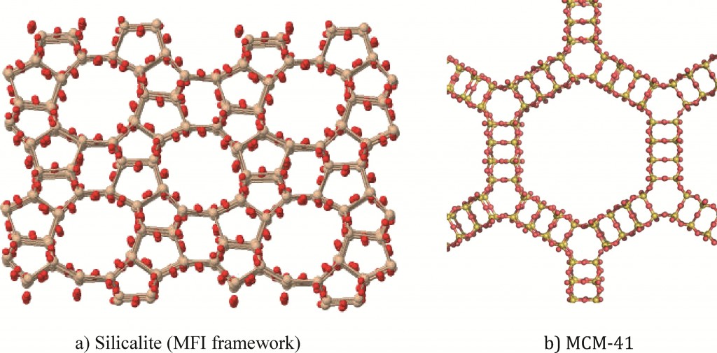
Zeolites and MSNs are silicate or aluminosilicate nanomaterials with well-defined pore networks; there are however some differences between the two porous nanomaterials.

Properties of zeolites:

  • Crystalline aluminosilicates (or silicates)
  • Regular arrangements of micropores
  • High surface areas
  • Exchangeable cations

Properties of MSN:

  • Amorphous silica materials
  • Regualr arrangement of mesopores
  • Very high surface area

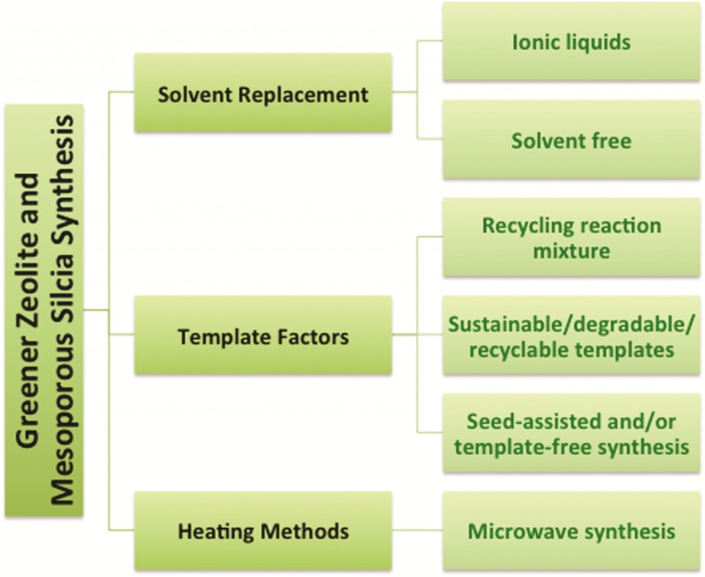
The first area discussed in this review is the synthesis of zeolite and MSNs using green synthetic routes. The green strategies can be organized into three main categories: solvent, template and heating. The diagram below demonstrates the strategies for the greener synthesis of zeolites and mesoporous silica.

Greener synthesis of zeolites and mesoporous silica

*Access is free through a registered RSC account – click here to register

Digg This
Reddit This
Stumble Now!
Share on Facebook
Bookmark this on Delicious
Share on LinkedIn
Bookmark this on Technorati
Post on Twitter
Google Buzz (aka. Google Reader)

Mass Spectrometry Proteomics Characterize Nanoparticle Protein Corona

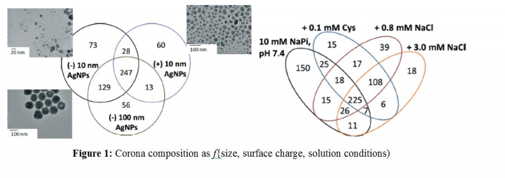
The adventitious protein adsorption on to nanoparticles (NPs), commonly known as the NP-protein corona is in the limelight for many good reasons including its influence on the NP uptake, accumulation and ultimate cellular fate. The consequences of this phenomenon can work in different ways. NP-protein corona can lower the toxicity of NPs when compared to the “bare” NPs but at the same time facilitate crossing biological barriers that can trigger activation of specific regulatory pathways. The particle size, composition and surface properties of NPs are known to influence the composition of the NP-protein corona. Thus, Korin Wheeler, from Santa Clara University, and coworkers investigate the protein populations across the NP-protein corona using silver nanoparticles (AgNPs) in the presence of Yeast (Saccharomyces cerevisiae) proteins (YPE) with the aid of mass spectrometry (MS) proteomics.”

Experiments were designed to probe the effects of AgNP size, surface charge and solution conditions. Two different sizes of AgNPs (10 nm vs 100 nm) with anionic (citrate) and cationic (branched polyethyleneimmine – BPEI) coatings were selected. Protein corona formation was conducted under 0.8 mM NaCl, modeled after freshwater salinity; 3.0 mM NaCl, mimicking mitochondrial salinity; and 0.1 mM Cys, which was shown to mediate NP toxicity. After incubating AgNPs with the YPE, bound and unbound proteins were separated via centrifugation, digested with trypsin followed by LC-MS/MS analysis.

Over 500 different proteins were identified as bound and unbound but no trends were reported linking the molecular weight, pI, length, or the amino acid composition of proteins to their enrichment in the corona. However, AgNP surface charge played a stronger role with those having similar coatings sharing majority of corona population. This highlighted electrostatic modulation of protein affinity for AgNPs under low ionic strength conditions. Further studies with NaCl and cys to simulate more environmentally and biologically relevant conditions revealed more changes in the protein corona populations and modifications in their solution phase behavior in terms of aggregation and sedimentation. However, under all the variable factors there is a population of ubiquitous proteins that get enriched in the corona as shown in the Figure below.

The findings of this research is expected to contribute towards better understanding the bio physicochemical factors mediating NP-corona formation, their characterization and the development of predictive models within the environment.

To access the full article, download a copy for free* by clicking the link below.

Silver nanoparticle protein corona composition compared across engineered particle properties and environmentally relevant reaction conditions

Richard Eigenheer, Erick R. Castellanos, Meagan Y. Nakamoto, Kyle T. Gerner, Alyssa M. Lampe and Korin E. Wheeler
DOI: 10.1039/C4EN00002A

*Access is free through a registered RSC account – click here to register

Digg This
Reddit This
Stumble Now!
Share on Facebook
Bookmark this on Delicious
Share on LinkedIn
Bookmark this on Technorati
Post on Twitter
Google Buzz (aka. Google Reader)

Tracing Silver Nanoparticles

To understand the fate of silver nanoparticles (AgNPs) in the environment they need to be traced in complex natural samples. Often, studies that interrogate the environmental fate of AgNPs require AgNP concentrations that exceed realistic environmental levels. Potentially, this produces unreliable results, as high particle concentration could induce and identify distinct patterns of particle behaviour that is not relevant for ‘realistic’ environmental concentrations.  Adam Laycock, from Imperial College London, and colleagues investigate whether the same effects are also observed at low, environmentally relevant AgNP concentrations.

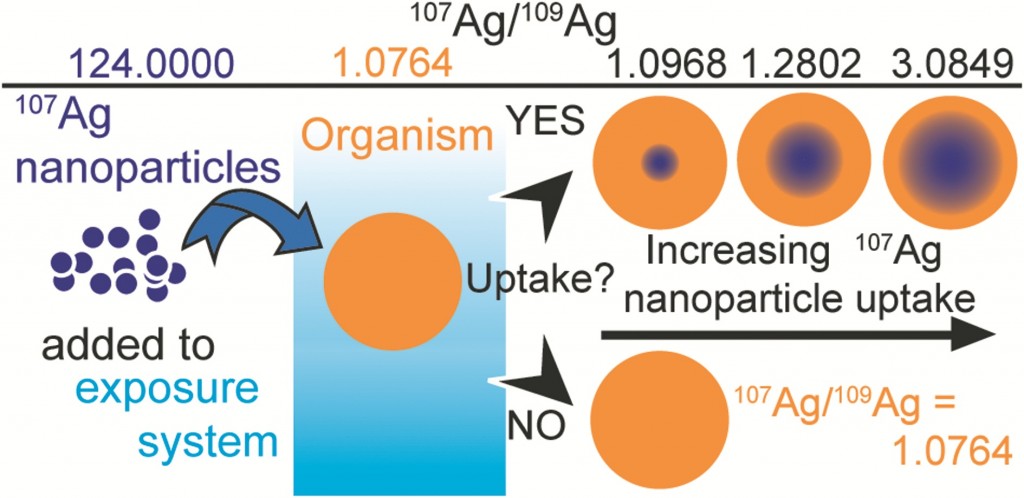
Stable isotope labelling is an attractive method of labelling which relies on the detection of changes when a contaminant is introduced to a system. Previous methods of tracing nanomaterials have had significant drawbacks. For example, fluorescent coatings can affect the surface chemistry of the nanomaterials causing dissociation. Radiolabelling, another technique used, involves the use of specialist equipment and licenses are required for the handling of radioactive material. Recent studies have shown that stable isotope tracing can be applied to nanomaterials – but first, the labelled NPs must be specifically prepared from a single, highly enriched stable isotope form of the elements.

In this study existing protocols were examined to develop techniques for the optimized preparation of isotopically labelled AgNPs. Three protocols were applied to produce particles with a variety of target sizes, with enriched 107Ag and natural Ag. The results show that the methods are suitable for small scale synthesis of stable labeled AgNPs at yields of approximately 80%. The labelling process does not generate unusual particle properties, demonstrating that isotopically modified AgNPs are equivalent to AgNPs with a natural isotope composition.

The authors finalise their study by presenting a series of calculations which reveal that stable isotope labelling can increase the detection sensitivity of AgNPS by at least a factor of 40, and possibly by up to 4000x in comparison to commonly employed bulk Ag concentration measurements. This approach of tracing nanomaterials is highly versatile, the label cannot be lost by dissociation or degradation, the element remains traceable and the use of enriched stable isotopes provides an extremely selective and sensitive means of elemental tracing, even in the presence  of high natural background levels.

To read the full experiment for free*, download the paper now:

Synthesis and characterization of isotopically labeled silver nanoparticles for tracing studies
Adam Laycock, Björn Stolpe, Isabella Römer, Agnieszka Dybowska, Eugenia (Éva) Valsami-Jones, Jamie R Lead and Mark Rehkamper
DOI: 10.1039/C3EN00100H

*Access is free through a registered RSC account – click here to register

Digg This
Reddit This
Stumble Now!
Share on Facebook
Bookmark this on Delicious
Share on LinkedIn
Bookmark this on Technorati
Post on Twitter
Google Buzz (aka. Google Reader)