3D in vitro modeling of Alzheimer’s disease using electrospun microfiber scaffolds

Alzheimer’s disease (AD) presently occupies the topmost position among the most commonly diagnosed neurodegenerative diseases worldwide with the number of affected people forecasted to reach 100 million by 2050. It is characterized by progressive memory loss, impairment of cognitive function, and inability to perform activities of daily life. The key to understanding AD lies in developing effective models which should ideally recapitulate all aspects of the disease. Furthermore, high inaccessibility to the human brain makes it desirable to study neuronal function and degeneration using appropriate in vivo or in vitro model systems of brain cultures. Increasing evidence indicates the superiority of three-dimensional (3D) in vitro cell culture platforms over conventional two-dimensional (2D) monolayer cultures in mimicking native in vivo microenvironments.

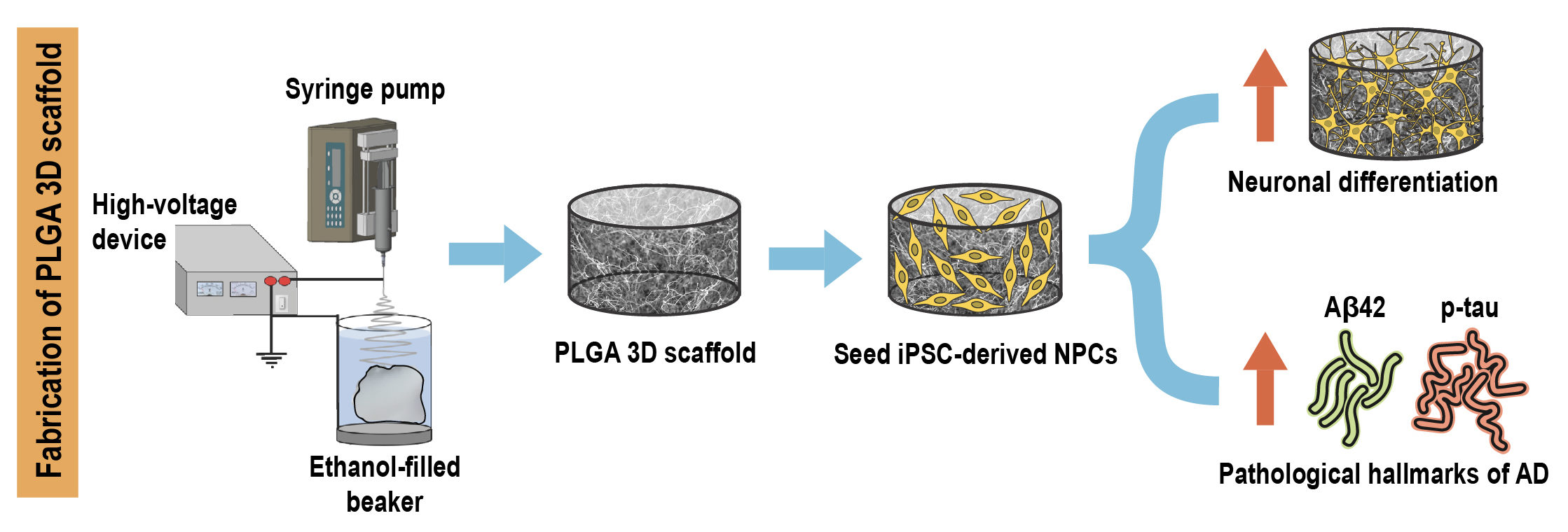
Researchers from Singapore have recently developed a novel 3D in vitro model of AD by encapsulating patient induced pluripotent stem cell (iPSC) derived neural progenitors in poly(lactic-co-glycolic acid) (PLGA) microtopographic scaffolds fabricated using wet electrospinning. They demonstrate that 3D culture robustly recapitulates and accelerates early-stage AD pathogenesis compared with Petri dish-based 2D monolayer controls.

Schematic showing fabrication of PLGA 3D scaffold

First, they achieved deep cellular infiltration and uniform distribution inside the 3D microfibrous scaffold by optimizing various parameters such as fiber diameter, pore size, porosity and hydrophilicity. The stiffness of the microfiber scaffold was found to be comparable to the elasticity of native brain tissue, indicating its capability to promote realistic physiological responses.

Next, they compared key neural stem cell features including viability, proliferation and differentiation in 3D culture with 2D monolayer controls. The 3D microfibrous substrate reduced cell proliferation and significantly accelerated neuronal differentiation within just seven days of culture.

Finally, they demonstrated that 3D scaffold-based culture spontaneously enhanced pathogenic amyloid-beta 42 (Aβ42) and phospho-tau levels in differentiated neurons carrying familial AD (FAD) mutations compared with age-matched healthy controls. More importantly, recapitulation of both pathologies was more pronounced and consistent in 3D culture compared with the same cell lines in 2D monolayer culture conditions.

Taken together, the results indicate that the tunable scaffold-based 3D neuronal culture platform serves as a suitable in vitro model that robustly recapitulates and accelerates pathogenic characteristics of FAD-iPSC derived neurons. It can also be extended to model other complex neurodegenerative diseases and to evaluate prospective therapeutic candidates.

To find out more please read:

A microfiber scaffold-based 3D in vitro human neuronal culture model of Alzheimer’s disease

Vivek Damodar Ranjan, Lifeng Qiu, Jolene Wei-Ling Lee, Xuelong Chen, Se Eun Jang, Chou Chai, Kah-Leong Lim, Eng-King Tan, Yilei Zhang, Wei Min Huang and Li Zeng

Biomaterials Science, 2020, DOI: 10.1039/D0BM00833H

Digg This
Reddit This
Stumble Now!
Share on Facebook
Bookmark this on Delicious
Share on LinkedIn
Bookmark this on Technorati
Post on Twitter
Google Buzz (aka. Google Reader)

A novel biomaterial implant for repair of spinal cord injury

Spinal cord injury (SCI) can be categorized as traumatic (90% of cases) or non-traumatic based on its origin. Traumatic SCI occurs when the primary injury is an external mechanical force (arising from traffic accidents, sports, violence etc.,), which damages the spinal cord and initiates a cascade of multiple secondary complications including neuronal/glial death with very slim chances of recovery. Current treatments for SCI are mainly palliative; however, studies involving surgical interventions for reconstructing injured sites via cell implantation have shown promise. Moreover, incorporating cells within engineered biomaterial substrates which act as extracellular matrix (ECM) substitutes not only lowers cell density requirements but also enables more accurate localised transplantation. Both natural and synthetic biomaterials are being investigated in this regard.

Researchers from the UK have recently developed Proliferate®, a macroporous and biodegradable polymer based on cross-linked poly-ε-lysine (pεK) as a biomaterial candidate for SCI implantation. They demonstrate the biocompatibility of the material with CNS cells via in vitro and in vivo studies, both in the original form and on incorporating functional ECM peptides.

First, they synthesized the polymer in two formats: (i) as inserts suspended in 24-well plate culture wells for in vitro studies and (ii) in tubular form with parallel channels facilitating cell guidance for in vivo studies. The material exhibited a beaded, heterogeneous 3D topography with the porosity capable of being tuned by varying the degree of cross-linking.

Next, they cultured astrocytes on the Proliferate® inserts in vitro and compared  cell morphology with controls grown on PLL-coated coverslips. Staining results showed that the astrocytes adopt a fibrous, ramified morphology typical of in vivo conditions when cultured on the inserts. In addition, the polymer supported differentiation, neuronal survival as well as neurite extension in myelinating cultures; however, myelination was slightly delayed in comparison with coverslip-based controls.

Finally, they implanted the tubular form of the biomaterial into adult rat contusion SCI for in vivo assessment at two timepoints i.e. 7 weeks and 6 months post-implantation. The Proliferate®  implants induced extensive vascularisation and cellular infiltration with no significant difference being observed in microglial response surrounding non-implanted injury cavities and construct-implanted injuries. Although, construct-tissue borders were permissive to astrocyte growth and migration, most cell guidance channels were observed to disintegrate with time and organized axonal growth seen only in intact channels.

Taken together, the results indicate the potential of this novel material, both as a solo implant as well as a substrate for delivery of essential biomolecules to the injury site for facilitation of axonal regeneration following SCI.

To find out more please read:

A novel poly-ε-lysine based implant, Proliferate®, for promotion of CNS repair following spinal cord injury

Sara Hosseinzadeh, Susan L. Lindsay, Andrew G. Gallagher, Donald A. Wellings, Mathis O. Riehle, John S. Riddell and Susan C. Barnett

Biomater. Sci., 2020, 8, 3611-3627, DOI: 10.1039/D0BM00097C

Digg This
Reddit This
Stumble Now!
Share on Facebook
Bookmark this on Delicious
Share on LinkedIn
Bookmark this on Technorati
Post on Twitter
Google Buzz (aka. Google Reader)

Culturing stem-cell derived neurons on a “bed-of-nails” substrate

Interfacing living cells with inorganic nanowire (NW) array substrates is one of the latest areas of exploration in life sciences with potential applications in electrical stimulation, biosensors, cell injection, axonal guidance and so on. A growing body of evidence has identified the role of substrate nanotopography in regulating various cellular phenotypes including cell morphology, adhesion, proliferation, differentiation and intracellular signaling. However, cellular interactions with high surface area vertical nanowires are relatively unexplored and further studies are necessary to fully reveal the correlations between NW array geometry and stem cell behavior.

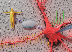
Researchers from Germany have recently interfaced human induced pluripotent stem cell (hiPSC)-derived neurons with tailor-made silicon nitride NW array substrates, achieving highly efficient neuronal differentiation and generating electrophysiologically mature neurons within 4 weeks of culture.

Figurative demonstration of the interface between stem cells and a person

First, they fabricated NW arrays using a top-down dry reactive ion etching (RIE) approach in 3 different arrangements – random, hexagonal and rectangular. The NW lengths were fixed to 1.2 μm with pitches of 1.8 μm and 4 μm, resulting in low density (LD) and high density (HD) arrangements respectively. The cells were transferred onto the NW substrates after 14 days in vitro (DIV) and cultured for another 14-16 DIV before performing functional characterization.

Next, they assessed viability of cells cultured on NW substrates and found that both material used as well as substrate topology had no negative impact on cell viability compared with controls cultured on glass coverslips. Furthermore, on studying cellular outgrowth and morphology, they observed that cells rested on NW tips in the case of HD arrays whereas they encapsulated the NWs in LD arrays, thus indicating the effect of NW density on regulating the settling regime of the cells.

Finally, they tested the electrophysiological integrity of the hiPSC-derived neurons via patch clamping and observed that the neurons cultured on the NW substrates fired characteristic action potentials and demonstrated no significant differences in electrophysiological parameters compared with controls.

Taken together, the results indicate the potential of this platform in stem cell research and regenerative medicine for interfacing human stem cell-derived neurons with tailor-made nanostructured substrates to achieve desired cell behaviors.

To find out more please read:

Interfacing human induced pluripotent stem cell-derived neurons with designed nanowire arrays as a future platform for medical applications

Jann Harberts, Undine Haferkamp, Stefanie Haugg, Cornelius Fendler, Dennis Lam, Robert Zierold, Ole Pless and Robert H. Blick

Biomater. Sci., 2020, 8, 2434-2446, DOI: 10.1039/D0BM00182A

Digg This
Reddit This
Stumble Now!
Share on Facebook
Bookmark this on Delicious
Share on LinkedIn
Bookmark this on Technorati
Post on Twitter
Google Buzz (aka. Google Reader)

Camouflaging tumor targeting nanoparticles with red blood cell membrane for pretargeted multimodal imaging of cancer

Managing cancer requires visualization of tumors using a plethora of imaging modalities such as positron-emission tomography (PET), magnetic resonance imaging (MRI), computed tomography (CT), photoacoustic tomography and optical imaging. Upconversion nanoparticles (UCNPs), a new generation of optical nanomaterials which convert near-infrared (NIR) radiation to visible light by a process called “upconversion luminescence” (UCL), are garnering a lot of attention in cancer diagnostics due to their ability to selectively label cancer cells.

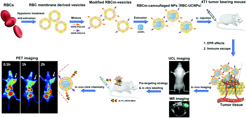
Researchers from Suzhou, China have recently coated tumor targeting UCNPs with red blood cell (RBC) membranes to render them stealthy, effectively preventing them from immune attack and clearance by the host system. Subsequently, they assessed the utility of these RBC-UCNPs for targeted multimodality imaging of 4T1 breast cancer, a triple-negative breast cancer.

First, they isolated cell membranes from the RBCs, reconstructing them into vesicles which were used to encapsulate UCNPs via extrusion. Folic acid (FA) molecules were inserted into the surface of these RBC-UCNPs to assess the tumor-targeting ability of nanoparticles. Upconversion fluorescence imaging revealed that RBC-FA-UCNPs intravenously injected into mice bearing 4T1 subcutaneously transplanted tumors exhibited quick accumulation, long-term retention and reduced uptake by the immune system.

Next, they investigated the feasibility of using these biomimetic nanoparticles in MRI and PET imaging for the detection of tumors in vivo. They found that the MR signal was significantly enhanced by the FA-RBC-UCNPs, indicating the increased circulation time of particles at the tumor site. Furthermore, a combination of pre-targeting strategy and in vivo click chemistry was utilized to mediate PET imaging, which indicated that the biomimetic nanoparticles displayed a higher tumor uptake of the tracer compared with controls, on application of a short half-life radionuclide.

Finally, they conducted in vivo toxicity studies in mice over a span of 30 days, to assess cytotoxicity of the nanoparticles. Blood chemistry, hematology, and histological analyses indicated non-significant induction of toxicity and organ damage, in turn demonstrating the biocompatibility of the biomimetic nanoparticles and their suitability for clinical utilization.

Taken together, the results indicate the potential of this platform for further applications in realizing early diagnosis, bioimaging and treatment of tumors, especially for deep-seated lesions.

To find out more please read:

Red blood cell membrane-coated upconversion nanoparticles for pretargeted multimodality imaging of triple-negative breast cancer

Mengting Li, Hanyi Fang, Qingyao Liu, Yongkang Gai, Lujie Yuan, Sheng Wang, Huiling Li, Yi Hou, Mingyuan Gao and Xiaoli Lan

Biomater. Sci., 2020,8, 1802-1814, DOI: 10.1039/d0bm00029a

Digg This
Reddit This
Stumble Now!
Share on Facebook
Bookmark this on Delicious
Share on LinkedIn
Bookmark this on Technorati
Post on Twitter
Google Buzz (aka. Google Reader)

2019 Biomaterials Science Outstanding Student Paper Award Winner

We are pleased to introduce the Biomaterials Science Outstanding Student Paper Award. This new annual award recognises outstanding work published in the journal, for which a substantial component of the research was conducted by a student. Read below for more information.

Our 2019 Winner 

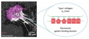
The inaugural recipient of the 2019 Biomaterials Science Outstanding Student Paper award is Ms Jenna Graham, who has now completed her PhD within the Vogel group at ETH Zürich, for her contributions towards the paper titled ‘Fibrillar fibronectin plays a key role as nucleator of collagen I polymerization during macromolecular crowding-enhanced matrix assembly’ (DOI: 10.1039/ C9BM00868C).

Article graphicIn this paper, the authors investigate the underpinning mechanisms how macromolecular crowding, a property of the native extracellular environment that tissue engineers mimic in in vitro cell culture, accelerates the assembly of a tissue matrix environment by the cultured cells themselves. The authors demonstrate that crowding increases the deposition of low-tension fibronectin to the substrate surface, which then acts as a scaffold for collagen matrix assembly. Although previous studies showed that crowding enhances the enzymatic cleavage of collagen and collagen polymer assembly, this work found that fibroblast cells must first build fibronectin fibers before collagen matrix can be assembled, and that this process is also accelerated by crowding. In fact, if cells are not able to harvest fibronectin from their environment and build fibers due to, for instance, chemical crosslinking, the enhancing effect of crowding on matrix assembly is abolished. These findings identify fibronectin as a key component in tissue engineering systems and demonstrate that adding supplemental fibronectin in the form of an adsorbed surface coating can further accelerate extracellular matrix assembly in a crowded cell culture environment.

Read the full article here now!

Eligibility

In order to be eligible for this award, the nominee must:

  • Have been a student at the time the research was conducted.
  • Be first author of a research article published in 2019 in Biomaterials Science.

Selection Process

In order to choose the winner of the 2019 Outstanding Student Paper Award, a shortlist of articles that were published throughout the year were selected by the editorial office and then subsequently assessed by the journal’s Editorial Board members. The winner was selected based upon the significance, impact and quality of the research.

Prize

The winner of the Outstanding Student Paper Award will receive an engraved plaque and a travel bursary of £500 to use towards a meeting of their choice.

***

To have your paper considered for the 2020 Biomaterials Science Outstanding Student Award, simply indicate upon submission if the first author of the paper fulfils this criteria.

Digg This
Reddit This
Stumble Now!
Share on Facebook
Bookmark this on Delicious
Share on LinkedIn
Bookmark this on Technorati
Post on Twitter
Google Buzz (aka. Google Reader)

Outstanding Reviewers for Biomaterials Science in 2019

We would like to highlight the Outstanding Reviewers for Biomaterials Science in 2019, as selected by the editorial team, for their significant contribution to the journal. The reviewers have been chosen based on the number, timeliness and quality of the reports completed over the last 12 months.

We would like to say a big thank you to those individuals listed here as well as to all of the reviewers that have supported the journal. Each Outstanding Reviewer will receive a certificate to give recognition for their significant contribution.

Dr Peng Huang, Shenzhen University, ORCID: 0000-0003-3651-7813

Dr Yongzhuo Huang, Shanghai Institute of Materia Medica, Chinese Academy of Sciences, ORCID: 0000-0001-7067-8915

Dr Yang Li, Harvard Medical School, ORCID: 0000-0001-8186-2435

Dr Jiao Jiao Li, University of Sydney, ORCID: 0000-0002-3584-6765

Prof. Kui Luo, Sichuan University, ORCID: 0000-0002-3536-1485

Prof. Li Tang, EPFL (École polytechnique fédérale de Lausanne), ORCID: 0000-0002-6393-982X

Prof. Guping Tang, Zhejiang University, ORCID: 0000-0003-3256-740X

Dr Yun-Long Wu, Xiamen University, ORCID: 0000-0001-6426-6340

Dr Zhen Yang, Houston Methodist Research Institute, ORCID: 0000-0001-9763-6766

Dr Bingran Yu, Beijing University of Chemical Technology, ORCID: 0000-0003-4912-5632

We would also like to thank the Biomaterials Science board and the biomaterials community for their continued support of the journal, as authors, reviewers and readers.


If you would like to become a reviewer for our journal, just email us with details of your research interests and an up-to-date CV or résumé. You can find more details in our author and reviewer resource centre.

Digg This
Reddit This
Stumble Now!
Share on Facebook
Bookmark this on Delicious
Share on LinkedIn
Bookmark this on Technorati
Post on Twitter
Google Buzz (aka. Google Reader)

Enhanced optical imaging agents to guide surgical removal of brain tumors

Obtaining real-time visual information and feedback is vital for surgeons while removing tumors located in the brain, where a lack of precision can lead to catastrophic surgical complications and reduced life expectancy. During surgery, the human eye can identify only anatomical structures and is unable to detect features at the molecular level, which makes it challenging for surgeons to differentiate tumor tissue from surrounding normal brain tissue. Fluorescence-guided surgery attempts to overcome this limitation and relies on the administration of a fluorescent dye which accumulates within the tumor and produces light which in turn is captured and visualized using a camera.

Researchers from Guangzhou, China have recently engineered microglial cells into optical imaging agent vehicles, achieving more accurate brain tumor imaging for fluorescence-guided surgery compared with the commercially used tracer 5-aminolevulinic acid (5-ALA).

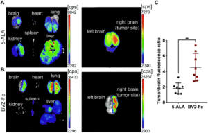
Fluorescence images of BV2-Fe accumulation at tumor sites in vivo

First, they activated BV2 microglial cells with citric-acid coated iron oxide nanoparticles (CIONPs) and loaded them with near-infrared fluorescent dye DiD (DiDBV2-Fe). Priming the cells with iron oxide nanoparticles downregulated M2 markers associated with the immunoresponse, and upregulated expression levels of genes that promote transportation of cells across the blood–brain barrier (BBB), thus achieving a two-fold favorable effect.

Next, they assessed the administration of DiDBV2-Fe in glioblastoma-bearing mice models via two routes:. intravenous and intracarotid artery injection. The latter route resulted in more efficient accumulation of activated cells in the brain tumor, 2.2 times higher than that of 5-ALA, 8 hours after application. Maximum fluorescence intensity images of brain tissues acquired at various timepoints from 2 to 24 hours using near-infrared imaging revealed clear tumor border demarcation. Confocal microscopy of harvested brain tumor sections showed noticeable co-localization of DiDBV2-Fe with the Ki67 positive tumor cells along with a significantly higher tumor-to-brain fluorescence ratio compared with 5-ALA (4.54 vs. 1.81).

Finally, they evaluated the in vivo preliminary safety of DiDBV2-Fe in comparison to 5-ALA. Administering DiDBV2-Fe did not induce acute liver injury, phototoxic or hypersensitivity reactions until a certain threshold was reached. In addition, the engineered microglia did not induce gene expression changes of the detected immunoregulatory proteins, unlike 5-ALA which induced both phototoxic and photoallergic reactions.

Taken together, the results indicate that these engineered microglial cells can serve as biological homing vehicles – in seeking out tumors and delivering optical imaging agents, which in turn can help surgeons navigate and identify tumor tissue via fluorescence during surgery.

To find out more please read:

Engineering microglia as intraoperative optical imaging agent vehicles potentially for fluorescence-guided surgery in gliomas

Ling Guo, Xiaochen Zhang, Runxiu Wei, Gaojie Li, Bingzhi Sun, Hongbo Zhang, Dan Liu, Cuifeng Wang and Min Feng

Biomater. Sci., 2020, 8, 1117-1126.

Digg This
Reddit This
Stumble Now!
Share on Facebook
Bookmark this on Delicious
Share on LinkedIn
Bookmark this on Technorati
Post on Twitter
Google Buzz (aka. Google Reader)

2020 Biomaterials Science Lectureship

The Biomaterials Science Lectureship is an annual award that honours an early-career researcher for their significant contribution to the biomaterials field. The recipient is selected by the Biomaterials Science Editorial Board from a list of candidates nominated by the community.

Dr Kanyi PuThis year we are delighted to award the Lectureship to Dr Kanyi Pu (Nanyang Technological University). He will present the Biomaterials Science lecture and receive his award at the 11th World Biomaterials Congress in Glasgow, UK in May 2020.

Dr. Pu is an Associate Professor in the School of Chemical and Biomedical Engineering (SCBE) at Nanyang Technological University. He is a highly cited researcher (2019 web of science), and the associate editor for ACS Applied Polymer Materials and Biomaterials Research, and Young Star Editor of Nano Research. He did his MS (2007) at Fudan University in China, his PhD (2011) at National University of Singapore in Singapore, and his postdoctoral training at Stanford University School of Medicine.

Dr. Pu has made significant contributions to the development of molecular imaging probes and technologies, particularly for photoacoustic imaging, chemiluminescence imaging and afterglow imaging. He is well known for his work on semiconducting polymer nanomaterials (SPNs) for molecular imaging, phototherapy and photoregulation. He is the inventor of the polymeric nanoreporters for molecular afterglow imaging and the molecular optical renal reporters for early diagnosis of acute kidney injury (AKI).

With a h-index of 65 (Jan 2020), he has won a number of awards for his creative work, including the distinguished lectureship award from the Chemistry Society of Japan, Wiley award for contribution in bioscience, young investigator travel award, and young innovator award in nanobiotechnology by Nano Research. He is the member of board of directors of Chinese American Society of Nanomedicine and Nanobiotechnology (CASNN). He also sits on the editorial advisory board of Advanced Functional Materials, Bioconjugate Chemistry, ACS Applied Bio Materials, Advanced Biosystems, Journal of Nanobiotechnology and ChemNanoMat.

To learn more about Kanyi’s research, have a look at his recent publications in Biomaterials Science and our sister journals:

Recent progress in the development of near-infrared organic photothermal and photodynamic nanotherapeutics
Houjuan Zhu, Penghui Cheng, Peng Chen and Kanyi Pu
Biomater. Sci., 2018, 6, 746-765

Near-infrared fluorescence probes to detect reactive oxygen species for keloid diagnosis
Penghui Cheng, Jianjian Zhang, Jiaguo Huang, Qingqing Miao, Chenjie Xu and Kanyi Pu
Chem. Sci., 2018, 9, 6340-6347

Near-infrared absorbing amphiphilic semiconducting polymers for photoacoustic imaging
Dong Cui, Chen Xie, Yan Lyu, Xu Zhen and Kanyi Pu
J. Mater. Chem. B, 2017, 5, 4406-4409

Multilayered semiconducting polymer nanoparticles with enhanced NIR fluorescence for molecular imaging in cells, zebrafish and mice
Houjuan Zhu, Yuan Fang, Xu Zhen, Na Wei, Yu Gao, Kathy Qian Luo, Chenjie Xu, Hongwei Duan, Dan Ding, Peng Chen and Kanyi Pu
Chem. Sci., 2016, 7, 5118-5125

Please join us in congratulating Kanyi on his award!

Digg This
Reddit This
Stumble Now!
Share on Facebook
Bookmark this on Delicious
Share on LinkedIn
Bookmark this on Technorati
Post on Twitter
Google Buzz (aka. Google Reader)

We are very pleased to welcome Professor Xinyuan Zhu to the Biomaterials Science Editorial Board. Read on to learn more about him!

Xinyuan Zhu is a full Professor of Polymer Science and Engineering at the School of Chemistry and Chemical Engineering, Shanghai Jiao Tong University, and Distinguished Professor of SJTU. Zhu focuses his research on the controlled preparation and biomedical applications of functional materials, including hyperbranched polymer synthesis, supramolecular polymer chemistry, drug and gene delivery, interactions between cells and polymers. He is a recipient of China National Funds for Distinguished Young Scientists (2010), and Cheung Kong Distinguished Professor (Ministry of Education of China, 2019). A number of his scientific research achievements have been applied in industries.

Read some of Xinyuan’s research below!

Methotrexate–Mn2+ based nanoscale coordination polymers as a theranostic nanoplatform for MRI guided chemotherapy
Yan Wu, Li Xu, Jiwen Qian, Leilei Shi, Yue Su, Youfu Wang, Dawei Li and Xinyuan Zhu
Biomater. Sci., 2020, 8, 712-719

Tirapazamine-embedded polyplatinum(iv) complex: a prodrug combo for hypoxia-activated synergistic chemotherapy
Dongbo Guo, Shuting Xu, Wumaier Yasen, Chuan Zhang, Jian Shen, Yu Huang, Dong Chen and Xinyuan Zhu
Biomater. Sci., 2020, 8, 694-701

Site-dependent fluorescence enhanced polymers with a self-restricted GFP chromophore for living cell imaging
Wenbin Fan, Hongping Deng, Lijuan Zhu, Chunlai Tu, Yue Su, Leilei Shi, Jiapei Yang, Linzhu Zhou, Li Xu and Xinyuan Zhu
Biomater. Sci., 2019, 7, 2421-2429

All these articles are currently FREE to read until 29th February 2020!

Digg This
Reddit This
Stumble Now!
Share on Facebook
Bookmark this on Delicious
Share on LinkedIn
Bookmark this on Technorati
Post on Twitter
Google Buzz (aka. Google Reader)

Welcome to our new Associate Editors Professor Lino Ferreira and Professor Khuloud Al-Jamal

We are very pleased to welcome Professor Lino Ferreira and Professor Khuloud Al-Jamal to the Biomaterials Science Editorial Board. Read more to learn all about them!

Lino Ferreira is the director of the Biomaterials and Stem Cell-Based Therapeutics research group at Center of Neurosciences and Cell Biology (CNC), at the University of Coimbra, Portugal, and the CNC coordinator of the MIT-Portugal Program. His current interests include the development of nanomateriais to track and manipulate exogenous and endogenous stem cells, and the development of 3D biomaterials to create synthetic stem cell niches in order to study and maximize the therapeutic potential of stem cells. For his work, he has recently received an European Research Council prize.

Read some of Lino’s research below!
A photodynamic antibacterial spray-coating based on the host–guest immobilization of the photosensitizer methylene blue
Tian-tian Yao, Jing Wang, Yun-fan Xue, Wei-jiang Yu, Qiang Gao, Lino Ferreira, Ke-Feng Ren and Jian Ji
J. Mater. Chem. B, 2019,7, 5089-5095

Intracellular delivery of more than one protein with spatio-temporal control
Miguel M. Lino, Susana Simões, Sónia Pinho and Lino Ferreira
Nanoscale, 2017,9, 18668-18680

Inflammatory modulation of stem cells by Magnetic Resonance Imaging (MRI)-detectable nanoparticles
Sezin Aday, Jose Paiva, Susana Sousa, Renata S. M. Gomes, Susana Pedreiro, Po-Wah So, Carolyn Ann Carr, Lowri Cochlin, Ana Catarina Gomes, Artur Paivad and Lino Ferreira
RSC Adv., 2014,4, 31706-31709


Professor Khuloud T. Al-Jamal is a Chair of Drug Delivery & Nanomedicine, King’s College London. She is also a registered pharmacist at the General Pharmaceutical Council. She started her academic career as a lecturer at King’s College London in 2011. She completed her pre-registration pharmacy training at The University College London Hospital and was awarded the Overseas Research Award Scheme (ORSA) Scholarship from The University of London (2000-2004) to complete her PhD in Drug Delivery from UCL-School of Pharmacy. She was awarded the prestigious CW Maplethorpe Research and Teaching Postdoctoral Fellowship from The University of London (2005-2007) to explore the use of cationic dendrimers as anti-angiogenic agents for growth inhibition of solid and metastatic tumours.

She has developed an extensive experience in designing and developing novel nanoscale delivery systems including dendrimers, liposomes, quantum Dots (QDs), polymers, viral vectors, chemically functionalised carbon nanotubes and graphene oxide. Her current work involves pre-clinical translation of novel nanomaterials designed specifically for drug, protein, nucleic acids and radionuclide delivery for therapeutic or diagnostic applications.

Learn more about Khuloud by reading some of her research articles below!

Design of antibody-functionalized carbon nanotubes filled with radioactivable metals towards a targeted anticancer therapy
Cinzia Spinato, Aritz Perez Ruiz de Garibay, Magdalena Kierkowicz, Elzbieta Pach, Markus Martincic, Rebecca Klippstein, Maxime Bourgognon, Julie Tzu-Wen Wang, Cécilia Ménard-Moyon, Khuloud T. Al-Jamal, Belén Ballesteros, Gerard Tobias and Alberto Bianco
Nanoscale, 2016,8, 12626-12638

Synthesis of double-clickable functionalised graphene oxide for biological applications
Kuo-Ching Mei, Noelia Rubio, Pedro M. Costa, Houmam Kafa, Vincenzo Abbate, Frederic Festy, Sukhvinder S. Bansal, Robert C. Hider and Khuloud T. Al-Jamal
Chem. Commun., 2015,51, 14981-14984

Cellular uptake mechanisms of functionalised multi-walled carbon nanotubes by 3D electron tomography imaging
Khuloud T. Al-Jamal, Hannah Nerl, Karin H. Müller, Hanene Ali-Boucetta, Shouping Li, Peter D. Haynes, Joerg R. Jinschek, Maurizio Prato, Alberto Bianco, Kostas Kostarelos and Alexandra E. Porter
Nanoscale, 2011,3, 2627-2635


As Biomaterials Science Associate Editors, Lino and Khuloud will be handling submissions to the journal. Why not submit your next paper to their Editorial Office?

Digg This
Reddit This
Stumble Now!
Share on Facebook
Bookmark this on Delicious
Share on LinkedIn
Bookmark this on Technorati
Post on Twitter
Google Buzz (aka. Google Reader)