Creating a blood substitute with polymers

Biomaterials Science web writer, Debanti Sengupta, highlights a HOT article from the journal

Researchers create a ‘blood substitute’ using hemoglobin and polymers that mimics the behavior of blood in rats.

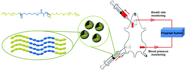
Graphical abstract: Asymmetric copolymer vesicles to serve as a hemoglobin vector for ischemia therapy

Blood transfusions are crucial for the successful treatment of many diseases and injuries. However, as we all know, donated blood can often be in short supply. Further, donated blood must always be screened for carrier diseases. In this study, the researchers aim to develop an encapsulation system for hemoglobin (a key component of blood transfusions) which can one day be used as a substitute for donated blood in the clinic.

The researchers used PEG (polyethylene-glycol) and PCL (poly caprolactone) as starting materials to build what they term ‘polymersomes’, or triblock copolymers consisting of a water-insoluble PCL segment and two water-soluble PEG chains. The hemoglobin-polymer aggregates were mixed into blood plasma substitute solutions. Once encapsulated, the hemoglobin-containing particles were studied to determine that the hemoglobin continued to be reactive. Most importantly, the researchers demonstrated that the encapsulated hemoglobin could continue to bind oxygen. Ascorbic acid was added to prevent hemoglobin from being oxidized to methemoglobin, an unreactive form of hemoglobin.

The researchers then demonstrated that hemoglobin could be contained in these polymersomes for up to 1 week with only 7% leakage of hemoglobin. The hemoglobin-polymersome particles were almost completely nontoxic to cells outside of the body, and did not interfere with regular blood components. Finally, animal studies were conducted where rats were first subjected to severe blood loss, followed by the introduction of hemoglobin-containing polymersomes. When hemoglobin-containing polymersomes were introduced, the animals recovered in a manner that was comparable to recovery after a blood transfusion. Further, the animals suffered only minimal inflammation after the polymersome treatment as compared to the introduction of non-encapsulated hemoglobin. This work is therefore an important step in demonstrating the feasibility of using similar ‘blood substitutes’ in clinical therapy in the future.

Asymmetric copolymer vesicles to serve as a hemoglobin vector for ischemia therapy
Bin Li, Yanxin Qi, ShaSha He, Yupeng Wang, Zhigang Xie, Xiabin Jing and Yubin Huang
Biomater. Sci., 2014, 2, 1254-1261 DOI: 10.1039/C4BM00123K

Debanti Sengupta recently completed her PhD in Chemistry from Stanford University.  She is currently a Siebel postdoctoral scholar at the University of California, Berkeley.

Follow the latest journal news on Twitter @BioMaterSci or go to our Facebook page.

Digg This
Reddit This
Stumble Now!
Share on Facebook
Bookmark this on Delicious
Share on LinkedIn
Bookmark this on Technorati
Post on Twitter
Google Buzz (aka. Google Reader)

Biomaterials Science 2013 Immediacy Index is 0.89

Biomaterials Science coverWe are pleased to announce that Biomaterials Science‘s first official Immediacy Index is 0.890– fifth highest for all the journals in the Materials Science, Biomaterials category, according to the 2013 Journal Citation Reports®.

Immediacy Index is a measure of how quickly articles in a journal are cited, and is calculated as the average number of citations articles receive in the year they are published.  Our high number and ranking indicate that, even as a new journal, Biomaterials Science is making a splash in the community.  We would like to thank all our readers, authors and board members for their contribution to the early success of the journal.

Biomaterials Science will receive its first (partial) Impact Factor next year in the 2014 Journal Citation Reports®. For the latest Impact Factors of other Royal Society of Chemistry journals, take a look at this blog post.

We recommend these highly cited Biomaterials Science articles

Montserrat Colilla, Blanca González and María Vallet-Regí
Yanan Yue and Chi Wu

Interested in publishing your own high impact paper with Biomaterials Science? Submit online today, or contact the Editorial Office.

Digg This
Reddit This
Stumble Now!
Share on Facebook
Bookmark this on Delicious
Share on LinkedIn
Bookmark this on Technorati
Post on Twitter
Google Buzz (aka. Google Reader)

Nanovehicles for targeted bone metastasis chemotherapy

The cover story for the July 2014 issue of Biomaterials Science highlights the use of nanovehicle particles for the targeted delivery of chemotherapy drugs to tumor cells in bone.

Bone metastasis, or the spreading of tumor cells from the initial tumor site to bone tissue, marks the stage of cancer progression where the diseased is deemed incurable. Currently, the main method to treat bone metastasis is to utilize aggressive chemotherapy drugs and destroy metastatic tumor cells.  Unfortunately, untargeted chemotherapy treatments result in death of both cancerous and healthy tissue. Treatments could be significantly improved with the technology to specifically target the destruction of tumor cells without affecting the surrounding bone tissue.

Researchers at the University of Utah, University of Iowa, and Wuhan University in China are making this desired technology a reality. Wang et al. have developed a nanovehicle capable of targeting tumor cells within bone tissue.  Nanovehicles (NVs) are essentially nano-sized cargo containers capable of delivering drugs to a desired destination. The cleverly designed NVs are designed to target diseased bone tissue to deliver chemotherapy drugs at a specific site.

The NVs developed in this study have three design features:

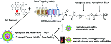
(1) The NVs core is made of hydrophilic and hydrophobic block co-polymers able to form a shell-like structure known as a micelle. The micelle serves as the cargo vessel that contains the chemotherapy drug doxorubicin.

(2) The outermost segments of the NVs are negatively charged bone-targeting peptides. The peptide is responsible for the accumulation of NVs in skeletal tissue, and the negative charge prevents cellular uptake of the NVs into healthy tissue.

(3) For uptake into cancer cells, the particle must be positively charged. Therefore, a positively charged peptide linker was added in between the micelle core and the negatively charged bone-targeting region. The peptide linker is cleavable by cathepsin K enzyme (CTSK), which is overexpressed in bone metastatic lesions. When the NV enters a metastatic lesion, the CTSK cleaves off the negatively charged peptides and exposes the positively charged peptides. This charge reversal allows for tumor cells present in the metastatic lesion to uptake the NV.

After extensively characterizing the nanoparticles with NMR and zeta potential measurements, the NVs were used to observe decreased tumor cell viability under CTSK-rich conditions in vitro. More strikingly, in vivo tests of the NVs in a mouse model of bone metastasis showed significant increases in mouse survival and decreases in overall tumor burden.

Collectively, these ingeniously designed nanovehicles show great promise in providing an effective targeted therapy for bone metastasis.

Peptide decoration of nanovehicles to achieve active targeting and pathology-responsive cellular uptake for bone metastasis chemotherapy
X. Wang, Y. Yang, H. Jia, W. Jia, S. Miller, B. Bowman, J. Feng, and F. Zhan
Biomater. Sci., 2014, Advance Article DOI: 10.1039/C4BM00020J

Brian Aguado is currently a Ph.D. Candidate and NSF Fellow in the Biomedical Engineering department at Northwestern University. He holds a B.S. degree in Biomechanical Engineering from Stanford University and a M.S. degree in Biomedical Engineering from Northwestern University. Read more about Brian’s research publications here.

Follow the latest journal news on Twitter @BioMaterSci or go to our Facebook page.

Digg This
Reddit This
Stumble Now!
Share on Facebook
Bookmark this on Delicious
Share on LinkedIn
Bookmark this on Technorati
Post on Twitter
Google Buzz (aka. Google Reader)

Protein corona key to safe, personalized nanomedicine

Biomaterials Science web writer, Robert van Lith, highlights a HOT article from the journal

This study introduces the concept of the “personalized protein corona” as a determinant factor in nano-biomedicine, specifically regarding the biological identity and fate of nanoparticles.

Graphical abstract: Personalized protein coronas: a “key” factor at the nanobiointerfaceThe concept of personalized medicine has become increasingly relevant in healthcare over the past decade. Ever since the human genome was unraveled, the idea of tailoring therapies specific to the individual patient has been at the forefront of medicine. Diagnosis, prognosis and most importantly treatment are thought to eventually be adapted to each individual’s genetic and pathophysiological makeup. Nonetheless, biomaterials for invasive use (e.g. implantation or injection) are still largely being designed for general use. This study argues for a much more specific approach of biomaterials design, looking at the nanoparticles (NPs) in particular.

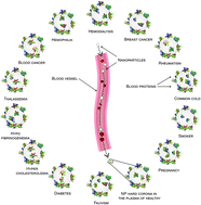
A collaborative effort headed by Dr. Mahmoudi from Stanford University, US, and the University of Medical Sciences in Tehran, Iran, Hajipour et al. evaluated effects of the altered protein composition of human plasma in different disease states on NP corona formation. Two NP materials were chosen for their analysis, hydrophobic polystyrene and hydrophilic silica, which are widely investigated for biomedical material approaches and currently evaluated for safety purposes. 

 NPs were exposed to plasma solutions of different patient populations to form hard protein coronas, after which the composition of NP protein coronas was analyzed using SDS-page, densitometry and zeta-potential measurements. Interestingly, the authors found that changes in plasma protein composition due to underlying conditions alter the protein decoration on NPs, resulting in an unpredictable biologic identity of the NPs and thus their biological fate. The severity of the condition was also found to impact protein deposition. Intriguingly, even among healthy subjects there was considerable variation in corona composition, creating even more of an impetus for personalized NP design.

Collectively, the researchers found that NP protein corona distinctly depends on particular condition, severity of condition and combination of conditions, with relatively low variation between subjects with similar health profiles. Moving forward, identifying the personalized protein corona for individual patients or at least the expected protein corona in specific patient populations may become an integral aspect of nanoparticle-based biomaterial therapies.

Personalized protein coronas: a “key” factor at the nanobiointerface 
M. J. Hajipour, S. Laurent, A. Aghaie, F. Rezaee and M. Mahmoudi
Biomater. Sci., 2014, Advance Article DOI: 10.1039/c4bm00131a

Robert van Lith


Robert van Lith is currently a Ph.D. Candidate in the Biomedical Engineering department at Northwestern University, working on novel biomaterials to modulate oxidative stress in tissues. He received an American Heart Association Fellowship and Society for Biomaterials award for his work. He holds B.S. and M.S. degrees in Biomedical Engineering from Eindhoven University of Technology, the Netherlands. Read more about Robert’s research publications here.

Digg This
Reddit This
Stumble Now!
Share on Facebook
Bookmark this on Delicious
Share on LinkedIn
Bookmark this on Technorati
Post on Twitter
Google Buzz (aka. Google Reader)

NanoBio Australia 2014 Biomaterials Science Poster Prize Winners

Biomaterials Science was delighted to award Poster Prizes at the NanoBio Australia 2014 conference held at  The University of Queensland, St. Lucia, Australia on 6th – 10th July 2014. The winner was Young-Seon Ko and the Runner-up was Ms Liyu Chen.
Young-Seon Ko and Liyu Chen receiving their poster prize.
Digg This
Reddit This
Stumble Now!
Share on Facebook
Bookmark this on Delicious
Share on LinkedIn
Bookmark this on Technorati
Post on Twitter
Google Buzz (aka. Google Reader)

High throughput evaluation of surfaces for stem cell culture

Polymer microarrays for stem cell adhesion studies

This study identifies new surfaces optimized for human pluripotent stem cell expansion using a high-throughput polymer microarray chip.

The biomaterials community is moving toward using high-throughput tools to evaluate cell-material interactions at an unprecedented rate. For instance, polymer microarrays have become popular for identifying candidate surfaces that elicit a desired biological response. Using a single microarray chip, low volumes of polymer solution are deposited in a grid-like format using robotic ink-jet or contact printing. The chip is polymerized to manufacture a polymer microarray. After fabricating the microarray, cells are deposited directly onto the spots of the chip and cultured to determine materials that support a specific cell phenotype.

At the Laboratory of Biophysics and Surface Analysis at the University of Nottingham, Celiz et al. evaluated human pluripotent stem cell growth on a polymer microarray containing 141 varieties of (meth)acrylate and (meth)acrylamide photo-curable polymers. To maximize the diversity of the microarray, monomers containing a variety of nitrogen, fluorine, oxygen, aromatic, and aliphatic side chains were utilized.  Monomers were selected on their ability to be photo-polymerized by UV irradiation. The surface chemistry after polymerization was assessed using ToF-SIMS, and water contact angle measurements determined the surface wettability of each spot.

Partial least squares analysis (PLS) was utilized to correlate surface chemistry and wettability to identify surfaces that would yield high human pluripotent stem cell (hPSC) adhesion. Of the 141 surfaces screened in the array, 47 polymers supported hPSC attachment. Interestingly, no relationship was observed between surface wettability and cell adhesion, indicating wettability of a surface is not sufficient to predict hPSC adhesion. PLS analysis subsequently identified correlations between polymer surface chemistry and experimental hPSC adhesion. Additional experiments confirmed that increased protein adsorption on specific polymer spots was a contributor to cell adhesion to the polymer surface.

Collectively, the polymer array developed in this study was able to operate as a high-throughput tool to identify surfaces amenable to hPSC adhesion. Moving forward, polymer microarrays have the potential to identify a broad library of surfaces capable of supporting sustained hPSC growth and pluripotency.

Chemically diverse polymer microarrays and high throughput surface characterization: a method for discovery of materials for stem cell culture
A. D. Celiz, J. G. W. Smith, A. K. Patel, R. Langer, D. G. Anderson, D. A. Barrett, L. E. Young, M. C. Davies, C. Denning and M. R. Alexander
Biomater. Sci., 2014, Advance Article DOI: 10.1039/C4BM00054D

Brian Aguado is currently a Ph.D. Candidate and NSF Fellow in the Biomedical Engineering department at Northwestern University. He holds a B.S. degree in Biomechanical Engineering from Stanford University and a M.S. degree in Biomedical Engineering from Northwestern University. Read more about Brian’s research publications here.

Follow the latest journal news on Twitter @BioMaterSci or go to our Facebook page.

Digg This
Reddit This
Stumble Now!
Share on Facebook
Bookmark this on Delicious
Share on LinkedIn
Bookmark this on Technorati
Post on Twitter
Google Buzz (aka. Google Reader)

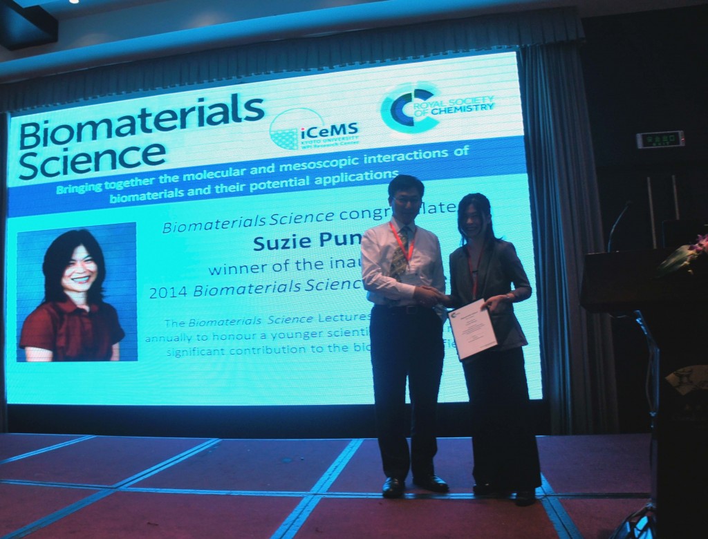
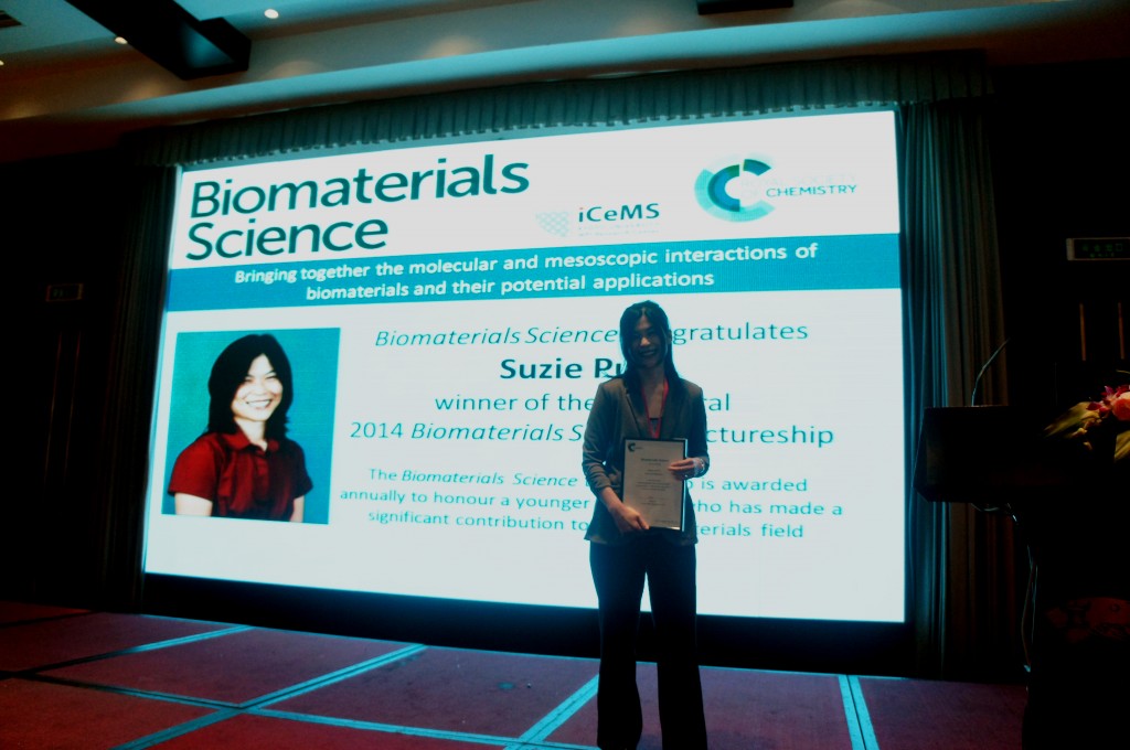
2014 Biomaterials Science Lectureship Awarded to Suzie Pun

It is with great pleasure that we announce Professor Suzie Pun (University of Washington) as the recipient of the inaugural Biomaterials Science Lectureship.

This award, which will be an annual event for the journal,  honours a younger scientist who has made a significant contribution to the biomaterials field. The recipient is selected by the Biomaterials Science Editorial Board from a list of candidates nominated by the community.

More about Suzie…

Suzie H. Pun received her Chemical Engineering Ph.D. degree in 2000 from the California Institute of Technology.  She then worked as a senior scientist at Insert Therapeutics/Calando Pharmaceuticals for 3 years developing polymeric drug delivery systems before joining the Department of Bioengineering at University of Washington (UW).  She is currently the Robert J Rushmer Associate Professor of Bioengineering, an Adjunct Associate Professor of Chemical Engineering, and a member of the Molecular Engineering and Sciences Institute at UW.  Her research focus area is in drug and gene delivery systems and she has published over 70 research articles in this area.  Current application areas for her group include biologics delivery to the central nervous system and cancer.  For this work, she was recognized with a Presidential Early Career Award for Scientists and Engineers in 2006. 

Take a look at this paper for an example of Suzie’s recent research:

Comparative study of guanidine-based and lysine-based brush copolymers for plasmid delivery Peter M. Carlson, Joan G. Schellinger, Joshuel A. Pahang, Russell N. Johnson and Suzie H. Pun  
Biomater. Sci., 2013, 1, 736-744 DOI: 10.1039/C3BM60079C 

We would like to thank everybody who nominated a candidate for the Lectureship- we received many excellent nominations, and the Editorial Board had a difficult task in choosing between some outstanding candidates.

We invite you to join us in congratulating Suzie in the comments below.

Digg This
Reddit This
Stumble Now!
Share on Facebook
Bookmark this on Delicious
Share on LinkedIn
Bookmark this on Technorati
Post on Twitter
Google Buzz (aka. Google Reader)

Top 10 Most-accessed Biomaterials Science articles – Q1 2014

This month sees the following articles in Biomaterials Science that are in the top ten most accessed from January – March:

Stimuli-responsive functionalized mesoporous silica nanoparticles for drug release in response to various biological stimuli
Xin Chen, Xiaoyu Cheng, Alexander H. Soeriyadi, Sharon M. Sagnella, Xun Lu, Jason A. Scott, Stuart B. Lowe, Maria Kavallaris and J. Justin Gooding
Biomater. Sci., 2014,2, 121-130
DOI: 10.1039/C3BM60148J

Hyaluronic acid hydrogel stiffness and oxygen tension affect cancer cell fate and endothelial sprouting
Yu-I Shen, Hasan E. Abaci, Yoni Krupski, Lien-Chun Weng, Jason A. Burdick and Sharon Gerecht
Biomater. Sci., 2014,2, 655-665
DOI: 10.1039/C3BM60274E

Hydrogel scaffolds as in vitro models to study fibroblast activation in wound healing and disease
Megan E. Smithmyer, Lisa A. Sawicki and April M. Kloxin
Biomater. Sci., 2014,2, 634-650
DOI: 10.1039/C3BM60319A

Fabrication of zeolite–polymer composite nanofibers for removal of uremic toxins from kidney failure patients
Koki Namekawa, Makoto Tokoro Schreiber, Takao Aoyagi and Mitsuhiro Ebara
Biomater. Sci., 2014,2, 674-679
DOI: 10.1039/C3BM60263J

Enzyme responsive materials: design strategies and future developments
Mischa Zelzer, Simon J. Todd, Andrew R. Hirst, Tom O. McDonald and Rein V. Ulijn
Biomater. Sci., 2013,1, 11-39
DOI: 10.1039/C2BM00041E

Mesoporous silica nanoparticles for the design of smart delivery nanodevices
Montserrat Colilla, Blanca González and María Vallet-Regí
Biomater. Sci., 2013,1, 114-134
DOI: 10.1039/C2BM00085G

Smart hydrogels as functional biomimetic systems
Han L. Lim, Yongsung Hwang, Mrityunjoy Kar and Shyni Varghese
Biomater. Sci., 2014,2, 603-618
DOI: 10.1039/C3BM60288E

Non-eroding drug-releasing implants with ordered nanoporous and nanotubular structures: concepts for controlling drug release
Moom Sinn Aw, Mima Kurian and Dusan Losic
Biomater. Sci., 2014,2, 10-34
DOI: 10.1039/C3BM60196J

Fracture-based micro- and nanofabrication for biological applications
Byoung Choul Kim, Christopher Moraes, Jiexi Huang, M. D. Thouless and Shuichi Takayama
Biomater. Sci., 2014,2, 288-296
DOI: 10.1039/C3BM60276A

Nanoscale semiconductor devices as new biomaterials
John Zimmerman, Ramya Parameswaran and Bozhi Tian
Biomater. Sci., 2014,2, 619-626
DOI: 10.1039/C3BM60280J

Why not take a look at the articles today and blog your thoughts and comments below.

Fancy submitting an article to Biomaterials Science? Then why not submit to us today!

Digg This
Reddit This
Stumble Now!
Share on Facebook
Bookmark this on Delicious
Share on LinkedIn
Bookmark this on Technorati
Post on Twitter
Google Buzz (aka. Google Reader)

Cancer therapy with nanoparticles

Biomaterials Science web writer, Debanti Sengupta, highlights a HOT article from the journal

Researchers have demonstrated that controlling nanoparticle shape and packing can impact how cancer therapeutics interact with cells.

Photodynamic therapy is an innovative emerging therapy for cancer.  This therapeutic modality consists of introducing light-activated molecules known as ‘photosensitizers’ into cancer cells.  When activated with light, these photosensitizers are able to produce toxic molecules within the cells (also known as reactive oxygen species or ROS) that can eventually cause cancer cell death.  Photosensitizers are often introduced into the cells using nanoparticle carriers. In order for effective cell death to occur, ROS must be formed within the nanoparticles and then diffuse out of the nanoparticles into the cells.

In this article, Chu et al. have designed silica-based nanocarriers that can control how photosensitizers interact with cells. They previously found that when photosensitizer-loaded nanoparticles were packed more densely, the amount of ROS that was released decreased.  In contrast, more loosely packed nanoparticles allowed for greater release of ROS into cells. In this study, the researchers designed a third type of nanoparticle consisting of a gold nanorod coated with dense silica. They found that this third type of nanoparticle released photosensitizers much more efficiently than the either densely packed or loosely packed silica-only particles. They also discovered that the nanorod-based particles produced a different distribution of ROS as compared to the loose or dense silica-only particles. When cells were treated with these nanoparticles, loosely-packed silica particles and the nanorod-based particles demonstrated dramatic decreases in cell-viability due to the action of the photosensitizers. Further, while the silica-based loose and dense particles caused cells to die via a programmed cell death (apoptotic) route, the nanorod-based particles caused cell death via cell injury resulting in premature cell death (necrosis).

These experiments suggest that controlling the composition and shape of the nanoparticles that carry photosensitizers to cancer cells can alter both the number of cancer cells killed as well as the mechanism by which the cells die. This work has implications for future photodynamic therapeutic strategies.

Zhiqin Chu, Silu Zhang, Chun Yin, Ge Lin and Quan Li
Biomater. Sci., 2014, Advance Article DOI: 10.1039/C4BM00024B

Debanti Sengupta recently completed her PhD in Chemistry from Stanford University.  She is currently a Siebel postdoctoral scholar at the University of California, Berkeley.

Follow the latest journal news on Twitter @BioMaterSci or go to our Facebook page.

Digg This
Reddit This
Stumble Now!
Share on Facebook
Bookmark this on Delicious
Share on LinkedIn
Bookmark this on Technorati
Post on Twitter
Google Buzz (aka. Google Reader)

Cerebellar neuron development on hybrid matrix constructs

The cerebellum is a region in the brain responsible for regulating motor control and cognitive functions such as attention and language. During development of the cerebellum (and of other tissues), cells interact with the surrounding microenvironment known as the extracellular matrix (ECM). These neuron-ECM interactions regulate neuronal differentiation, growth, formation of synapses, and neurite outgrowth. Specifically, ECM components including collagen and laminin-1 (lam-1) are known to regulate the alignment, migration, and neurite outgrowth of Purkinje cells (PCs). However, the dual signaling roles of collagen and laminin-1 during cerebellar tissue development have not been fully explored.

Dr. Shantanu Sur, a postdoctoral fellow in Prof. Samuel Stupp’s laboratory at Northwestern University, has collaborated with Dr. Thomas Launey at the RIKEN Brain Science Institute in Japan to explore cerebellar tissue development using biomaterial hydrogels. Sur has developed an artificial matrix consisting of collagen and synthetic peptide amphiphile (PA) molecules presenting IKVAV, the peptide on laminin-1 responsible for cell adhesion and neurite outgrowth. Sur et al. evaluated the spatiotemporal expression of lam-1 and collagen in rat cerebellums during PC development (embryonic to post-natal) using histology and immunostaining. Given the changes in the ratio of lam-1 to collagen during PC formation and growth, these results suggest the dynamic involvement of these ECM proteins in forming the neural architecture of the cerebellum.

Using a biomimetic approach to mimic the critical lam-1 to collagen ratio, Sur proceeded to model the dynamic nature of the ECM during PC development with a synthetic collagen-PA hydrogel. Collagen (types I-V) and IKVAV-PA molecules were mixed together in solution at varying concentrations and gelled using ammonia vapor. This simple method allowed Sur to evaluate the density of PC growth, axon guidance, and dendrite morphology in gels using a wide array of collagen and IKVAV-PA concentrations. Strikingly, effects on PC phenotype were observed as a function of the collagen:IKVAV-PA ratio and not the absolute concentrations of each ECM component within the matrix.

Sur comments that the hybrid matrix provides an easily tunable environment to enable the in vitro testing of the role of ECM signals on neuronal maturation. “Our study shows that the optimal ECM-derived cues for neurons change at specific stages of development,” Sur says. “This observation will drive us to work on the design of a dynamic matrix where the extracellular signals delivered to the neuron can be tuned spatiotemporally.” Additionally, the collagen/IKVAV-PA gels may be used to identify cell-ECM interactions during the development of other tissue types, given the simplicity of the technique.

Collectively, this study demonstrates the exciting use of engineered matrices to evaluate spatiotemporal cell-ECM interactions, with the hopes to further elucidate mechanisms of tissue development.

Synergistic regulation of cerebellar Purkinje neuron development by laminin epitopes and collagen on an artificial hybrid matrix construct
Shantanu Sur, Mustafa O. Guler, Matthew J. Webber, Eugene T. Pashuck, Masao Ito, Samuel I. Stupp, and Thomas Launey
Biomater. Sci., 2014, Advance Article, DOI: 10.1039/C3BM60228A

Brian Aguado is currently a Ph.D. Candidate and NSF Fellow in the Biomedical Engineering department at Northwestern University. He holds a B.S. degree in Biomechanical Engineering from Stanford University and a M.S. degree in Biomedical Engineering from Northwestern University. Read more about Brian’s research publications here.

To keep up-to-date with all the latest research, sign-up to our RSS feed or Table of contents

Digg This
Reddit This
Stumble Now!
Share on Facebook
Bookmark this on Delicious
Share on LinkedIn
Bookmark this on Technorati
Post on Twitter
Google Buzz (aka. Google Reader)