Growing replacement bones – is biomaterial geometry important?

Biomaterials Science web writer, Debanti Sengupta, highlights a popular article from the journal

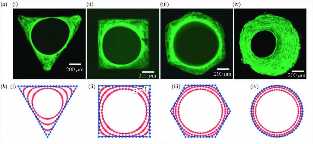
Staining of actin stress fibers

Staining of actin stress fibers to visualize the tissue formed in vitro and to study the effects of curvature (a). The predicted tissue regeneration based on a linear curvature-dependent theoretical model is depicted in subfigure (b). Theoretical predictions match these in vitro experimental observations.

In this paper, Professor Amir Zadpoor reviews the role of biomaterial scaffold geometries on regenerating bone tissue.  Scaffold curvature, pore size, and pore shape are all shown to be important in stimulating more bone growth.

In the case of large bone injuries, the role of scaffolds in regenerating bones becomes increasingly critical.  The ideal bone scaffolds need to be biocompatible, mechanically strong, and contain pores that allow the transport of nutrients and new cell growth.

The geometry of the scaffold plays a very important role in regenerating bone tissue, and is explored in this paper.  Broadly speaking, the curvature of the scaffold, the shape, and the size of the pores are all key components that influence how well the bone is structured and grows.

Strikingly, it has been found that curvature of the surface proportionally impacts the rate of tissue regeneration. Curvature of the surface can be much smaller than, on the scale of, or much larger than cells that grow on the surface. Smaller curvatures can impact individual cell focal adhesions, through which cells can both sense the underlying surface and apply force to it. Larger curvatures can impact cell stress fibers as well as the overall forces on the growing bone tissue.

Pore size can also dramatically impact bone regeneration.  It is already known that a limited amount of inflammation is actually good for bone growth.  Pore size can affect inflammation – for example, larger pores and wider scaffold geometry angles can increase local inflammation.  Pore size can also dictate the amount of oxygen and nutrients reaching the cells, and whether cartilage or bone is formed first.

Pore shape is also extremely important – for example, it has been shown that when cells are grown in scaffolds shaped like parallelograms, alkaline phosphatase activity is increased. Since alkaline phosphatase is a byproduct of bone growth, this data suggests a role for the shape of scaffold pores in bone generation.

However, there are significant caveats to be considered when studying bone regeneration in the lab. Since bone grows in stages, a short in vitro study may not capture all nuances of bone growth that occur in vivo. It is important to study in vitro and in vivo bone generation in parallel to reconcile any contradictory data. Further, it must be noted that it is often difficult to isolate the independent impact of one specific property of a scaffold without affecting another property.  For example, changing pore size can also change the overall mechanical properties of the scaffold.

Despite these limitation, it is clear that the geometrical properties of the scaffold can significantly impact bone growth.  Computational studies and modeling are also now being used to optimize many scaffold properties, and have the potential to drive further research to elucidate the role of scaffold geometry in clinically valuable bone growth.

Check out the full article:

Bone tissue regeneration: the role of scaffold geometry by Amir A. Zadpoor


Web writer Debanti Sengupta

Web writer Debanti Sengupta


Debanti Sengupta completed her PhD in Chemistry in 2012 from Stanford University.  She was previously a Siebel postdoctoral scholar at the University of California, Berkeley, and is currently a postdoctoral scholar in Radiation Oncology at Stanford University. Follow her on Twitter @yoginiscientist.
Follow the latest journal news on Twitter @BioMaterSci or go to our Facebook page.

Digg This
Reddit This
Stumble Now!
Share on Facebook
Bookmark this on Delicious
Share on LinkedIn
Bookmark this on Technorati
Post on Twitter
Google Buzz (aka. Google Reader)

12th International Conference on Materials Chemistry (MC12) – call for abstracts

We are delighted to announce that 12th International Conference on Materials Chemistry (MC12) will be held at the University of York on 20 – 23 July 2015. Abstracts are now invited for this event so submit today and take advantage of this excellent opportunity to present your work alongside scientists from across the globe.

This cutting edge international research conference is organised around four exciting and diverse areas of the application of materials chemistry. One prominent theme at MC12 is Biomaterials, encompassing materials for tissue engineering, biomaterials for healthcare, green biomaterials and advanced synthesis methods of biomaterials. Visit http://rsc.li/mc12 to find out more and submit your abstract today.

Digg This
Reddit This
Stumble Now!
Share on Facebook
Bookmark this on Delicious
Share on LinkedIn
Bookmark this on Technorati
Post on Twitter
Google Buzz (aka. Google Reader)

Biomaterials Science Scope and Standards

Visitors to the Biomaterials Science website may have noticed that we have made some changes to the journal’s scope statement.  These changes are the result of conversations with researchers about how the journal can best serve a dynamic, multi-disciplinary and still relatively young research field.

Over its first two years of publication, Biomaterials Science has begun to establish itself as a home for research which provides insight into the fundamental science of biomaterials.  With our updated scope statement, we hope to emphasise what distinguishes the journal from others in the field.

To help authors get a better feel of the scope and standards of Biomaterials Science, we have included a list of papers that we believe are particularly characteristic of the journal. This list will be regularly updated as we continue to publish cutting edge biomaterials science research.

Our updated scope statement:

Biomaterials Science is an international, high impact journal exploring the underlying science behind the function, interactions and design of biomaterials. Its scope encompasses insights into the chemistry, biology and materials science underpinning biomaterials research, new concepts in biomaterials design, and using materials to answer fundamental biological questions.

The journal is a collaborative venture between the Royal Society of Chemistry and the Institute for Integrated Cell-Material Sciences, Kyoto University, Japan.  It publishes primary research and review-type articles which advance fundamental understanding in areas including:

Molecular design of biomaterials, including proof of concept studies

How to optimize binding of coated nanoparticles: coupling of physical interactions, molecular organization and chemical state
R. J. Nap and I. Szleifer

Incorporation of sulfated hyaluronic acid macromers into degradable hydrogel scaffolds for sustained molecule delivery
Brendan P. Purcell, Iris L. Kim, Vanessa Chuo, Theodore Guenin, Shauna M. Dorsey and Jason A. Burdick

Science of cells and materials at the mesoscale  

Mesoscopic science, where materials become life and life inspires materials
Norio Nakatsuji

Sub-100 nm patterning of TiO2 film for the regulation of endothelial and smooth muscle cell functions
R. Muhammad, S. H. Lim, S. H. Goh, J. B. K. Law, M. S. M. Saifullah, G. W. Ho and E. K. F. Yim

Changing ligand number and type within nanocylindrical domains through kinetically constrained self-assembly – impacts of ligand ‘redundancy’ on human mesenchymal stem cell adhesion and morphology
Haiqing Li and Justin J. Cooper-White

Materials as model systems for stem cell biology

Biomaterial arrays with defined adhesion ligand densities and matrix stiffness identify distinct phenotypes for tumorigenic and non-tumorigenic human mesenchymal cell types
Tyler D. Hansen, Justin T. Koepsel, Ngoc Nhi Le, Eric H. Nguyen, Stefan Zorn, Matthew Parlato, Samuel G. Loveland, Michael P. Schwartz and William L. Murphy

Artificial microniches for probing mesenchymal stem cell fate in 3D
Yujie Ma, Martin P. Neubauer, Julian Thiele, Andreas Fery and W. T. S. Huck

Synthetic hydrogel platform for three-dimensional culture of embryonic stem cell-derived motor neurons
Daniel D. McKinnon, April M. Kloxin and Kristi S. Anseth

A high-throughput polymer microarray approach for identifying defined substrates for mesenchymal stem cells
Cairnan R. E. Duffy, Rong Zhang, Siew-Eng How, Annamaria Lilienkampf, Guilhem Tourniaire, Wei Hu, Christopher C. West, Paul de Sousa and Mark Bradley

Materials for tissue engineering and regenerative medicine

A microstereolithography resin based on thiol-ene chemistry: towards biocompatible 3D extracellular constructs for tissue engineering
Ian A. Barker, Matthew P. Ablett, Hamish T. J. Gilbert, Simon J. Leigh, James A. Covington, Judith A. Hoyland, Stephen M. Richardson and Andrew P. Dove

Evaluation of MMP substrate concentration and specificity for neovascularization of hydrogel scaffolds
S. Sokic, M. C. Christenson, J. C. Larson, A. A. Appel, E. M. Brey and G. Papavasiliou

Heparin-induced conformational changes of fibronectin within the extracellular matrix promote hMSC osteogenic differentiation
Bojun Li, Zhe Lin, Maria Mitsi, Yang Zhang and Viola Vogel

Materials and systems for therapeutic delivery

Cargo delivery to adhering myoblast cells from liposome-containing poly(dopamine) composite coatings
Martin E. Lynge, Boon M. Teo, Marie Baekgaard Laursen, Yan Zhang and Brigitte Städler

“Nail” and “comb” effects of cholesterol modified NIPAm oligomers on cancer targeting liposomes
Wengang Li, Lin Deng, Basem Moosa, Guangchao Wang, Afnan Mashat and Niveen M. Khashab

Protein–polymer therapeutics: a macromolecular perspective
Yuzhou Wu, David Y. W. Ng, Seah Ling Kuan and Tanja Weil

Interactions at the biointerface

Fibronectin-matrix sandwich-like microenvironments to manipulate cell fate
J. Ballester-Beltrán, D. Moratal, M. Lebourg and M. Salmerón-Sánchez

Biophysical properties of nucleic acids at surfaces relevant to microarray performance
Archana N. Rao and David W. Grainger

Biologically inspired and biomimetic materials, including bio-inspired self-assembly systems and cell-inspired synthetic tools

DNA origami technology for biomaterials applications
Masayuki Endo, Yangyang Yang and Hiroshi Sugiyama

Quantitative study on the antifreeze protein mimetic ice growth inhibition properties of poly(ampholytes) derived from vinyl-based polymers
Daniel E. Mitchell, Mary Lilliman, Sebastian G. Spain and Matthew I. Gibson

Next-generation tools and methods for biomedical applications

Application of biomaterials for the detection of amyloid aggregates
Tamotsu Zako and Mizuo Maeda

Alteration of epigenetic program to recover memory and alleviate neurodegeneration: prospects of multi-target molecules
Ganesh N. Pandian, Rhys D. Taylor, Syed Junetha, Abhijit Saha, Chandran Anandhakumar, Thangavel Vaijayanthi and Hiroshi Sugiyama

Nanoscale semiconductor devices as new biomaterials
John Zimmerman, Ramya Parameswaran and Bozhi Tian

Digg This
Reddit This
Stumble Now!
Share on Facebook
Bookmark this on Delicious
Share on LinkedIn
Bookmark this on Technorati
Post on Twitter
Google Buzz (aka. Google Reader)

Bioprinting vascular networks for tissue-engineered organs

Robert van Lith highlights a hot article in Biomaterials Science

Ex vivo engineering of 3D organs for transplantation purposes has made tremendous strides in recent years, yet complex tissues remain challenging due to their need for vascularization. A new study takes great steps towards solving this issue using a new, simple bioprinting process to create branched vascular networks capable of effective media exchange. The introduced methodology provides a pathway towards successful incorporation of a branched vasculature into tissue-engineered organs.


In vitro study of directly bioprinted perfusable vasculature conduits

The concept of tissue engineering – combining cells with biomaterials to create living, functional tissues – to provide a solution to the lack of suitable organs for transplantation has been tremendously popular for several decades now, and for good reason. It harnesses the potential to not only tailor organ characteristics to individual patients and reduce transplant rejection risks, but also to virtually erase waiting time for patients in need. The combined efforts of numerous researchers have already led to great success in engineering simple tissues such as skin, yet the evolution towards engineering more complex tissues has proven to be highly challenging. A major roadblock has been, and continues to be, the need for nutrient delivery and media exchange for living tissues to survive, let alone thrive. The incorporation of a vascular network is required for this, yet current methods for creating these networks can either not generate efficient, perfusable systems with the necessary mechanical properties, or are too complex to effectively utilize in thick tissues.

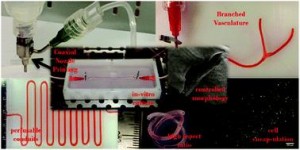
To address the current challenges, Ibrahim Ozbolat’s research group at the University of Iowa designed a novel system for bioprinting vascular conduits. Bioprinting allows for precise 3D fabrication of cell constructs, usually using a sacrificial support biomaterial. In his study, Dr. Ozbolat’s system consists of a coaxial nozzle in which cell-loaded alginate is dispensed through the sheath, and a crosslinking calcium chloride solution through the core, to allow for instantaneous formation of hollow fiber conduits without the need for post-fabrication procedures. The nozzle is under precise robotic control, allowing fabrication of conduits of desired dimension and geometry.

In the current work, the applicability of the bioprinting unit is demonstrated using human umbilical vein smooth muscle cells (HUVSMCs) embedded in alginate as the vessel wall material. By varying alginate and calcium chloride concentrations, conduit properties could be controlled. Relevant properties reported here include vascular lumen and wall dimensions, burst pressures and wall permeability to allow nutrient diffusion. Especially the latter is of paramount importance for long-term functioning of complex engineered tissues. Importantly, this report also shows that despite significant loss of viability during the bioprinting process, alginate-encapsulated cell recovered completely and proliferated well, laying down extracellular matrix throughout the vessel wall as evidenced by histology. Perhaps most intriguing is the demonstrated formation of branched vascular conduits using Dr. Ozbolat’s system.

The straight-forward bioprinting system reported here, allowing for tight control of vascular conduit dimensions, mechanical and perfusion properties, represents a highly promising platform for incorporating effective media exchange and nutrient transport in 3D engineered tissues. Especially the unique lack of post-fabrication requirements and capability for printing branched vessels increase the applicability of this particular design.

In vitro study of directly bioprinted perfusable vasculature conduits
Yahui Zhang, Yin Yu, Adil Akkouch, Amer Dababneh, Farzaneh Dolati and Ibrahim T. Ozbolat

Biomater. Sci., 2015, Advance Article DOI: 10.1039/C4BM00234B


Biomaterials Science web writer Robert van LithRobert van Lith is currently a Post-Doc in the Biomedical Engineering department at Northwestern University, developing intrinsically antioxidant biomaterials. He recently received his Ph.D. from Northwestern University for his work on citrate-based antioxidant polyesters, receiving an American Heart Association Fellowship and Society for Biomaterials award for his work. He was trained in the Netherlands, holding an M.S. degree in Biomedical Engineering from Eindhoven University of Technology. Read more about Robert’s research publications here.

Digg This
Reddit This
Stumble Now!
Share on Facebook
Bookmark this on Delicious
Share on LinkedIn
Bookmark this on Technorati
Post on Twitter
Google Buzz (aka. Google Reader)

Issue 12 is now online

Biomaterials Science Issue 12 Issue 12 of Biomaterials Science is now available to read online.

View the issue here

On the cover:

Daniel E. Mitchell, Mary Lilliman, Sebastian G. Spain and Matthew I. Gibson

Antifreeze (glyco) proteins (AF(G)Ps) from the blood of polar fish species are extremely potent ice recrystallization inhibitors (IRI), but are difficult to synthesise or extract from natural sources. Here, poly(ampholytes), which contain a mixture of cationic and anionic side chains are quantitatively evaluated for their IRI activity.

Also featuring:

The development, characterization, and cellular response of a novel electroactive nanostructured composite for electrical stimulation of neural cells D. Depan and R. D. K. Misra

Combination of magnetic field and surface functionalization for reaching synergistic effects in cellular labeling by magnetic core–shell nanospheres Tina Gulin-Sarfraz, Jixi Zhang, Diti Desai, Jarmo Teuho, Jawad Sarfraz, Hua Jiang, Chunfu Zhang, Cecilia Sahlgren, Mika Lindén, Hongchen Gu and Jessica M. Rosenholm

Digg This
Reddit This
Stumble Now!
Share on Facebook
Bookmark this on Delicious
Share on LinkedIn
Bookmark this on Technorati
Post on Twitter
Google Buzz (aka. Google Reader)

Themed collection for Michael Sefton’s 65th birthday

Nicholas Peppas (University of Texas) introduces our latest themed collection, put together in celebration of Michael Sefton’s 65th birthday.

I am delighted to express my thoughts on the occasion of this special collection in honor of Michael V. Sefton of the University of Toronto. Michael has been a friend for 44 years and has been a source of inspiration for several generations of biomaterials scientists, biomedical engineers, chemical engineers and polymer scientists.  He has been a leader in the fields of biomaterials, regenerative medicine and tissue engineering for the past 40 years.  Michael is recognized for seminal contributions to biomaterials science, regenerative medicine and tissue engineering, for development of novel methods for diabetes treatment and for visionary international leadership of the field of biomedical engineering.

Michael Sefton was born 65 years ago, on October 20, 1949, in London, United Kingdom. At a young age, the family left the UK and came to Canada where Michael, his brother and sister grew up in a loving family, always excelling. He entered the Chemical Engineering Department of the University of Toronto in 1967 and had the fortune to be educated by leading scientists in polymer science and artificial organs. This combination of the two areas led to his decision to pursue a graduate degree in chemical engineering, concentrating on biomaterials. So, we both arrived to the Massachusetts Institute of Technology (MIT) in August 1971 and we started working in the Chemical Engineering Department, he as a research assistant of Ken Smith, I as a volunteer in Ed Merrill’s laboratory. As all loyal students working on biomaterials those days did, we took courses such as 10.68 “Physical Chemistry of Polymers”, 10.64 “Structure and Properties of Polymers” and 10.69 “Polymerization Reactions”, along with 2.905 “Biomaterials” and the famous 10.56 “Chemical Engineering in Medicine”, the legendary course introduced to the curriculum 50 years ago by Ed Merrill and taught by his former PhD student (and our academic brother), the young Clark Colton.

Digg This
Reddit This
Stumble Now!
Share on Facebook
Bookmark this on Delicious
Share on LinkedIn
Bookmark this on Technorati
Post on Twitter
Google Buzz (aka. Google Reader)

Stem cell-materials interactions themed issue now online

Graphical abstract: Front coverWe hope you enjoy reading our latest themed issue on stem cell—materials interactions, Guest Edited by Matthias Lutolf (EPFL) and Jason Burdick (University of Pennsylvania).

Stem cells have an enormous potential in regenerative medicine and drug discovery but the development of stem cell based therapies and models in these fields has been slow. This is largely due to the difficulty of maintaining functional stem cells in a culture dish or controlling their directed differentiation. Naturally, stem cells reside in highly complex microenvironments (termed ‘niches’) that regulate their behavior.

This themed issue focuses on emerging efforts to engineer these niches to better control and probe stem cell fate in culture and in vivo, including the development of new biomaterials, the better understanding of stem cell and biomaterial interfaces, and the implementation of biomaterials and bioreactors together.

Take a look at these themed issue highlights:

Nanotopography – potential relevance in the stem cell niche Lesley-Anne Turner and Matthew J. Dalby

Biophysical regulation of hematopoietic stem cells C. Lee-Thedieck and J. P. Spatz

Stem cell culture using cell-derived substrates Binata Joddar, Takashi Hoshiba, Guoping Chen and Yoshihiro Ito

Chemically diverse polymer microarrays and high throughput surface characterisation: a method for discovery of materials for stem cell culture A. D. Celiz, J. G. W. Smith, A. K. Patel, R. Langer, D. G. Anderson, D. A. Barrett, L. E. Young, M. C. Davies, C. Denning and M. R. Alexander

Dual-stage growth factor release within 3D protein-engineered hydrogel niches promotes adipogenesis Midori Greenwood-Goodwin, Eric S. Teasley and Sarah C. Heilshorn

Artificial microniches for probing mesenchymal stem cell fate in 3DYujie Ma, Martin P. Neubauer, Julian Thiele, Andreas Fery and W. T. S. Huck

Download more articles here

Digg This
Reddit This
Stumble Now!
Share on Facebook
Bookmark this on Delicious
Share on LinkedIn
Bookmark this on Technorati
Post on Twitter
Google Buzz (aka. Google Reader)

Self assembling substrates probe impact of ligands on stem cell fate

By precisely controlling the number of various adhesive ligands in integrin-sized nanodomains, this study provides important insights about the impact of local ligand redundancy on mesenchymal stem cell adhesion and phenotype.

Graphical abstract: Changing ligand number and type within nanocylindrical domains through kinetically constrained self-assembly – impacts of ligand ‘redundancy’ on human mesenchymal stem cell adhesion and morphology

In regenerative medicine, one of the limiting factors has been the number, complex isolation and limited lifespan of patients’ differentiated cell types for seeding scaffolds and subsequent cultivation of a functional tissue. The use of patient-specific stem cell populations, which have a prolonged life-span and can potentially be differentiated into any cell type desired, has emerged as the prevailing tissue engineering paradigm in the past decade. Despite its tremendous potential, steering stem cell differentiation towards a specific phenotypical outcome has been challenging. Polymer surfaces have been used extensively to elucidate the permissive cues required to drive selective differentiation, such as surface functionalities and adhesive molecules. Especially the impact of surface distribution and concentration of adhesive ligands remains a question mark. Yet, it is challenging to tether multiple distinct functionalities in a controllable manner and assess their respective effects on stem cell adhesion and differentiation.

Based at the University of Queensland, Justin Cooper-White and Haiqing Li sought to increase the understanding of surface ligand-dependent stem cell fate by precisely controlling the concentration and spatial organization of various ligands. To this end, they analyzed the effects of two well-known cell adhesion ligands, IKVAV and RGD, on mesenchymal stem cell adhesion and morphology.

Polystyrene-polyethyleneoxide (PS-PEO) block copolymers were shown to self-assemble into polymer films with a uniform distribution of cylindrical nanodomains of PEO, approximately the size of adhesion-controlling cell integrin pairs. By adding varying percentages of azide- or aminooxy-terminated PEO, orthogonal click chemistry was used to sequentially functionalize those cylindrical nanodomains with a controllable local density of grafted adhesion sequences IKVAV and RGD. Then, the researchers assessed the effects of varying local IKVAV and RGD densities in the nanodomains on human mesenchymal stem cell adhesion, spreading morphology and focal adhesion complex formation. They found that with increasing IKVAV or RGD ligand density, leading to ligand ‘redundancy’ for integrin pairs, stem cells showed increased attachment, spreading and stress fiber formation. Moreover, an increase in ratio between RGD and IKVAV densities increased stem cell adhesion.

Together, the researchers found that PS-PEO block co-polymers with functional end-groups, permitting orthogonal chemistry, allowed tight control of ligand decoration within cylindrical nanodomains. Furthermore, using IKVAV and RGD as exemplars, they showed the effect of varying ligand redundancy within nanodomains on stem cell fate. The reported self-assembling substrates offer a highly flexible platform technology to investigate and elucidate the impact of various ligands and their density on integrin binding, which also determines cell phenotype.

Changing ligand number and type within nanocylindrical domains through kinetically constrained self-assembly – impacts of ligand ‘redundancy’ on human mesenchymal stem cell adhesion and morphology
H. Li and J. J. Cooper-White
Biomater. Sci., 2014, Advance Article DOI: 10.1039/C4BM00109E

Robert van Lith is currently a Ph.D. Candidate in the Biomedical Engineering department at Northwestern University, working on novel biomaterials to modulate oxidative stress in tissues. He received an American Heart Association Fellowship and Society for Biomaterials award for his work. He holds B.S. and M.S. degrees in Biomedical Engineering from Eindhoven University of Technology, the Netherlands. Read more about Robert’s research publications here.

Digg This
Reddit This
Stumble Now!
Share on Facebook
Bookmark this on Delicious
Share on LinkedIn
Bookmark this on Technorati
Post on Twitter
Google Buzz (aka. Google Reader)

Suzie Pun accepts Biomaterials Science Lectureship Award at SIPCD 2014

Suzie Pun, recipient of the inaugural Biomaterials Science lectureship, accepted her award at the 3rd Symposium on Innovative Polymers for Controlled Delivery (SIPCD 2014), which took place in Suzhou, China on 16-19th September and is where Suzie delivered her first Award Lecture. The award was presented by  Biomaterials Science Editorial Board member Jun Wang.

Suzie Pun accepts Biomaterials Science Lectureship Award

As part of the Lectureship, Suzie, a Professor in the bioengineering department at University of Washington, will also be presenting her Award Lecture at the 1st International Symposium on Immunobiomaterials in Tianjin, China, and NanoDDS 2014 in North Carolina, US.


Read Suzie Pun’s latest Biomaterials Science article

D. S. Chu, D. L. Sellers, M. J. Bocek, A. E. Fischedick, P. J. Horner and S. H. Pun
Polymers conjugated to multiple pendant bivalirudin peptides via MMP9-sensitive linkages were synthesized for localized thrombin inhibition.  Localized delivery of the polymers in an injectable hydrogel resulted in decreased cell proliferation and reduced astrogliosis after spinal cord injury.
Digg This
Reddit This
Stumble Now!
Share on Facebook
Bookmark this on Delicious
Share on LinkedIn
Bookmark this on Technorati
Post on Twitter
Google Buzz (aka. Google Reader)

Top 10 Most-accessed Biomaterials Science articles – Q2 2014

This month sees the following articles in Biomaterials Science that are in the top ten most accessed from April – June:

Electrospinning and Additive Manufacturing: Converging Technologies
Paul D. Dalton, Cédryck Vaquette, Brooke L. Farrugia, Tim R. Dargaville, Toby D. Brown and Dietmar W. Hutmacher
Biomater. Sci., 2013,1, 171-185
DOI: 10.1039/C2BM00039C

Smart Hydrogels as Functional Biomimetic Systems
Han L. Lim, Yongsung Hwang, Mrityunjoy Kar and Shyni Varghese
Biomater. Sci., 2014,2, 603-618
DOI: 10.1039/C3BM60288E

Stimuli-Responsive Functionalized Mesoporous Silica Nanoparticles for Drug Release in Response to Various Biological Stimuli
Xin Chen, Xiaoyu Cheng, Alexander H. Soeriyadi, Sharon M. Sagnella, Xun Lu, Jason A. Scott, Stuart B. Lowe, Maria Kavallaris and J. Justin Gooding
Biomater. Sci., 2014,2, 121-130
DOI: 10.1039/C3BM60148J

Personalized Protein Corona: “Key” Factor at the NanoBio Interface
Mohammad J. Hajipour, Sophie Laurent, Afsaneh Aghaie, Farhad Rezaee and Morteza Mahmoudi
Biomater. Sci., 2014,2, 1210-1221
DOI: 10.1039/C4BM00131A

Magnetic Iron Oxide-Fluorescent Carbon Dots Integrated Nanoparticles for Dual-modal Imaging, Near-infrared Light-responsive Drug Carrier and Photothermal Therapy
Hui Wang, Jing Shen, Yingyu Li, Zengyan Wei, Guixin Cao, Zheng Gai, Kunlun Hong, Probal Banerjee and Shuiqin Zhou
Biomater. Sci., 2014,2, 915-923
DOI: 10.1039/C3BM60297D

Hyaluronic acid hydrogel stiffness and oxygen tension affect cancer cell fate and endothelial sprouting
Yu-I Shen, Hasan E. Abaci, Yoni Krupski, Lien-Chun Weng, Jason A. Burdick and Sharon Gerecht
Biomater. Sci., 2014,2, 655-665
DOI: 10.1039/C3BM60274E

Enzyme responsive materials: Design strategies and future developments
Mischa Zelzer, Simon J. Todd, Andrew R. Hirst, Tom O. McDonald and Rein V. Ulijn
Biomater. Sci., 2013,1, 11-39
DOI: 10.1039/C2BM00041E

Mesoporous silica nanoparticles for the design of smart delivery nanodevices
Montserrat Colilla, Blanca González and María Vallet-Regí
Biomater. Sci., 2013,1, 114-134
DOI: 10.1039/C2BM00085G

A peptide functionalized poly(ethylene glycol) (PEG) hydrogel for investigating the influence of biochemical and biophysical matrix properties on tumor cell migration
Samir P. Singh, Michael P. Schwartz, Justin Y. Lee, Benjamin D. Fairbanks and Kristi S. Anseth
Biomater. Sci., 2014,2, 1024-1034
DOI: 10.1039/C4BM00022F

Gelatin hydrogels formed by orthogonal thiol-norbornene photochemistry for cell encapsulation
Zachary Mũnoz, Han Shih and Chien-Chi Lin
Biomater. Sci., 2014,2, 1063-1072
DOI: 10.1039/C4BM00070F

Why not take a look at the articles today and blog your thoughts and comments below.

Fancy submitting an article to Biomaterials Science? Then why not submit to us today!

Digg This
Reddit This
Stumble Now!
Share on Facebook
Bookmark this on Delicious
Share on LinkedIn
Bookmark this on Technorati
Post on Twitter
Google Buzz (aka. Google Reader)