13th International Nanomedicine & Drug Delivery Symposium, nanoDDS15

nanoDDS15

We are delighted to be sponsoring a poster session at the 13th International Nanomedicine & Drug Delivery Symposium (nanoDDS15) which will be held at the University of Washington in Seattle, USA on 16th-18th September 2015. Professors Pat Stayton, member of the Biomaterials Science Editorial Board, and Suzie Punn, member of the Biomaterials Science Advisory Board, are co-chairing this exciting meeting.

If you are developing next-generation delivery vehicles to make diagnostics more sensitive and drugs more effective, then nanoDDS15 is the place for you!

Dates and deadlines:

Registration for nanoDDS15 is open, but abstracts for poster presentation are no longer being accepted.

Who’s speaking?

Key speakers include Biomaterials Science Editorial Board member Jun Wang, and Advisory Board members Darrell Irvine and Kazunori Kataoka. The full symposium schedule is now live so why not take a look and plan your visit now! For a full list of confirmed speakers click here.

See the nanoDDS15 website for full details.

Digg This
Reddit This
Stumble Now!
Share on Facebook
Bookmark this on Delicious
Share on LinkedIn
Bookmark this on Technorati
Post on Twitter
Google Buzz (aka. Google Reader)

Top 10 Most-accessed Biomaterials Science articles – Q1 2015

This month sees the following articles in Biomaterials Science that are in the top ten most accessed from January – March 2015:

Protein–polymer therapeutics: a macromolecular perspective
Yuzhou Wu, David Y. W. Ng, Seah Ling Kuan and Tanja Weil
Biomater. Sci., 2015,3, 214-230
DOI: 10.1039/C4BM00270A

Fracture-based micro- and nanofabrication for biological applications
Byoung Choul Kim, Christopher Moraes, Jiexi Huang, M. D. Thouless and Shuichi Takayama
Biomater. Sci., 2014,2, 288-296
DOI: 10.1039/C3BM60276A

Cell-derived matrices for tissue engineering and regenerative medicine applications
Lindsay E. Fitzpatrick and Todd C. McDevitt
Biomater. Sci., 2015,3, 12-24
DOI: 10.1039/C4BM00246F

Mesoporous silica nanoparticles for the design of smart delivery nanodevices
Montserrat Colilla, Blanca González and María Vallet-Regí
Biomater. Sci., 2013,1, 114-134
DOI: 10.1039/C2BM00085G

Stimuli-responsive functionalized mesoporous silica nanoparticles for drug release in response to various biological stimuli
Xin Chen, Xiaoyu Cheng, Alexander H. Soeriyadi, Sharon M. Sagnella, Xun Lu, Jason A. Scott, Stuart B. Lowe, Maria Kavallaris and J. Justin Gooding
Biomater. Sci., 2014,2, 121-130
DOI: 10.1039/C3BM60148J

Polymeric 3D nano-architectures for transport and delivery of therapeutically relevant biomacromolecules
G. Gunkel-Grabole, S. Sigg, M. Lomora, S. Lörcher, C. G. Palivan and W. P. Meier
Biomater. Sci., 2015,3, 25-40
DOI: 10.1039/C4BM00230J

Electrospinning and additive manufacturing: converging technologies
Paul D. Dalton, Cédryck Vaquette, Brooke L. Farrugia, Tim R. Dargaville, Toby D. Brown and Dietmar W. Hutmacher
Biomater. Sci., 2013,1, 171-185
DOI: 10.1039/C2BM00039C

Enzyme responsive materials: design strategies and future developments
Mischa Zelzer, Simon J. Todd, Andrew R. Hirst, Tom O. McDonald and Rein V. Ulijn
Biomater. Sci., 2013,1, 11-39
DOI: 10.1039/C2BM00041E

Design of thiol–ene photoclick hydrogels using facile techniques for cell culture applications
Lisa A. Sawicki and April M. Kloxin
Biomater. Sci., 2014,2, 1612-1626
DOI: 10.1039/C4BM00187G

Hydrogel scaffolds as in vitro models to study fibroblast activation in wound healing and disease
Megan E. Smithmyer, Lisa A. Sawicki and April M. Kloxin
Biomater. Sci., 2014,2, 634-650
DOI: 10.1039/C3BM60319A

Why not take a look at the articles today and blog your thoughts and comments below.

Fancy submitting an article to Biomaterials Science? Then why not submit to us today!

Digg This
Reddit This
Stumble Now!
Share on Facebook
Bookmark this on Delicious
Share on LinkedIn
Bookmark this on Technorati
Post on Twitter
Google Buzz (aka. Google Reader)

2015 Biomaterials Science Lectureship awarded to Joel Collier

We are delighted to announce Associate Professor Joel Collier (University of Chicago) as the 2015 Biomaterials Science Lectureship winner.

The Biomaterials Science Lectureship is an annual award that honours an early-stage career researcher for their significant contribution to the biomaterials field. The recipient is selected by the Biomaterials Science Editorial Board from a list of candidates nominated by the community.

Read on to find out more about Joel…

2015 Biomaterials Science Lectureship awarded to Joel Collier

Joel H. Collier, PhD is an Associate Professor at the University of Chicago. His primary appointment is in the Department of Surgery, with appointments on the Committee on Immunology, the Committee on Molecular Medicine, and the Graduate Program in Biophysical Sciences. He is a Fellow of the Institute for Molecular Engineering. His research focuses on designing novel biomolecular materials for applications within immunotherapies, three-dimensional cell culture systems, and strategies for tissue repair. He received his undergraduate degree in Materials Science from Rice University and his PhD in Biomedical Engineering from Northwestern University. He has won several awards, including the 2012 Distinguished Junior Investigator in the University of Chicago’s Biological Sciences Division.

Take a look at Joel’s Biomaterials Science article to find out more about his current research:

Controllably degradable β-sheet nanofibers and gels from self-assembling depsipeptides
Ye F. Tian, Gregory A. Hudalla, Huifang Han and Joel H. Collier
Biomater. Sci., 2013, 1, 1037-1045

Keep your eyes peeled for Joel’s upcoming Biomaterials Science article in honour of the Lectureship award.

We would like to thank everybody who nominated a candidate for the Lectureship; we received many excellent nominations, and the Editorial Board had a difficult task in choosing between some outstanding candidates.

Please join us in congratulating Joel in his fantastic achievements by adding your comments below.

Digg This
Reddit This
Stumble Now!
Share on Facebook
Bookmark this on Delicious
Share on LinkedIn
Bookmark this on Technorati
Post on Twitter
Google Buzz (aka. Google Reader)

Society for Biomaterials 2015 Annual Meeting, SFB 2015

Society for Biomaterials 2015

The Society for Biomaterials will hold its 2015 Annual Meeting (SFB 2015) in Charlotte, NC, USA on 15th-18th April 2015 at the Charlotte Convention Centre.

Dates and deadlines:

Registration is open, sign up now to avoid disappointment.

The abstract submission deadline for poster presentations has passed and abstracts are no longer being accepted.

Browse the programme:

The programme for SFB 2015 is live, so why not take a look. Key speakers include: Ashutosh Chilkoti, Duke University and Stuart B. Goodman, Stanford University. For a full list of speakers visit the website.

Themes:

The theme of this year’s meeting is Driving Innovation and the Race to Translation; a tribute to the city’s rich motor racing history. In keeping with that theme, the meeting will be organised into five thematic areas:

1. Translation: Focused on translating research from the lab to the clinic, advancing biomaterials in their product development life cycle

2. Biofabrication/Biomanufacturing: Manufacturing techniques e.g. nanoprinting, rapid prototyping, microfluidic based printing, etc.

3. Multi-Functional Biomaterial Design: Drug/gene delivery, biosensing, and complex tissue regeneration, etc.

4. Biocompatibility and Immune Engineering: How to harness/modulate the immune response (innate and adaptive) for biomaterial design or technology translation

5. Other topics of interest to the biomaterials community

For full details visit the SFB 2015 website.

Digg This
Reddit This
Stumble Now!
Share on Facebook
Bookmark this on Delicious
Share on LinkedIn
Bookmark this on Technorati
Post on Twitter
Google Buzz (aka. Google Reader)

To crosslink or not to crosslink? – How polymeric micelles can target tumors effectively

Robert van Lith highlights a hot article in Biomaterials Science

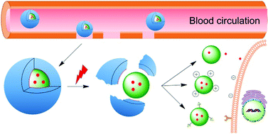
Although big strides have been made in the development of anti-cancer drugs, a major hindrance in their development is the lack of appropriate models to test their efficacy. 2D models are poor representations for real-life situations, while animal models present ethical issues. 3D cell-based tumour models would be a major improvement, yet transport modes in 3D models are poorly understood, impeding targeted strategies. A new study published in Biomaterials Science by Lu et al. elucidates how polymeric micelles, a main cancer-treatment platform, are taken up and transported, allowing for improved development of strategies to effectively deliver drug payloads to tumours.

Despite the plethora of anti-cancer drugs developed in the past decades, testing efficacy prior to human trials remains suboptimal at best. 2D cell models poorly reflect the real-life situation, with different pharmacokinetics and nutrient availability, leading to misleading observations and faulty conclusions. Better representations are animal tumour models, yet – separate from obvious ethical concerns – animal metabolism is not necessarily comparable to humans. The development of a more representative 3D multicellular tumour spheroid (MCTS) model to investigate treatment modalities would be a big step towards alleviating those issues, yet the mode of drug carrier transport in 3D models is poorly understood, impeding targeted strategies.

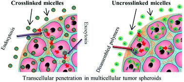
To address the current caveats in MCTS knowledge, Dr. Martina Stenzel’s research group at the University of New South Wales investigated how polymeric micelles are taken up and transported through the outer layers of MCTS’s. Her group created both crosslinked and uncrosslinked polymeric micelles (called CKM and UCM, respectively) and delivered them to a pancreatic MCTS’s. The penetration was monitored using a fluorescent payload, Nile Red, and various modes of endocytosis, as well as exocytosis, were blocked. Their results indicate that it is both caveolae-mediated endocytosis and exocytosis mechanisms are required for good penetration depth of micelles into MCTS’s. Taken together, this evidence points toward transcellular mechanisms as the primary mode of transport for drug-loaded polymeric micelles.

Dr. Stenzel’s group further shows that UCM micelles could not penetrate as far as CKM micelles. The rapid release of their toxic payload doxorubicin creates an apoptotic peripheral cell layer, leading to cessation of additional transcellular transport. Perhaps the most captivating aspect of her research, though understated in the main article, is that the mode of micellar transport seems to be identical in other tumour models.

How polymer micelles are transported in tumor models


The primary mechanism for micellar penetration in MCTS models shown in this article creates important guidance to other researchers investigating anti-cancer drug delivery to tumours. An essential insight is that micelles need to be capable of retaining their structural integrity long enough to prevent payload-induced penetration limitations. Intriguingly, there are indications that the shown penetration mechanisms are extrapolatable to other tumours as well. This study therefore represents a great step forward towards creating better utilization of in vitro tumour models.

Check out the full article:
H. Lu, R.H. Utama, U. Kitiyotsawat, K. Babiuch, Y. Jiang and M.H. Stenzel
Biomater. Sci., 2014, Advance Article, DOI: 10.1039/C4BM00323C


Biomaterials Science web writer Robert van Lith

Robert van Lith (@RvLith) is currently a Post-Doc in the Biomedical Engineering department at Northwestern University, developing intrinsically antioxidant  biomaterials. He recently received his Ph.D. from Northwestern University for his work on citrate-based antioxidant polyesters, receiving an American Heart Association Fellowship and Society for Biomaterials award for his work. He was trained in the Netherlands, holding an M.S. degree in Biomedical Engineering from Eindhoven University of Technology. Read more about Robert’s research publications here.


Follow the latest journal news on Twitter @BioMaterSci or go to our Facebook page.

Digg This
Reddit This
Stumble Now!
Share on Facebook
Bookmark this on Delicious
Share on LinkedIn
Bookmark this on Technorati
Post on Twitter
Google Buzz (aka. Google Reader)

Size of Internalized Calcium Phosphate Particles Plays a Critical Role in Cell Fate

Ellen Tworkoski highlights a recent hot article in Biomaterials Science

Calcium phosphate (CaP) based materials have long been popular choices for a range of medical applications including bone replacement and drug delivery.  However, recent studies indicate a need for a closer look at how cells react to small, degraded CaP particles that find their way into the cell’s interior.  In a recent study, a research team from the University of Birmingham demonstrated that when CaP particles with a diameter larger than 1.5μm penetrated the interior of the cell but were not sequestered by the cell’s lysosomes, a series of events eventually leading to cell death could be observed.

Within the past few years, an increased focus on the end results of CaP degradation have shown that the eventual cytotoxicity of these materials is heavily dependent on the total volume of internalized material.  A study by Motskin et al. in 2009 illustrated that many of these degraded particles are localized to the lysosomes, cellular structures whose acidic environments are responsible for the degradation of waste products which, in the case of CaP, results in the generation of calcium ions.  It was suggested that the generation of a surplus of these ions could interfere with the cell’s signalling pathways, potentially triggering apoptosis.

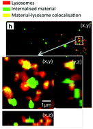
Intracellular Distribution of CaP showing co-localization with lysosomes

In a study published recently in Biomaterials Science, Williams et al. quantified the volume and size distribution of CaP material within cells by chemically grafting a fluorescent probe to the surface of silicon-substituted hydroxyapatite (SiHA), and then exposing a mouse osteoblast precursor cell line to the labelled particles.  Following exposure, the group observed changes in cell behaviour that were indicative of the onset of cell death including alterations in cell morphology, an increase in the number of lysosomes, and cellular detachment from the underlying substrate.  As the cells transitioned from the early stages of cell death (morphology changes) to later stages (detachment from the substrate), there was a marked increase in the amount of SiHA within the cytoplasm but outside of the lysosomes.  Moreover, the onset of cell death was correlated with SiHA aggregates within the cytoplasm that were 1.5μm in diameter or larger.

The group hypothesized that this may have been due to a destabilization of the lysosome membrane which prevented undissolved CaP from remaining within the lysosomes or, alternatively, the destabilization of endosomes which were responsible for delivering particles to the lysosomes.  Regardless, this study proves a need for additional research into the size-dependent effects of CaP particles on cell health and suggests a new design concern for CaP based medical materials.

Check out the full article here:
Quantification of volume and size distribution of internalised calcium phosphate particles and their influence on cell fate
by Richard L. Williams, Isaac Vizcaíno-Castón, and Liam M. Grover


Ellen Tworkoski is a Web Writer for Biomaterials Science and a graduate student in the department of Biomedical Engineering at Northwestern University.

Digg This
Reddit This
Stumble Now!
Share on Facebook
Bookmark this on Delicious
Share on LinkedIn
Bookmark this on Technorati
Post on Twitter
Google Buzz (aka. Google Reader)

Making it personal – Engineered tissues for neural regeneration

Brian Aguado highlights a recent hot article in Biomaterials Science

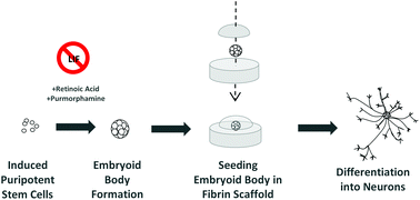
The “holy grail” of neural tissue engineering is to develop functional tissue constructs in an effort to reconnect nerves that have been previously disconnected from disease or injury (i.e. spinal cord injury).  One technique for developing personalized regenerative cell therapies is to use adult somatic cells from a patient (such as skin cells) and re-program them to an induced pluripotent stem cell (iPSC) state. After isolating iPSCs, patient-specific neurons may be generated and transplanted back into the patient, with the hope that the newly formed neurons will form connections in the tissue and restore function.

At the University of Victoria, Montgomery et al. have developed a method to differentiate mouse iPSCs into neurons within a fibrin gel. Fibrin has been regarded as a traditional biomaterial to effectively differentiate embryonic stem cells into neurons. In this study, fibrin was utilized to evaluate the efficiency of two iPS differentiation protocols. The first protocol tested was the traditional “4-/4+” method, involving no treatment to iPS embryoid bodies for 4 days of culture, then adding retinoic acid (RA) for the subsequent 4 days of culture. The second protocol tested was the newer “2-/4+” method, involving no treatment to iPS embryoid bodies for 2 days of culture, then adding RA and purmorphamine for the subsequent 4 days of culture. Individual embryoid bodies were then isolated and placed within a 3D fibrin gel to observe differentiation of neurons.


Interestingly, the overall differentiation efficiency was increased using the 6 day 2-/4+ method compared to using the traditional 4-/4+ method after seeding the treated iPS cells in 3D fibrin gels. Phenotypes were characterized using immunofluorescence staining and RT-PCR, with increases in expression of the neuronal markers TUJ1 and nestin at Day 14 of culture using the 2-/4+ method compared to the 4-/4+ method. The SOX2 pluripotent marker remained low at Day 14, indicating cells were differentiating to a neuronal state.

Taken together, this study demonstrates the ability to differentiate neurons from mouse iPSCs using simple differentiation and encapsulation protocols. Future work will need to be conducted to see if these protocols can be implemented to achieve neuronal differentiation using human iPSCs.

Check out the full article:
Engineering personalized neural tissue by combining induced pluripotent stem cells with fibrin scaffolds, by Amy Montgomery, Alix Wong, Nicole Gabers and Stephanie M. Willerth


Brian Aguado

Brian Aguado (@BrianAguado) is currently a Ph.D. Candidate and NSF Fellow in the Biomedical Engineering department at Northwestern University. He holds a B.S. degree in Biomechanical Engineering from Stanford University and a M.S. degree in Biomedical Engineering from Northwestern University. Read more about Brian’s research publications here.


Follow the latest journal news on Twitter @BioMaterSci or go to our Facebook page.

Digg This
Reddit This
Stumble Now!
Share on Facebook
Bookmark this on Delicious
Share on LinkedIn
Bookmark this on Technorati
Post on Twitter
Google Buzz (aka. Google Reader)

Joint Biomaterials Science and Soft Matter ‘Silk and silk-inspired materials’ Web Collection

Take a look at the ‘Silk and silk-inspired materials’ web collection, a joint venture by Biomaterials Science and Soft Matter.

Are you interested in why spider silk is so strong? Or maybe you’re intrigued to find out how silk can be utilised in cell delivery? Whatever your curiosity be sure to check out the ‘Silk and silk-inspired materials’ web collection and find out why this growing area of research is proving so popular!

The web collection features articles from both Biomaterials Science and Soft Matter by leading authors from around the world. The collection contains a range of article types which cover the properties and rheology of silk-inspired materials as well as investigations into the surface properties of spider silk particles. Please follow the link to read all the articles in this popular area of research.

Digg This
Reddit This
Stumble Now!
Share on Facebook
Bookmark this on Delicious
Share on LinkedIn
Bookmark this on Technorati
Post on Twitter
Google Buzz (aka. Google Reader)

Biomaterials Science Lectureship: Nominations now open

Do you know someone who deserves recognition for their contribution to the biomaterials field?

Now is your chance to propose they receive the accolade they deserve.

Biomaterials Science is pleased to announce that nominations are now being accepted for its Biomaterials Science Lectureship 2015. This annual award was established in 2014 to honour an early-stage career scientist who has made a significant contribution to the biomaterials field.

Suzie Pun was the winner of the 2014 Biomaterials Science Lectureship. Suzie is part of the Department of Bioengineering at the University of Washington.

Qualification

To be eligible for the Biomaterials Science Lectureship, the candidate should be in the earlier stages of their scientific career, typically within 15 years of attaining their doctorate or equivalent degree, and will have made a significant contribution to the field.

Description

The recipient of the award will be asked to present a lecture three times, one of which will be located in the home country of the recipient. The Biomaterials Science Editorial Office will provide the sum of £1000 to the recipient for travel and accommodation costs.

The award recipient will be presented with the award at one of the three award lectures. They will also be asked to contribute a lead article to the journal and will have their work showcased on the back cover of the issue in which their article is published.

Selection

The recipient of the award will be selected and endorsed by the Biomaterials Science Editorial Board.

Nominations

Those wishing to make a nomination should send details of the nominee, including a brief C.V. (no longer than 2 pages A4) together with a letter (no longer than 2 pages A4) supporting the nomination, to the Biomaterials Science Editorial Office by 6th March 2015. Self-nomination is not permitted.

Digg This
Reddit This
Stumble Now!
Share on Facebook
Bookmark this on Delicious
Share on LinkedIn
Bookmark this on Technorati
Post on Twitter
Google Buzz (aka. Google Reader)

Take 1…minute for chemistry in health

Can you explain the importance of chemistry to human health in just 1 minute? If you’re an early-career researcher who is up to the challenge, making a 1 minute video could win you £500.

The chemical sciences will be fundamental in helping us meet the healthcare challenges of the future, and we are committed to ensuring that they contribute to their full potential. As part of our work in this area, we are inviting undergraduate and PhD students, post-docs and those starting out their career in industry to produce an original video that demonstrates the importance of chemistry in health.Take 1... minute for chemistry in health

We are looking for imaginative ways of showcasing how chemistry helps us address healthcare challenges. Your video should be no longer than 1 minute, and you can use any approach you like.

The winner will receive a £500 cash prize, with a £250 prize for second place and £150 prize for third place up for grabs too.

Stuck for inspirationLast year’s winning video is a good place to start. John Gleeson’s video was selected based on the effective use of language, dynamic style, creativity and its accurate content.

The closing date for entries to be submitted is 30 January 2015. Our judging panel will select the top five videos. We will then publish the shortlisted videos online and open the judging to the public to determine the winner and the runners up.

For more details on how to enter the competition and who is eligible, join us at the Take 1… page.

Good luck!

Digg This
Reddit This
Stumble Now!
Share on Facebook
Bookmark this on Delicious
Share on LinkedIn
Bookmark this on Technorati
Post on Twitter
Google Buzz (aka. Google Reader)