Archive for the ‘Uncategorized’ Category

Paper of the month: Magnetic delivery of Fe3O4@polydopamine nanoparticle-loaded natural killer cells suggest a promising anticancer treatment

Natural killer (NK) cells have the intrinsic ability to recognise and eliminate cancer cells along with the potential to inhibit metastasis. NK cells utilize a variety of ways to kill tumor cells, such as stimulating cytokine release, direct cytotoxicity and activating targeted cells apoptosis. Non-small cell lung cancer (NSCLC) has shown significant response to NK cell based immunotherapy in clinical settings. Recently, researchers have begun investigating ways to augment the recruitment and infiltration of NK cells into tumors for improved theranostics. Hence, it is vital to develop non-invasive methods for in vivo control and for the monitoring of the administered NK cells with tissue targeting ability. FDA approved superparamagnetic iron oxide nanoparticles (SPIONs) based magnetic resonance imaging (MRI) contrast agents have been proven biocompatible delivery vehicles and imaging probes for NK cells.

c8bm00588e

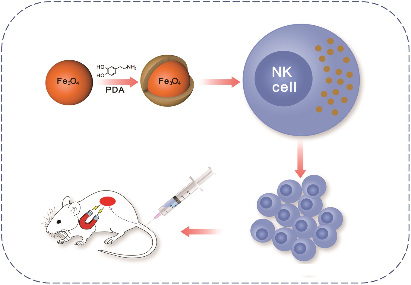
In the present work Jiang et al have synthesized nanoparticles (NPs) composed of a Fe3O4 core and polydopamine (PDA) shell, for tumor theranostics. The aim of this study was to develop magnetic NPs for an immune-cell delivery system to target NSCLC cells. The system stimulated the accumulation of NK cells at the tumor site via the placement of a tiny external magnetic device inside animals. The NK cells actively took up the Fe3O4@PDA NPs due to its physiological stability, while the biology of NK cells was not affected, owing to its biocompatible nature. In vivo studies demonstrated the reduced expression of Ki-67 and the elevated apoptosis of A549 cancer cells upon treatment with Fe3O4@PDA NP-labeled NK cells. Though there are some limitations associated with the invasive approach, the magnetic delivery of NP-NKs can be of promising value in clinical applications.

Tips/comments from the authors:

  • Coating of magnetic NPs with PDA played a vital role in its cellular uptake, as surface modification is an essential factor in the determination of the biocompatibility and the cellular absorption of magnetic NPs.
  • Due to the biocompatible nature of magnetic NPs, even high concentrations (100 μg/mL) did not induce apoptosis of NK cells.
  • With improved retention over time the delivery of Fe3O4@PDA NP-labeled NK cells can be expedited to the tumor via application of local magnetic field.
  • The position of the tumor and the implanted magnetic field should be close enough, while sufficient time should be given to the magnetic field for the achievement of potent therapeutic effect.
  • The non-invasive nature of three-dimensional (3-D) rotating magnetic fields or high gradient magnetic fields can enhance the magnetic strength in a central point for the effective accumulation of NPs.


Read the full article here: Magnetic delivery of Fe3O4@polydopamine nanoparticle-loaded natural killer cells suggest a promising anticancer treatment Biomater. Sci., 2018,6, 2714-2725


About the web writer

Muhammad OvaisMuhammad Ovais is a Web Writer for Biomaterials Science. Currently, he is a PhD candidate in Prof. Chunying Chen Lab at CAS Center for Excellence in Nanoscience, CAS Key Laboratory for Biomedical Effects of Nanomaterials and Nanosafety, National Center for Nanoscience and Technology (NCNST), Beijing. His research interest lies in the development of novel nano-delivery systems for cancer immunotherapy. He has published a total of ~30 research/review articles. You can find or contact him on ResearchGate, LinkedIn and Chunying Chen’s lab

Contact Email: movais@bs.qau.edu.pk

Twitter: https://twitter.com/OVAISBiotec

Digg This
Reddit This
Stumble Now!
Share on Facebook
Bookmark this on Delicious
Share on LinkedIn
Bookmark this on Technorati
Post on Twitter
Google Buzz (aka. Google Reader)

Focus on: Nanoparticle Delivery Systems

Developing nanoparticle formulations that can deliver drugs more effectively to the target sites with enhanced efficacy and reduced side effects has been an overarching goal in the field of nanobiotechnology. Dendrimers, micelles, and liposomes represent three major classes of nanoparticles that have shown promising results in drug delivery and bio-sensing.  Each type of nanoparticle has its own strengths and limitations in terms of the desirable payload, site of action, duration of action, release profile, and dosing frequency. Therefore, it is imperative to engineer these classical nanoparticle delivery systems for specific drug delivery application.

Nanoparticle delivery system

This month we focus on four articles published in Biomaterials Science reporting the recent advances in leveraging those different nanoparticle delivery systems for efficient, controlled, and targeted delivery of therapeutic agents.

1. Nucleobase-modified polyamidoamine-mediated miR-23b delivery to inhibit the proliferation and migration of lung cancer
Haobo Han, Jiebing Yang, Yudi Wang, Wenqi Chen, Jiawen Chen, Yan Yang and Quanshun Li
Biomater. Sci., 2017, 5, 2268. DOI: 10.1039/c7bm00599g

In the current study, the authors aimed to further improve the transfection efficiency and biocompatibility of conventional polyamidoamine (PAMAM) dendrimers. To this end, the surface of PAMAM was chemically modified with 2-amino-6-chloropurine. This modification further enhanced the carrier/DNA interaction via the fine balance of hydrogen bonding and electrostatic interaction. Compared to the prototype PAMAM, the modified PAMAM demonstrated higher transfection efficiency. In an in vitro model, this gene carrier delivered miR-23b, a potent anti-proliferative and anti-invasive agent, more efficiently into A549 cancer cells, indicating the potential of this carrier in cancer nanotherapy.

2. Novel poly(vinyl alcohol)-based amphiphilic nanogels by non-covalent boric acid crosslinking of polymeric micelles
Hen Moshe, Yuval Davizon, Maya Menaker Raskin and Alejandro Sosnik
Biomater. Sci., 2017, 5, 2295. DOI: 10.1039/c7bm00675f

Poor physical stability often presents as a major drawback for polymeric micelles. The authors addressed this issue by non-covalent crosslinking of a poly(vinyl alcohol) (PVA) based polymeric micelles system with boric acid. Compared to the non-crosslinked control, this novel micelles demonstrated improved physical stability under harsh environment. More interestingly, these micelles could be spray-dried and efficiently consolidated into dry powders which were able to regenerate back into the original nanoparticles upon re-dispersion. This non-covalently crosslinked micelles also maintained good mucoadhesiveness and cytocompatibility.

3. Codelivery of sorafenib and GPC3 siRNA with PEI-modified liposomes for hepatoma therapy
Weitong Sun, Yong Wang, Mingyue Cai, Liteng Lin, Xiaoyan Chen, Zhong Cao, Kangshun Zhu and Xintao Shuai
Biomater. Sci., 2017, 5, 2468. DOI: 10.1039/c7bm00866j

Combination therapy using chemotherapeutic drugs and siRNA represents a promising strategy that can potentially induce and/or enhance the synergistic anticancer effects. To overcome the individual drawbacks of sorafenib and gene therapy, the authors developed a PEI based liposome system which allow the co-delivery of GPC3 siRNA and hydrophobic sorafenib molecule. The drug loaded liposomal delivery system displayed enhanced anticancer effects by suppressing the expression of the anti-apoptotic GPC3 gene and the proliferative cyclin D1 gene simultaneously in human hepatocellular carcinoma (HCC) HepG2 cells. Further, the improved therapeutic effects of this delivery system was demonstrated in an in vivo xenograft model.

4. Dimeric camptothecin-loaded RGD-modified targeted cationic polypeptide-based micelles with high drug loading capacity and redox-responsive drug release capability
Zhaopei Guo, Xingzhi Zhou, Mengze Xu, Huayu Tian, Xuesi Chen and Meiwan Chen
Biomater. Sci., 2017, 5, 2501. DOI: 10.1039/c7bm00791d

To tackle the low bioavailability problem of camptothecin, the authors devised a novel polymeric micelles system composed of cationic polypeptide poly-lysine-block-poly-leucine, polyethylene glycol (PEG), and arginine-glycine-aspartic acid (RGD) peptide. The micelles system increased drug encapsulation efficiency, drug loading capacity, and physical stability of camptothecin. The RGD moiety further enhanced the intracellular uptake of micelles due to the cellular targeting capability of RGD sequence. Importantly, the drug loaded micelles effectively inhibited the proliferation of malignant MDA-MB-231 breast cancer cells by inducing cellular apoptosis and decreasing mitochondrial membrane potential.

Read these articles for free until 10 January 2018

About the webwriterYingfei Xue

Yingfei Xue is a web writer for Biomaterials Science. Currently, he is a PhD candidate and graduate student researcher in Dr. Shilpa Sant lab at the University of Pittsburgh, USA.  His research focus on nano-/micro-technology in novel heart valve therapy. Find him on Twitter: @Phil_Xue or connect with him on ResearchGate

Digg This
Reddit This
Stumble Now!
Share on Facebook
Bookmark this on Delicious
Share on LinkedIn
Bookmark this on Technorati
Post on Twitter
Google Buzz (aka. Google Reader)

Mapping Oxygen Gradients in 3D Cell Cultures

Microenvironmental oxygen levels and gradients within three-dimensional (3D) tissue cultures directly influence cellular behavior and function, dictating the mode of proliferation, metabolism and interaction of cells with each other and their environment. While advances and prevalence of in vitro generated 3D cultures have spurred new techniques and systems for biological interrogation, it is necessary to develop and implement parallel systems to monitor and characterize the oxygen microenvironment within the tissue cultures and around them in the vessel used for the cultures. Conventional oxygen evaluation platforms can be ill-suited for continuous oxygen evaluation in custom tissue cultures. The Takayama group was able to robustly evaluate multiple 3D culture platforms by combining the use of phase-fluorimetry and lab-fabricated dispersible oxygen responsive microparticles. Oxygen microsensors were used to evaluate two spheroid culture vessels, hanging-drop and low-adhesion microwell plates, to highlight the variations in the oxygen levels peripheral to the spheroids in the two culture techniques. Dramatic differences can be seen in the steady state oxygen levels between the two culture techniques because of the difference in distance between the spheroids and the air-liquid interface in these two vessel types. These results highlighted the importance of minding the gas exchange location as compared to the cell culture to ensure appropriate tissue culture microenvironments.

Figure 1

Furthermore, these microsensors were used to map radial oxygen distribution across a circular, cell-patterned hydrogel by dispersing the microsensors within the culture. Coupling the spatial oxygen mapping to computational models of oxygen diffusion, the authors were able to estimate oxygen uptake behavior of the tissue culture. While 3D tissue culture platforms leverage the in vitro tissue architecture to produce more physiologically similar phenomena, integrated design and analysis of these 3D cell cultures from both biomaterial and oxygen supply aspects will be paramount in enabling researchers to effectively recreate some of the complexities present within both healthy and diseased tissues.

Tips from the authors:

  1. When fabricating oxygen microsensing beads, infusion with Dichloromethane enabled large amount of Ruthenium caging within the PDMS microspheres, while leaving them oxygen sensitive. While other solvents swell PDMS more readily and enabled higher efficiency infusion of ruthenium, these solvents resulted in oxygen unresponsive ruthenium loaded PDMS beads.
  2. Microsensors cannot be effectively integrated in the multicellular spheroids we tried with HEK293T, HS-5 and MDA-MB-231 cells; as the spheroids contract microsensors are ejected out of the spheroids.
  3. The only limitation of phase-fluorimetry for the oxygen measurements is sufficient signal output that it can be detected by the photodiode, or other detection system. This was generally not a problem with beads greater than 80 microns assuming the culture systems was less than 1-mm thick. However, we were unable to effectively infuse beads under 80 microns with enough ruthenium to have enough output signals from the microsensors to get robust readings with cultures > 1 mm.

Article Link:

Dispersible oxygen microsensors map oxygen gradients in three-dimensional cell cultures Biomater. Sci., 2017,5, 2106-2113

About the WDr. Sudip Mukherjee ebwriter:

Dr. Sudip Mukherjee is a Web Writer for Biomaterials Science. He is currently a Postdoctoral Research Associate working alongside Dr. Omid Veiseh at the Department of Bioengineering at the Rice University. His research is involved in the development of advanced nanomaterials for drug/gene delivery in cancer theranostics, immunomodulatory applications & angiogenesis. He published a total of ~30 research articles/patents. He serves as International Advisory Board Member for ‘Materials Research Express’, IOP Sciences. He is an associate member (AMRSC) of RSC, UK. He serves as reviewer for several international journals like Chem Comm, J Mater Chem A, J Mater Chem B, Journal of Biomedical Nanotechnology, RSC Advances, IOP Nanotechnology etc.

Digg This
Reddit This
Stumble Now!
Share on Facebook
Bookmark this on Delicious
Share on LinkedIn
Bookmark this on Technorati
Post on Twitter
Google Buzz (aka. Google Reader)

Polymeric biomaterials for cancer nanotechnology themed issue now online

We are delighted to announce that the Polymeric biomaterials for cancer nanotechnology themed issue is now available online.

Polymeric biomaterials for cancer nanotechnology

Guest Edited by Jianjun Cheng (University of Illinois at Urbana-Champaign, USA) and Suzie H. Pun (University of Washington, USA), this themed issue highlights the latest discoveries and innovations in polymeric biomaterials for cancer nanotechnology.

Polymeric biomaterials have been extensively used in nanomedicine formulations for cancer therapy. Preclinical and clinical studies have in general revealed that polymeric nanocarriers, when used for chemotherapeutic drug delivery, reduce systemic toxicity and thus mitigate adverse side effects of the drug. This themed issue contains reviews and research articles in the areas of: (i) expanding the available suite of polymeric biomaterials that can be reproducibly and controllably manufactured at a suitable scale, (ii) designing carriers with improved biodistribution to tumour sites, (iii) increasing tumour distribution and penetration of polymeric nanocarriers, and (iv) controlling efficient drug release at a desired location and with optimal kinetics.

Read all the themed issue papers here

A few articles from the themed issue are highlighted below:

Drug-free macromolecular therapeutics – a new paradigm in polymeric nanomedicines
Te-Wei Chu and Jindřich Kopeček
Biomater. Sci., 2015,3, 908-922

Lipid-coated polymeric nanoparticles for cancer drug delivery
Sangeetha Krishnamurthy, Rajendran Vaiyapuri, Liangfang Zhang and Juliana M. Chan
Biomater. Sci., 2015, 3, 923-936

Enhanced transcellular penetration and drug delivery by crosslinked polymeric micelles into pancreatic multicellular tumor spheroids
Hongxu Lu, Robert H. Utama, Uraiphan Kitiyotsawat, Krzysztof Babiuch, Yanyan Jiang and Martina H. Stenzel
Biomater. Sci., 2015, 3, 1085-1095

Polymeric assembly of hyperbranched building blocks to establish tunable nanoplatforms for lysosome acidity-responsive gene/drug co-delivery
Hui-Zhen Jia, Wei Zhang, Xu-Li Wang, Bin Yang, Wei-Hai Chen, Si Chen, Gang Chen, Yi-Fang Zhao, Ren-Xi Zhuo, Jun Feng and Xian-Zheng Zhang
Biomater. Sci., 2015,3, 1066-1077

Digg This
Reddit This
Stumble Now!
Share on Facebook
Bookmark this on Delicious
Share on LinkedIn
Bookmark this on Technorati
Post on Twitter
Google Buzz (aka. Google Reader)

Biomaterials Science celebrates its first (partial) Impact Factor

Biomaterials Science is pleased to announce its first (partial) Impact Factor of 3.831


Biomaterials Science is committed to publishing exceptional articles that explore the underlying science behind the function, interactions and design of biomaterials. Its impressive first (partial) Impact Factor of 3.831 is a strong assurance that research published in our new journal is highly visible and relevant to the biomaterials community. Take a look at these popular Biomaterials Science articles below:

Sustained delivery of bioactive neurotrophin-3 to the injured spinal cord
Irja Elliott Donaghue, Charles H. Tator and Molly S. Shoichet
Biomater. Sci., 2015, 3, 65-72

Hyperbranched PEG-based supramolecular nanoparticles for acid-responsive targeted drug delivery
Xiaofei Chen, Xuemei Yao, Chunran Wang, Li Chen and Xuesi Chen
Biomater. Sci., 2015, 3, 870-878

Angiopoietin-1 peptide QHREDGS promotes osteoblast differentiation, bone matrix deposition and mineralization on biomedical materials
Nicole T. Feric, Calvin C. H. Cheng, M. Cynthia Goh, Vyacheslav Dudnyk, Val Di Tizio and Milica Radisic
Biomater. Sci., 2014, 2, 1384-1398

A novel hanging spherical drop system for the generation of cellular spheroids and high throughput combinatorial drug screening
A. I. Neto, C. R. Correia, M. B. Oliveira, M. I. Rial-Hermida, C. Alvarez-Lorenzo, Ruis L. Reis and Joao F. Mano
Biomater. Sci., 2015, 3, 581-585

In vitro model alveoli from photodegradable microsphere templates
Katherine J. R. Lewis, Mark W. Tibbitt, Yi Zhao, Kelsey Branchfield, Xin Sun, Vivek Balasubramaniam and Kristi S. Anseth
Biomater. Sci., 2015, 3, 821-832

Noninvasive theranostic imaging of HSV-TK/GCV suicide gene therapy in liver cancer by folate-targeted quantum dot-based liposomes
Dan Shao, Jing Li, Yue Pan, Xin Zhang, Xiao Zheng, Zheng Wang, Ming Zhang, Hong Zhang and Li Chen
Biomater. Sci., 2015, 3, 833-841

Publishing your research in Biomaterials Science means that your article will be read and cited by your colleagues.

Our unique combination of high quality articles, outstanding Editorial and Advisory Board, free colour and flexible manuscript format make it clear to see why Biomaterials Science is a leading journal within the biomaterials field.

Our fast times to publication ensure that your research is reviewed and announced to the community rapidly.

From receipt, your research papers will be published in 68 days. Communications articles will be published in 53 days. (Data taken from average manuscript handling times between July 2014 – January 2015)

So join the many leading scientists that have already chosen to publish in Biomaterials Science and submit your research today to be seen with the best!

Submit your research
Digg This
Reddit This
Stumble Now!
Share on Facebook
Bookmark this on Delicious
Share on LinkedIn
Bookmark this on Technorati
Post on Twitter
Google Buzz (aka. Google Reader)

Organ-on-a-Chip World Congress & 3D-Printing in the Life Sciences

Biomaterials Science is pleased to announce that the Organ-on-a-Chip World Congress & 3D-Printing will be held at Wyndham Boston Beacon Hill in Boston, USA on the 8th – 9th July 2015.

Deadlines and dates

Registration is open, so why not sign up for this fascinating meeting now!

If you are interesting in presenting a poster you must submit your abstract by 30th June 2015. Abstracts for oral presentations are also being accepted.

Themes and topics

The 3D-printing field is expanding exponentially and it is starting to impact the life sciences arena.  The current interest in this space is the use of various bioinks to “print” parts of tissues in the goal and hope to bioprint organs as well as body parts in the future for regenerative medicine and other medical applications.

This conference explores 3D-printing in the life sciences through presentations from academic researchers as well as industry participants. Several companies involved in bioprinting and bioinks will be exhibiting at this conference. The companion conference track explores Organ-on-a-Chip/Body-on-a-chip, which employs the use of microfluidics and lab-on-a-chip (LOAC) technologies to build “cell clusters in 3D-format” in functionally-relevant patterns.  These patterns enable cellular function to be recapitulated ex vivo and has wide-ranging potential for drug discovery and development applications in the pharmaceutical and biotechnology industries.

Please contact event organisers Karen Saunders or Enal Razvi if you have any queries.

Head to the conference website to find out more about this 2-track event.

Join the conversation on Twitter: #OOAC2015 and let us know if you’re going @BioMaterSci.


Digg This
Reddit This
Stumble Now!
Share on Facebook
Bookmark this on Delicious
Share on LinkedIn
Bookmark this on Technorati
Post on Twitter
Google Buzz (aka. Google Reader)

Nanocarriers for cancer drug delivery

Chen et al. discuss the emerging antitumor applications of extracellularly reengineered polymeric nanocarriers.


Chen et al. write an informative and interesting review on the methods by which nanoparticle drug delivery vehicles are engineered using diverse triggers that result in drug release.

In the field of drug delivery, particularly to sites of tumour, there are many different considerations – the drug must be delivered to the site of the tumour, it must be intact when delivered, and it must act to destroy cancerous tissue while remaining as nontoxic as possible to healthy tissue. As a result, much research has been devoted to the development of core-shell drug delivery structures that consist of the drug in the nanoparticle core surrounded by a protective shell. This protective shell may be removed using both internal and external triggers. Many nanocarriers use PEG (polyethylene glycol)-based shells for ease of solubility and in order to prevent proteins from being absorbed onto the surface of the shell. However, additional materials are also increasingly used for the development of these materials.

The authors review the materials as well as common strategies used to remove the shell. Specifically, they summarise literature that exploits changes in pH, since the acidity of the tumour microenvironment differs from its healthy surroundings. Charge-reversal nanocarriers with a positively charged core and a negatively charged shell are also used. In addition, enzymes can degrade the external shell. An enzyme family known as matrix metalloproteinases, or MMPs, is commonly used for this purpose, but other enzymes are also beginning to be explored. Finally, these nanocarriers can also be assembled or de-assembled using interactions between the nanocarrier and the host body.

Emerging antitumor applications of extracellularly reengineered polymeric nanocarriers by Jinjin Chen, Jianxun Ding, Chunsheng Xiao, Xiuli Zhuang and Xuesi Chen

Debanti Sengupta


Debanti Sengupta completed her PhD in Chemistry in 2012 from Stanford University.  She was previously a Siebel postdoctoral scholar at the University of California, Berkeley, and is currently a postdoctoral scholar in Radiation Oncology at Stanford University. Follow her on Twitter @debantisengupta.

Follow the latest journal news on Twitter @BioMaterSci or go to our Facebook page.

Digg This
Reddit This
Stumble Now!
Share on Facebook
Bookmark this on Delicious
Share on LinkedIn
Bookmark this on Technorati
Post on Twitter
Google Buzz (aka. Google Reader)

Recent Appointees in Materials Science 2015 Conference (RAMS2015)

Recent Appointees in Materials Science 2015 Conference

We are delighted to announce that the Recent Appointees in Materials Science 2015 Conference (RAMS2015) will be held at the University of Warwick on 16-17th September 2015.

Deadlines and dates

Registration will open shortly so be sure to sign up to this essential meeting before 1st September 2015! The cost of registration is £125 for accommodation and meals, including the conference banquet at Warwick Castle. A reduced rate of £70 is offered for those not requiring accommodation.

Abstract submissions are now being accepted for oral and poster presentation but make sure you submit your abstracts by the deadline on 30th June 2015.

Bursaries

A small number of bursaries are available for those with limited travel budgets and will be assessed on an individual basis. Enquire about bursaries here.

Keynote speakers

Biomaterials Science Advisory Board member Andrew Dove (University of Warwick) will be speaking along with other keynote speakers Aron Walsh (University of Bath) and Mary Ryan (Imperial College London). View the full list of invited speakers here.

For full details visit the RAMS2015 website. We hope you can join the materials science community for this fantastic event.

Digg This
Reddit This
Stumble Now!
Share on Facebook
Bookmark this on Delicious
Share on LinkedIn
Bookmark this on Technorati
Post on Twitter
Google Buzz (aka. Google Reader)

13th International Nanomedicine & Drug Delivery Symposium, nanoDDS15

nanoDDS15

We are delighted to be sponsoring a poster session at the 13th International Nanomedicine & Drug Delivery Symposium (nanoDDS15) which will be held at the University of Washington in Seattle, USA on 16th-18th September 2015. Professors Pat Stayton, member of the Biomaterials Science Editorial Board, and Suzie Punn, member of the Biomaterials Science Advisory Board, are co-chairing this exciting meeting.

If you are developing next-generation delivery vehicles to make diagnostics more sensitive and drugs more effective, then nanoDDS15 is the place for you!

Dates and deadlines:

Registration for nanoDDS15 is open, but abstracts for poster presentation are no longer being accepted.

Who’s speaking?

Key speakers include Biomaterials Science Editorial Board member Jun Wang, and Advisory Board members Darrell Irvine and Kazunori Kataoka. The full symposium schedule is now live so why not take a look and plan your visit now! For a full list of confirmed speakers click here.

See the nanoDDS15 website for full details.

Digg This
Reddit This
Stumble Now!
Share on Facebook
Bookmark this on Delicious
Share on LinkedIn
Bookmark this on Technorati
Post on Twitter
Google Buzz (aka. Google Reader)

To crosslink or not to crosslink? – How polymeric micelles can target tumors effectively

Although big strides have been made in the development of anti-cancer drugs, a major hindrance in their development is the lack of appropriate models to test their efficacy. 2D models are poor representations for real-life situations, while animal models present ethical issues. 3D cell-based tumour models would be a major improvement, yet transport modes in 3D models are poorly understood, impeding targeted strategies. A new study published in Biomaterials Science by Lu et al. elucidates how polymeric micelles, a main cancer-treatment platform, are taken up and transported, allowing for improved development of strategies to effectively deliver drug payloads to tumours.

Despite the plethora of anti-cancer drugs developed in the past decades, testing efficacy prior to human trials remains suboptimal at best. 2D cell models poorly reflect the real-life situation, with different pharmacokinetics and nutrient availability, leading to misleading observations and faulty conclusions. Better representations are animal tumour models, yet – separate from obvious ethical concerns – animal metabolism is not necessarily comparable to humans. The development of a more representative 3D multicellular tumour spheroid (MCTS) model to investigate treatment modalities would be a big step towards alleviating those issues, yet the mode of drug carrier transport in 3D models is poorly understood, impeding targeted strategies.

To address the current caveats in MCTS knowledge, Dr. Martina Stenzel’s research group at the University of New South Wales investigated how polymeric micelles are taken up and transported through the outer layers of MCTS’s. Her group created both crosslinked and uncrosslinked polymeric micelles (called CKM and UCM, respectively) and delivered them to a pancreatic MCTS’s. The penetration was monitored using a fluorescent payload, Nile Red, and various modes of endocytosis, as well as exocytosis, were blocked. Their results indicate that it is both caveolae-mediated endocytosis and exocytosis mechanisms are required for good penetration depth of micelles into MCTS’s. Taken together, this evidence points toward transcellular mechanisms as the primary mode of transport for drug-loaded polymeric micelles.

Dr. Stenzel’s group further shows that UCM micelles could not penetrate as far as CKM micelles. The rapid release of their toxic payload doxorubicin creates an apoptotic peripheral cell layer, leading to cessation of additional transcellular transport. Perhaps the most captivating aspect of her research, though understated in the main article, is that the mode of micellar transport seems to be identical in other tumour models.

How polymer micelles are transported in tumor models


The primary mechanism for micellar penetration in MCTS models shown in this article creates important guidance to other researchers investigating anti-cancer drug delivery to tumours. An essential insight is that micelles need to be capable of retaining their structural integrity long enough to prevent payload-induced penetration limitations. Intriguingly, there are indications that the shown penetration mechanisms are extrapolatable to other tumours as well. This study therefore represents a great step forward towards creating better utilization of in vitro tumour models.

Check out the full article:
H. Lu, R.H. Utama, U. Kitiyotsawat, K. Babiuch, Y. Jiang and M.H. Stenzel
Biomater. Sci., 2014, Advance Article, DOI: 10.1039/C4BM00323C


Biomaterials Science web writer Robert van Lith

Robert van Lith (@RvLith) is currently a Post-Doc in the Biomedical Engineering department at Northwestern University, developing intrinsically antioxidant  biomaterials. He recently received his Ph.D. from Northwestern University for his work on citrate-based antioxidant polyesters, receiving an American Heart Association Fellowship and Society for Biomaterials award for his work. He was trained in the Netherlands, holding an M.S. degree in Biomedical Engineering from Eindhoven University of Technology. Read more about Robert’s research publications here.


Follow the latest journal news on Twitter @BioMaterSci or go to our Facebook page.

Digg This
Reddit This
Stumble Now!
Share on Facebook
Bookmark this on Delicious
Share on LinkedIn
Bookmark this on Technorati
Post on Twitter
Google Buzz (aka. Google Reader)