Archive for the ‘Uncategorized’ Category

Size of Internalized Calcium Phosphate Particles Plays a Critical Role in Cell Fate

Calcium phosphate (CaP) based materials have long been popular choices for a range of medical applications including bone replacement and drug delivery.  However, recent studies indicate a need for a closer look at how cells react to small, degraded CaP particles that find their way into the cell’s interior.  In a recent study, a research team from the University of Birmingham demonstrated that when CaP particles with a diameter larger than 1.5μm penetrated the interior of the cell but were not sequestered by the cell’s lysosomes, a series of events eventually leading to cell death could be observed.

Within the past few years, an increased focus on the end results of CaP degradation have shown that the eventual cytotoxicity of these materials is heavily dependent on the total volume of internalized material.  A study by Motskin et al. in 2009 illustrated that many of these degraded particles are localized to the lysosomes, cellular structures whose acidic environments are responsible for the degradation of waste products which, in the case of CaP, results in the generation of calcium ions.  It was suggested that the generation of a surplus of these ions could interfere with the cell’s signalling pathways, potentially triggering apoptosis.

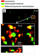
Intracellular Distribution of CaP showing co-localization with lysosomes

In a study published recently in Biomaterials Science, Williams et al. quantified the volume and size distribution of CaP material within cells by chemically grafting a fluorescent probe to the surface of silicon-substituted hydroxyapatite (SiHA), and then exposing a mouse osteoblast precursor cell line to the labelled particles.  Following exposure, the group observed changes in cell behaviour that were indicative of the onset of cell death including alterations in cell morphology, an increase in the number of lysosomes, and cellular detachment from the underlying substrate.  As the cells transitioned from the early stages of cell death (morphology changes) to later stages (detachment from the substrate), there was a marked increase in the amount of SiHA within the cytoplasm but outside of the lysosomes.  Moreover, the onset of cell death was correlated with SiHA aggregates within the cytoplasm that were 1.5μm in diameter or larger.

The group hypothesized that this may have been due to a destabilization of the lysosome membrane which prevented undissolved CaP from remaining within the lysosomes or, alternatively, the destabilization of endosomes which were responsible for delivering particles to the lysosomes.  Regardless, this study proves a need for additional research into the size-dependent effects of CaP particles on cell health and suggests a new design concern for CaP based medical materials.

Check out the full article here:
Quantification of volume and size distribution of internalised calcium phosphate particles and their influence on cell fate
by Richard L. Williams, Isaac Vizcaíno-Castón, and Liam M. Grover


Ellen Tworkoski is a Web Writer for Biomaterials Science and a graduate student in the department of Biomedical Engineering at Northwestern University.

Digg This
Reddit This
Stumble Now!
Share on Facebook
Bookmark this on Delicious
Share on LinkedIn
Bookmark this on Technorati
Post on Twitter
Google Buzz (aka. Google Reader)

Making it personal – Engineered tissues for neural regeneration

The “holy grail” of neural tissue engineering is to develop functional tissue constructs in an effort to reconnect nerves that have been previously disconnected from disease or injury (i.e. spinal cord injury).  One technique for developing personalized regenerative cell therapies is to use adult somatic cells from a patient (such as skin cells) and re-program them to an induced pluripotent stem cell (iPSC) state. After isolating iPSCs, patient-specific neurons may be generated and transplanted back into the patient, with the hope that the newly formed neurons will form connections in the tissue and restore function.

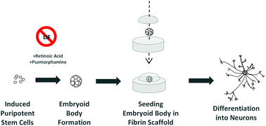
At the University of Victoria, Montgomery et al. have developed a method to differentiate mouse iPSCs into neurons within a fibrin gel. Fibrin has been regarded as a traditional biomaterial to effectively differentiate embryonic stem cells into neurons. In this study, fibrin was utilized to evaluate the efficiency of two iPS differentiation protocols. The first protocol tested was the traditional “4-/4+” method, involving no treatment to iPS embryoid bodies for 4 days of culture, then adding retinoic acid (RA) for the subsequent 4 days of culture. The second protocol tested was the newer “2-/4+” method, involving no treatment to iPS embryoid bodies for 2 days of culture, then adding RA and purmorphamine for the subsequent 4 days of culture. Individual embryoid bodies were then isolated and placed within a 3D fibrin gel to observe differentiation of neurons.


Interestingly, the overall differentiation efficiency was increased using the 6 day 2-/4+ method compared to using the traditional 4-/4+ method after seeding the treated iPS cells in 3D fibrin gels. Phenotypes were characterized using immunofluorescence staining and RT-PCR, with increases in expression of the neuronal markers TUJ1 and nestin at Day 14 of culture using the 2-/4+ method compared to the 4-/4+ method. The SOX2 pluripotent marker remained low at Day 14, indicating cells were differentiating to a neuronal state.

Taken together, this study demonstrates the ability to differentiate neurons from mouse iPSCs using simple differentiation and encapsulation protocols. Future work will need to be conducted to see if these protocols can be implemented to achieve neuronal differentiation using human iPSCs.

Check out the full article:
Engineering personalized neural tissue by combining induced pluripotent stem cells with fibrin scaffolds, by Amy Montgomery, Alix Wong, Nicole Gabers and Stephanie M. Willerth


Brian Aguado

Brian Aguado (@BrianAguado) is currently a Ph.D. Candidate and NSF Fellow in the Biomedical Engineering department at Northwestern University. He holds a B.S. degree in Biomechanical Engineering from Stanford University and a M.S. degree in Biomedical Engineering from Northwestern University. Read more about Brian’s research publications here.


Follow the latest journal news on Twitter @BioMaterSci or go to our Facebook page.

Digg This
Reddit This
Stumble Now!
Share on Facebook
Bookmark this on Delicious
Share on LinkedIn
Bookmark this on Technorati
Post on Twitter
Google Buzz (aka. Google Reader)

Take 1…minute for chemistry in health

The chemical sciences will be fundamental in helping us meet the healthcare challenges of the future, and we are committed to ensuring that they contribute to their full potential. As part of our work in this area, we are inviting undergraduate and PhD students, post-docs and those starting out their career in industry to produce an original video that demonstrates the importance of chemistry in health.Take 1... minute for chemistry in health

We are looking for imaginative ways of showcasing how chemistry helps us address healthcare challenges. Your video should be no longer than 1 minute, and you can use any approach you like.

The winner will receive a £500 cash prize, with a £250 prize for second place and £150 prize for third place up for grabs too.

Stuck for inspirationLast year’s winning video is a good place to start. John Gleeson’s video was selected based on the effective use of language, dynamic style, creativity and its accurate content.

The closing date for entries to be submitted is 30 January 2015. Our judging panel will select the top five videos. We will then publish the shortlisted videos online and open the judging to the public to determine the winner and the runners up.

For more details on how to enter the competition and who is eligible, join us at the Take 1… page.

Good luck!

Digg This
Reddit This
Stumble Now!
Share on Facebook
Bookmark this on Delicious
Share on LinkedIn
Bookmark this on Technorati
Post on Twitter
Google Buzz (aka. Google Reader)

Growing replacement bones – is biomaterial geometry important?

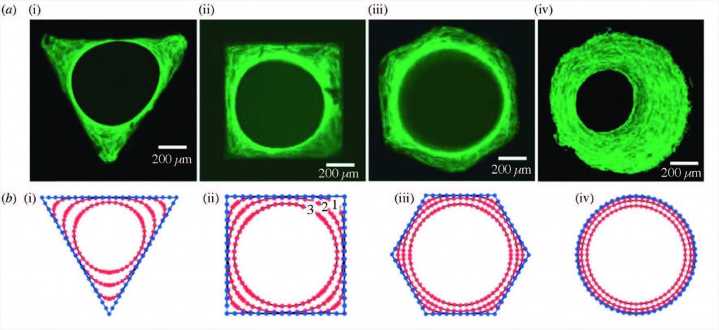
Staining of actin stress fibers

Staining of actin stress fibers to visualize the tissue formed in vitro and to study the effects of curvature (a). The predicted tissue regeneration based on a linear curvature-dependent theoretical model is depicted in subfigure (b). Theoretical predictions match these in vitro experimental observations.

In this paper, Professor Amir Zadpoor reviews the role of biomaterial scaffold geometries on regenerating bone tissue.  Scaffold curvature, pore size, and pore shape are all shown to be important in stimulating more bone growth.

In the case of large bone injuries, the role of scaffolds in regenerating bones becomes increasingly critical.  The ideal bone scaffolds need to be biocompatible, mechanically strong, and contain pores that allow the transport of nutrients and new cell growth.

The geometry of the scaffold plays a very important role in regenerating bone tissue, and is explored in this paper.  Broadly speaking, the curvature of the scaffold, the shape, and the size of the pores are all key components that influence how well the bone is structured and grows.

Strikingly, it has been found that curvature of the surface proportionally impacts the rate of tissue regeneration. Curvature of the surface can be much smaller than, on the scale of, or much larger than cells that grow on the surface. Smaller curvatures can impact individual cell focal adhesions, through which cells can both sense the underlying surface and apply force to it. Larger curvatures can impact cell stress fibers as well as the overall forces on the growing bone tissue.

Pore size can also dramatically impact bone regeneration.  It is already known that a limited amount of inflammation is actually good for bone growth.  Pore size can affect inflammation – for example, larger pores and wider scaffold geometry angles can increase local inflammation.  Pore size can also dictate the amount of oxygen and nutrients reaching the cells, and whether cartilage or bone is formed first.

Pore shape is also extremely important – for example, it has been shown that when cells are grown in scaffolds shaped like parallelograms, alkaline phosphatase activity is increased. Since alkaline phosphatase is a byproduct of bone growth, this data suggests a role for the shape of scaffold pores in bone generation.

However, there are significant caveats to be considered when studying bone regeneration in the lab. Since bone grows in stages, a short in vitro study may not capture all nuances of bone growth that occur in vivo. It is important to study in vitro and in vivo bone generation in parallel to reconcile any contradictory data. Further, it must be noted that it is often difficult to isolate the independent impact of one specific property of a scaffold without affecting another property.  For example, changing pore size can also change the overall mechanical properties of the scaffold.

Despite these limitation, it is clear that the geometrical properties of the scaffold can significantly impact bone growth.  Computational studies and modeling are also now being used to optimize many scaffold properties, and have the potential to drive further research to elucidate the role of scaffold geometry in clinically valuable bone growth.

Check out the full article:

Bone tissue regeneration: the role of scaffold geometry by Amir A. Zadpoor


Web writer Debanti Sengupta

Web writer Debanti Sengupta


Debanti Sengupta completed her PhD in Chemistry in 2012 from Stanford University.  She was previously a Siebel postdoctoral scholar at the University of California, Berkeley, and is currently a postdoctoral scholar in Radiation Oncology at Stanford University. Follow her on Twitter @yoginiscientist.
Follow the latest journal news on Twitter @BioMaterSci or go to our Facebook page.

Digg This
Reddit This
Stumble Now!
Share on Facebook
Bookmark this on Delicious
Share on LinkedIn
Bookmark this on Technorati
Post on Twitter
Google Buzz (aka. Google Reader)

Zeolite–polymer composite adsorbs uremic toxins

Scientists in Japan have developed a nanofibre mesh that can adsorb creatinine from blood with the hope that it can eventually be developed into a wearable blood-cleaning device for patients with kidney failure.

It is hoped the mesh can eventually be developed into a wearable blood-cleaning device

It is hoped the mesh can eventually be developed into a wearable blood-cleaning device

Kidney failure causes dangerous concentrations of waste products, such as potassium, urea and creatinine, to build-up in the body. Apart from having a kidney transplant, the next best solution for patients is dialysis. Dialysis, however, is far from ideal. It is time-consuming and relies on access to specialist equipment, clean water, electricity, dialysate, and, usually, a hospital. Often these requirements aren’t accessible in rural parts of developing countries and disaster areas.

Read the full article at Chemistry World.

Fabrication of zeolite–polymer composite nanofibers for removal of uremic toxins from kidney failure patients
Mitsuhiro Ebara  
Biomater. Sci., 2014, Advance Article
DOI: 10.1039/C3BM60263J

Digg This
Reddit This
Stumble Now!
Share on Facebook
Bookmark this on Delicious
Share on LinkedIn
Bookmark this on Technorati
Post on Twitter
Google Buzz (aka. Google Reader)

Happy holidays from Biomaterials Science!

All of us in the Biomaterials Science Editorial team would like to wish you all a merry Christmas and a happy new year! The Editorial office will be closed from 24th December 2013 and will reopen on 2nd January 2014.

We’re really looking forward to 2014, which will see more high quality articles from top international biomaterials scientists, some great themed issues and the first Biomaterials Science lectureship.  Thank you for all of your contributions which have helped make 2013 a great first year for Biomaterials Science.

Don’t miss out on all the journal news – follow us on twitter @BioMaterSci and like us on Facebook!

Digg This
Reddit This
Stumble Now!
Share on Facebook
Bookmark this on Delicious
Share on LinkedIn
Bookmark this on Technorati
Post on Twitter
Google Buzz (aka. Google Reader)

Happy holidays from Biomaterials Science!

All of us in the Biomaterials Science Editorial team would like to wish you all a merry Christmas and a happy new year! The Editorial office will be closed from 21 December 2012 and will reopen on 2 January 2013.

We’re really looking forward to 2013, which will see more high quality articles from top international biomaterials scientists and much more.

Don’t miss out on all the journal news – follow us on twitter @BioMaterSci and like us on Facebook!

Digg This
Reddit This
Stumble Now!
Share on Facebook
Bookmark this on Delicious
Share on LinkedIn
Bookmark this on Technorati
Post on Twitter
Google Buzz (aka. Google Reader)

Any questions? Hopefully this will help

Biomaterials Science front coverIf you have any questions about Biomaterials Science then please contact the editorial office  (that’s what we’re here for!). But if you’re not familiar with RSC Journals then here’s a few links that you might find helpful.

Don’t forget if you want to find out about the first articles once they’re published online you can sign up for the Biomaterials Science RSS feed or Table of Contents alert.

To keep up with the latest news you can Like us on Facebook or Follow us on Twitter.

Digg This
Reddit This
Stumble Now!
Share on Facebook
Bookmark this on Delicious
Share on LinkedIn
Bookmark this on Technorati
Post on Twitter
Google Buzz (aka. Google Reader)