Analysis of thyroid tissues by FT-IR microscopectroscopy

Jennifer A. Dougan is a guest web-writer for Analyst. She is currently a Post-Doctoral Research Associate at Imperial College London, UK.


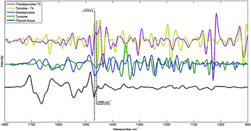
FTIR spectra of hormones triiodothyronine, thyroxine, diiodotyrosine, tyrosine and thyroid tissue

The combination of vibrational spectroscopy with mapping and imaging techniques to address important biochemical questions is an area of active and expanding research.  By subjecting adjacent tissue sections to standard histopathological screening and spectroscopic imaging, a greater understanding of the biochemical processes underlying tissue form and function can be achieved.

Researchers from Northeastern University, USA, and the Institute of Energy and Nuclear Research (IPEN), Sao Paolo, have used Fourier transform infrared (FTIR) microspectroscopy to examine healthy thyroid tissues. FTIR allows spatially resolved images, or maps, to be obtained by use of focal plane array detectors and spectral processing of the data then reveals chemical images.  In this HOT Analyst paper, Denise Zezell and coworkers present an example from their investigation of 80 different patient samples, which were analysed by transflection-mode FTIR mapping.  The approach was  applied to the study of healthy thyroid tissue and, with reference to thyroid specific hormones, iodination state. 

To know more about this research, click on the links below. This paper will be free to read for the next three weeks:

The characterization of normal thyroid tissue by micro-FTIR spectroscopy
Thiago M. Pereira, Denise M. Zezell, Benjamin Bird, Milos Miljković and Max Diem
Analyst, 2013,138, 7094-7100
DOI: 10.1039/C3AN00296A

Digg This
Reddit This
Stumble Now!
Share on Facebook
Bookmark this on Delicious
Share on LinkedIn
Bookmark this on Technorati
Post on Twitter
Google Buzz (aka. Google Reader)

Classifying myobacterial strains using surface enhanced Raman spectroscopy

Laura Ruvuna is guest web writer for Analyst. She is currently a PhD student at Northwestern University, Evanston, Illinois, USA.

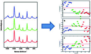
Spectroscopic analysis of mycolic acid profiles

In bacterial-based diagnostics, the bacteria can evolve over time and it is important to be able to differentiate between multiple strains. Mycobacterium tuberculosis, which is responsible for causing tuberculosis (TB), has multiple drug-resistant strains. The most accurate method to identify toxic strains of TB, high performance liquid chromatography (HPLC), requires fluorescent labeling and extraction of the mycolic acids, from the outer membrane of the bacteria.

Researchers at the University of Georgia in the United States wanted a more direct detection method and approached this problem in two important aspects. They began by identifying the high molecular weight lipids within the bacteria using HPLC, gas chromatography (GC), and nuclear magnetic resonance (NMR) for several TB bacterial strains. NMR in particular was very important in identifying the structures of mycolic acids. Using the information from these techniques and surface enhanced Raman spectroscopy (SERS), they chemically classified the mycolic acid from both TB and non-TB strains. The SERS spectra underwent further statistical analysis and accurately identified different strains of TB label-free and with high sensitivity.

To know more about this research, click on the link below. This paper will be free to read for the next three weeks.

Identification of mycobacteria based on spectroscopic analyses of mycolic acid profiles
Omar E. Rivera-Betancourt, Russell Karls, Benjamin Grosse-Siestrup, Shelly Helms, Frederick Quinn and Richard A. Dluhy
Analyst, 2013,138, 6774-6785
DOI: 10.1039/C3AN01157G

Digg This
Reddit This
Stumble Now!
Share on Facebook
Bookmark this on Delicious
Share on LinkedIn
Bookmark this on Technorati
Post on Twitter
Google Buzz (aka. Google Reader)

Probe and Chip Approaches to Cell Analysis Themed Issue: Submissions Open!

Harris et al., Analyst, 2013, 138, 4769-4776

We are delighted to announce a new themed issue of Analyst on

Probe and Chip Approaches to Cell Analysis

This issue, edited by Professor Luke Lee (University of California, Berkeley, USA) and Professor Jonathan Cooper (University of Glasgow, UK), aims to cover a wide range of aspects of cell analysis based on recent microfluidic and lab-on-a chip technologies by highlighting analytical developments and novel applications.

Why not submit today? Submission deadline: 9th January 2014. Email us at ANALYST-RSC@rsc.org for more information.

All articles will be peer-reviewed and, to be suitable for publication, must meet the usual quality and significance standards of the journal.

Please indicate in your covering letter that your article is to be considered for the Probe and Chip Approaches to Cell Analysis themed issue.

Digg This
Reddit This
Stumble Now!
Share on Facebook
Bookmark this on Delicious
Share on LinkedIn
Bookmark this on Technorati
Post on Twitter
Google Buzz (aka. Google Reader)

More HOT Articles in Analyst

Take a look at our new HOT articles just published in Analyst! These papers are all free to read for the next three weeks. Enjoy!

In vitro detection of calcium in bone by modified carbon dots
A. Shanti Krishna, C. Radhakumary and K. Sreenivasan
Analyst, 2013,138, 7107-7111
DOI: 10.1039/C3AN01239E, Paper

Measuring and modelling cell-to-cell variation in uptake of gold nanoparticles
J. Charles. G. Jeynes, Christopher Jeynes, Michael J. Merchant and Karen J. Kirkby
Analyst, 2013,138, 7070-7074
DOI: 10.1039/C3AN01406A, Communication

Adenosine capped QDs based fluorescent sensor for detection of dopamine with high selectivity and sensitivity
Qin Mu, Hu Xu, Yan Li, Shijian Ma and Xinhua Zhong
Analyst, 2013, Advance Article
DOI: 10.1039/C3AN01592K, Paper

A pyrene-based simple but highly selective fluorescence sensor for Cu2+ ions via a static excimer mechanism
Soma Sarkar, Swapnadip Roy, Anindita Sikdar, R. N. Saha and Sujit S. Panja
Analyst, 2013,138, 7119-7126
DOI: 10.1039/C3AN00928A, Paper

Whole organ cross-section chemical imaging using label-free mega-mosaic FTIR microscopy
Paul Bassan, Ashwin Sachdeva, Jonathan H. Shanks, Mick D. Brown, Noel W. Clarke and Peter Gardner
Analyst, 2013,138, 7066-7069
DOI: 10.1039/C3AN01674A, Communication

A highly sensitive LED-induced chemiluminescence platform for aptasensing of platelet-derived growth factor
Xinfeng Zhang, Hui Zhang, Shuxia Xu and Yonghua Sun
Analyst, 2013, Advance Article
DOI: 10.1039/C3AN01566A, Paper

Effects of surface charges of graphene oxide on neuronal outgrowth and branching
Qin Tu, Long Pang, Yun Chen, Yanrong Zhang, Rui Zhang, Bingzhang Lu and Jinyi Wang
Analyst, 2013, Advance Article
DOI: 10.1039/C3AN01796F, Paper

Paracrine signalling of inflammatory cytokines from an in vitro blood brain barrier model upon exposure to polymeric nanoparticles
Michelle Nic Raghnaill, Mattia Bramini, Dong Ye, Pierre-Olivier Couraud, Ignacio A. Romero, Babette Weksler, Christoffer Åberg, Anna Salvati, Iseult Lynch and Kenneth A Dawson
Analyst, 2013, Accepted Manuscript
DOI: 10.1039/C3AN01621H, Paper

Adenosine capped QDs based fluorescent sensor for detection of dopamine with high selectivity and sensitivity
Qin Mu, Hu Xu, Yan Li, Shijian Ma and Xinhua Zhong
Analyst, 2013, Advance Article
DOI: 10.1039/C3AN01592K, Paper

Digg This
Reddit This
Stumble Now!
Share on Facebook
Bookmark this on Delicious
Share on LinkedIn
Bookmark this on Technorati
Post on Twitter
Google Buzz (aka. Google Reader)

HOT Articles in Analyst

Take a look at our new HOT articles just published in Analyst and free for you for the next couple of weeks:

Combined in situ atomic force microscopy and infrared attenuated total reflection spectroelectrochemistry
Daniel Neubauer, Jochen Scharpf, Alberto Pasquarelli, Boris Mizaikoff and Christine Kranz
Analyst, 2013,138, 6746-6752
DOI: 10.1039/C3AN01169K, Paper

An iridium(III)-based lab-on-a-molecule for cysteine/homocysteine and tryptophan using triple-channel interrogation Kun Chen and Michael Schmittel
Analyst, 2013,138, 6742-6745
DOI: 10.1039/C3AN01530K, Communication

Determination of aerosol oxidative activity using silver nanoparticle aggregation on paper-based analytical devices
Wijitar Dungchai, Yupaporn Sameenoi, Orawon Chailapakul, John Volckens and Charles S. Henry
Analyst, 2013,138, 6766-6773
DOI: 10.1039/C3AN01235B, Paper

Biocatalytic analysis of biomarkers for forensic identification of ethnicity between Caucasian and African American groups
Friederike Kramer, Lenka Halámková, Arshak Poghossian, Michael J. Schöning, Evgeny Katz and Jan Halámek
Analyst, 2013,138, 6251-6257
DOI: 10.1039/C3AN01062G, Communication

Biocatalytic analysis of biomarkers for forensic identification of ethnicity between Caucasian and African American groups
Friederike Kramer, Lenka Halámková, Arshak Poghossian, Michael J. Schöning, Evgeny Katz and Jan Halámek
Analyst, 2013,138, 6251-6257
DOI: 10.1039/C3AN01062G, Communication

Analysis of free fractions for chiral drugs using ultrafast extraction and multi-dimensional high-performance affinity chromatography
Xiwei Zheng, Michelle J. Yoo and David S. Hage
Analyst, 2013,138, 6262-6265
DOI: 10.1039/C3AN01315D, Communication

Radical cascades in electron transfer dissociation (ETD) – implications for characterizing peptide disulfide regio-isomers
Lei Tan, Kirt L. Durand, Xiaoxiao Ma and Yu Xia
Analyst, 2013,138, 6759-6765
DOI: 10.1039/C3AN01333B, Paper

Enhanced peroxydisulfate electrochemiluminescence for dopamine biosensing based on Au nanoparticle decorated reduced graphene oxide
Yuting Yan, Qian Liu, Kun Wang, Ling Jiang, Xingwang Yang, Jing Qian, Xiaoya Dong and Baijing Qiu
Analyst, 2013,138, 7101-7106
DOI: 10.1039/C3AN01533E, Paper

Digg This
Reddit This
Stumble Now!
Share on Facebook
Bookmark this on Delicious
Share on LinkedIn
Bookmark this on Technorati
Post on Twitter
Google Buzz (aka. Google Reader)

Featured on the Covers of Issue 23

Stephenson-Brown et al., Analyst, 2013, 138, 7146-7151

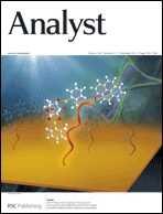
We are delighted to highlight the latest covers of Analyst, showing new research on the development and application of a sensory system to detect glucose.

John Fossey from the University Birmingham, UK, and collaborators at the University of Bath, UK, and at the East China University of Science and Technology, China, present two papers describing a new glucose sensor based on the principle of self-assembly and surface plasmon resonance (SPR) optical technique.

Wang et al., Analyst, 2013, 138, 7146-7151

The sensor is prepared modifying the surface of a gold electrode with a bis-boronic acid receptor. The receptor contains a glucose selective chemosensor unit and a surface anchoring unit, which allows selective detection of glucose. This system has the unique capacity to label-free and allows detection of molecular reactions in real time with highly sensitivity. According to this research, SPR could be an attractive sensing platform to monitor glucose and possibly other analytes.

To read more about this work, please access the links below. These two papers are free to read, so have a look now:

A bis-boronic acid modified electrode for the sensitive and selective determination of glucose concentrations
Hui-Chen Wang, Hao Zhou, Baoqin Chen, Paula M. Mendes, John S. Fossey, Tony D. James and   Yi-Tao Long
Analyst, 2013, 138, 7146-7151
DOI: 10.1039/C3AN01234D

Glucose selective Surface Plasmon Resonance-based bis-boronic acid sensor
Alex Stephenson-Brown, Hui-Chen Wang, Parvez Iqbal, Jon A. Preece, Yitao Long, John S. Fossey, Tony D. James and   Paula M. Mendes 
Analyst, 2013, 138, 7140-7145
DOI: 10.1039/C3AN01233F

Digg This
Reddit This
Stumble Now!
Share on Facebook
Bookmark this on Delicious
Share on LinkedIn
Bookmark this on Technorati
Post on Twitter
Google Buzz (aka. Google Reader)

Top ten most accessed Analyst articles from July to September 2013

Here are the top ten most accessed Analyst articles for July, August and September 2013.

Detection of calcium phosphate crystals in the joint fluid of patients with osteoarthritis – analytical approaches and challenges
Alexander Yavorskyy, Aaron Hernandez-Santana, Geraldine McCarthy and Gillian McMahon  
Analyst, 2008,133, 302-318
DOI: 10.1039/b716791a

Illuminating disease and enlightening biomedicine: Raman spectroscopy as a diagnostic tool
David I. Ellis, David P. Cowcher, Lorna Ashton, Steve O’Hagan and Royston Goodacre  
Analyst, 2013,138, 3871-3884
DOI: 10.1039/c3an00698k

Upconversion nanoparticles in biological labeling, imaging, and therapy
Feng Wang, Debapriya Banerjee, Yongsheng Liu, Xueyuan Chen and Xiaogang Liu
Analyst, 2010,135, 1839-1854
DOI: 10.1039/c0an00144a

Advances in nano-scaled biosensors for biomedical applications
Jianling Wang, Guihua Chen, Hui Jiang, Zhiyong Li and Xuemei Wang  
Analyst, 2013,138, 4427-4435
DOI: 10.1039/c3an00438d

A coumarin–indole-based near-infrared ratiometric pH probe for intracellular fluorescence imaging
Xiao-Dong Liu, Ying Xu, Ru Sun, Yu-Jie Xu, Jian-Mei Lu and Jian-Feng Ge  
Analyst, 2013,138, 6542-6550
DOI: 10.1039/c3an01033c

Analytical methods to assess nanoparticle toxicity
Bryce J. Marquis, Sara A. Love, Katherine L. Braun and Christy L. Haynes  
Analyst, 2009,134, 425-439
DOI: 10.1039/b818082b

Solid-state Forensic Finger sensor for integrated sampling and detection of gunshot residue and explosives: towards ‘Lab-on-a-finger’
Amay J. Bandodkar, Aoife M. O’Mahony, Julian Ramírez, Izabela A. Samek, Sean M. Anderson, Joshua R. Windmiller and Joseph Wang
Analyst, 2013,138, 5288-5295
DOI: 10.1039/c3an01179h

Selective fluorescence sensing of Cu(ii) and Zn(ii) using a new Schiff base-derived model compound: naked eye detection and spectral deciphering of the mechanism of sensory action
Aniruddha Ganguly, Bijan Kumar Paul, Soumen Ghosh, Samiran Kar and Nikhil Guchhait
Analyst, 2013,138, 6532-6541
DOI: 10.1039/c3an00155e

Carbon dots and chitosan composite film based biosensor for the sensitive and selective determination of dopamine
Qitong Huang, Shirong Hu, Hanqiang Zhang, Jianhua Chen, Yasan He, Feiming Li, Wen Weng, Jiancong Ni, Xiuxiu Bao and Yi Lin  
Analyst, 2013,138, 5417-5423
DOI: 10.1039/c3an00510k

Quantum dots for fluorescent biosensing and bio-imaging applications
Jingjing Li and Jun-Jie Zhu  
Analyst, 2013,138, 2506-2515
DOI: 10.1039/c3an36705c

We hope you enjoyed reading these articles, and if you have any thoughts or comments, please leave these in the comment box below.

Fancy submitting an article to Analyst? Why not submit to us here today or alternatively email us your suggestions!

Digg This
Reddit This
Stumble Now!
Share on Facebook
Bookmark this on Delicious
Share on LinkedIn
Bookmark this on Technorati
Post on Twitter
Google Buzz (aka. Google Reader)

Themed Issue on Molecular Analysis for Art, Archaeometry and Conservation now published

Courtesy of Shutterstock

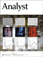
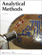
The new joint web themed Issue of Analyst and Analytical Methods on Molecular Analysis for Art, Archaeometry and Conservation has now been published online!

Take a look at this great collection of papers illustrating the most innovative research aimed at preserving our rich cultural heritage.

The themed issue highlights a great number of analytical techniques employed to study art and cultural objects at a molecular level, characterising their structure, properties and chemistry. 

Bertrand et al., Analyst, 2013, 138, 4463-4469

If you are interested in the study and preservation of art of the past and the present, discover the latest analytical breakthroughs described in our Web Collection.

“A deep connection to our past and shared cultural heritage must be preserved to foster a balanced society where all humanity can thrive”, from the Editorial of  Francesca Casadio and Richard P. Van Duyne, Guest Editors of the Issue.

In addition, below are some articles of the issue that you might be interested to read. These papers will be free to read for the next 4 weeks. Enjoy!

Looking beneath Dalí’s paint: non-destructive canvas analysis
Marta Oriola, Alenka Možir, Paul Garside, Gema Campo, Anna Nualart-Torroja, Irene Civil, Marianne Odlyha, May Cassar and Matija Strlič 
Anal. Methods, 2013, Advance Article
DOI: 10.1039/C3AY41094C

Caggiani et al., Anal. Methods, 2013, 5, 4345-4354

Alteration of Asian lacquer: in-depth insight using a physico-chemical multiscale approach
Anne-Solenn Le Hô, Chloé Duhamel, Céline Daher, Ludovic Bellot-Gurlet, Céline Paris, Martine Regert, Michel Sablier, Guilhem André, Jean-Paul Desroches and Paul Dumas 
Analyst, 2013, 138, 5685-5696
DOI: 10.1039/C3AN00608E

Analysis of cadmium based pigments with time-resolved photo-luminescence
Anna Cesaratto, Cosimo D’Andrea, Austin Nevin, Gianluca Valentini, Francesco Tassone, Roberto Alberti, Tommaso Frizzi and Daniela Comelli  
Anal. Methods, 2013, Accepted Manuscript
DOI: 10.1039/C3AY41585F

Quantifying degradation of collagen in ancient manuscripts: the case of the Dead Sea Temple
Scroll R. Schütz, L. Bertinetti, I. Rabin, P. Fratzl and A. Masic 
Analyst, 2013,138, 5594-5599
DOI: 10.1039/C3AN00609C

Please click here to access the full web collection.  

Digg This
Reddit This
Stumble Now!
Share on Facebook
Bookmark this on Delicious
Share on LinkedIn
Bookmark this on Technorati
Post on Twitter
Google Buzz (aka. Google Reader)

Royal Society of Chemistry Roadshow in Brazil this November

We are pleased to announce The Royal Society of Chemistry Brazil Roadshow that will take place in three amazing Brazilian locations this November.

The roadshow consists of three one-day scientific symposia in Campinas, Porto Alegre and Belo Horizonte, featuring lectures by some of the world’s leading international scientists in analytical science and sustainable chemistry.

The roadshow is free to attend and will bring together academics, students and industrial scientists in a stimulating and friendly environment. Join us to find out more about our journals and wider activities! Click here to register.

4th November: University of Campinas (IQ-UNICAMP), Campinas, Brazil

6th November: The Federal University of Rio Grande do Sul (IQ-UFRGS), Porto Alegre, Brazil

8th November: The Federal University of Minas Gerais (DQ-UFMG), Belo Horizonte, Brazil

Among the outstanding scientists presenting at the roadshow:
Frank Vanhaecke (Chair of the JAAS Editorial Board)

Craig Banks (Analytical Methods Associate Editor)

James Clark (Green Chemistry Advisory Board member)

Boris Mizaikoff, (Analyst Associate Editor)

Janet Scott (Green Chemistry Advisory Board member)

The Royal Society of Chemistry is proud to support and publish high-impact research from all over the globe, and a large amount of our content is from authors in Brazil. Below are some examples of the high-quality content published in Green Chemistry, Analyst, Analytical Methods, RSC Advances and JAAS by your Brazilian colleagues, please take a look at the papers listed below:

Glycerol as a recyclable solvent for copper-catalyzed cross-coupling reactions of diaryl diselenides with aryl boronic acids
Vanessa G. Ricordi, Camilo S. Freitas, Gelson Perin, Eder J. Lenardão, Raquel G. Jacob, Lucielli Savegnago and Diego Alves
Green Chem., 2012, 14, 1030–1034, DOI: 10.1039/C2GC16427B
Reversible polymerization of novel monomers bearing furan and plant oil moieties: a double click exploitation of renewable resources
Carla Vilela, Letizia Cruciani, Armando J. D. Silvestre and Alessandro Gandini
RSC Adv., 2012, 2, 2966–2974, DOI: 10.1039/C2RA20053H

Easy dual-mode ambient mass spectrometry with Venturi self-pumping, canned air, disposable parts and voltage-free sonic-spray ionization
Nicolas V. Schwab, Andreia M. Porcari, Mirela B. Coelho, Eduardo M. Schmidt, Jose L. Jara, Jesui V. Visentainer and Marcos N. Eberlin
Analyst, 2012, 137, 2537–2540, DOI: 10.1039/C2AN16312H
Capacitively coupled contactless conductivity detection on microfluidic systems—ten years of development
Wendell Karlos Tomazelli Coltro, Renato Sousa Lima, Thiago Pinotti Segato, Emanuel Carrilho, Dosil Pereira de Jesus, Claudimir Lucio do Lago and José Alberto Fracassi da Silva
Anal. Methods, 2012, 4, 25–33, DOI: 10.1039/C1AY05364G

Determination of trace metals in high-salinity petroleum produced formation water by inductively coupled plasma mass spectrometry following on-line analyte separation/preconcentration
Eliane Padua Oliveira, Lu Yang, Ralph E. Sturgeon, Ricardo Erthal Santelli, Marcos Almeida Bezerra, Scott N. Willie and Ramsés Capilla
J. Anal. At. Spectrom., 2011, 26, 578-585, DOI: 10.1039/C0JA00108B

Digg This
Reddit This
Stumble Now!
Share on Facebook
Bookmark this on Delicious
Share on LinkedIn
Bookmark this on Technorati
Post on Twitter
Google Buzz (aka. Google Reader)

LEDs Overcome the “Enemy” of Raman Spectroscopy

Polly-Anna Ashford is a guest web writer for Analyst. She is currently a PhD student at the University of East Anglia, UK.

LED-SERDS in Raman Spectroscopy

LED-SERDS in Raman Spectroscopy

Raman spectroscopy finds a range of uses in analytical chemistry. Like infra-red (IR) spectroscopy it can provide a vibrational fingerprint by which compounds may be identified. Unlike IR, Raman is a scattering technique that does not require complex sample preparation, and water can be used as a solvent. In some samples, however, the Raman spectrum is rendered useless by intense fluorescence signals.

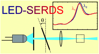
Chemical engineers Renata Adami (University of Salerno, Italy) and Johannes Kiefer (University of Aberdeen, UK) have developed a new approach to shifted-excitation Raman difference spectroscopy (SERDS) for suppression of fluorescence. SERDS experiments conventionally require expensive diode lasers, but in this Analyst paper Kiefer and Adami demonstrate the use of light-emitting diodes (LEDs) as an inexpensive light source. A dielectric bandpass filter narrows and stabilises the broad LED spectrum to make it usable for SERDS. While the resulting spectral resolution is low, different compounds and functional groups can be clearly distinguished using this method.

To read the full access, please click on the link below. This paper will be free to read for the next three weeks:

Light-emitting diode based shifted-excitation Raman difference spectroscopy (LED-SERDS)
Renata Adami and Johannes Kiefer
Analyst, 2013,138, 6258-6261
DOI: 10.1039/C3AN01367G
Digg This
Reddit This
Stumble Now!
Share on Facebook
Bookmark this on Delicious
Share on LinkedIn
Bookmark this on Technorati
Post on Twitter
Google Buzz (aka. Google Reader)