Biocomputer decides when to administer drugs

Scientists in the US have devised an enzymatic logic system that could be used for releasing drugs. This is the first report of a man-made biomolecular system that can process a series of physiological signals, without the use of electronics.

Biocomputer-based logic systems that process biomolecular signals could revolutionise drug administration. By harnessing signal-responsive electrode surfaces that respond to biochemical signals, widespread personalised medicine takes a step closer to reality.

Historically, drug-release systems have been plagued by slow and uncontrolled release. Various external triggers, including temperature, pH and biochemical species, have been used to stimulate drug release. Systems activated by biochemical signals are often complicated and limited, combining both the receptor and the release system. Physical separation of these two components on individual electrodes would simplify the process.

Expanding on their recent work with glucose sensing electrodes, Evgeny Katz and Shay Mailloux at Clarkson University, in collaboration with Jan Halámek from the State University of New York at Albany, have developed a logical biomolecule release system. An electrode covered by a redox-active, iron(III)-cross-linked alginate polymer film with physically entrapped biomolecules serves as the substance-releasing component and a pyrroloquinoline quinone (PQQ)-modified electrode serves as the biocatalytic electrode.

To read the full article, please visit Chemistry World

A model system for targeted drug release triggered by biomolecular signals logically processed through enzyme logic networks
Shay Mailloux, Jan Halámek and Evgeny Katz
Analyst, 2014, Advance Article
DOI: 10.1039/C3AN02162A, Communication

Digg This
Reddit This
Stumble Now!
Share on Facebook
Bookmark this on Delicious
Share on LinkedIn
Bookmark this on Technorati
Post on Twitter
Google Buzz (aka. Google Reader)

The Emerging Technologies competition open for applications

The Royal Society of Chemistry launches the 2014 UK Emerging Technologies Competition

Applications are invited from university researchers and small companies working in the UK in applied research in the chemical, life and materials sciences for a chance to turn their research into commercial success. The prize consists of ongoing mentoring and support from high profile multinational companies, up to £10,000 cash prize and more. We are proud to announce the mentor companies Procter and Gamble, GlaxoSmithKline, Catalent Applied Drug Delivery Institute, Croda and more will be joining soon.

Emerging Technologies Competition 2014

The applicants can be individuals or teams and they must submit a brief online application to the Royal Society of Chemistry. The entries will be judged by an independent panel of science and business experts and the shortlisted applications will be invited for the second round of the competition. Each team will pitch to a specialist panel at a public event and up to 5 teams will be crowned as winners. But everyone is a winner! The finalists will be able to access one to one FREE advice from business and finance specialists. In addition this is an excellent opportunity to practice pitching ideas to high profile multinational companies and to meet and network with fellow entrepreneurs, investors and business leaders.

Key dates:
First round closes on 1 March 2014
Second round takes place on 25 June 2014,
Chemistry Centre, London

Visit www.rsc.li/emerging-technologies for full details.
UK applications only please

If you have any questions please contact Aurora Antemir antemira@rsc.org.

Digg This
Reddit This
Stumble Now!
Share on Facebook
Bookmark this on Delicious
Share on LinkedIn
Bookmark this on Technorati
Post on Twitter
Google Buzz (aka. Google Reader)

Crime scene chemistry – Analyst articles on Chemistry World 2013

What discoveries caused the biggest buzz in chemistry labs in 2013? With the help of an expert panel of journal editors Chemistry World reviews the ground breaking research and important trends in this year’s crop of chemical science papers.

Find out which Analyst articles have been featured in Chemistry World this year:

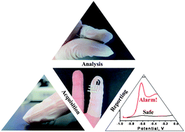
Crime scene chemistry Graphical abstract: Solid-state Forensic Finger sensor for integrated sampling and detection of gunshot residue and explosives: towards ‘Lab-on-a-finger’

Improvements in forensic techniques have also featured on our pages this year. A team in the US developed a device that investigators can wear on their fingertips to rapidly identify traces of explosives and gunshot residue. The sensor consists of an electrode screen-printed onto a stretchable sheath worn on the index finder, and a sheath for the thumb coated with a solid-state ionogel electrolyte. To analyse a sample the investigator simply squeezes their finger and thumb together after swiping a surface, completing the electrochemical cell. A portable analyser then reads the voltammetric signal, identifying distinct peaks for explosives or gunshot residue. The process takes just a few minutes, cutting down the lengthy practice of sample collection and lab analysis.

Another group has developed a bioassay that can be used to analyse blood samples on-site to give investigators an early indication of a suspect’s ethnicity. Evgeny Katz at Clarkson University, US, in collaboration with Jan Halámek, now at the State University of New York at Albany, analysed levels of two biomarkers – creatine kinase and lactate dehydrogenase – in the blood of people of Caucasian and African American ethnicity. They then developed a bioassay to amplify the differences in these levels. The test could successfully distinguish between ethnicities in real human blood samples, as well as samples a day old, as could well be the case at a crime scene.

To find out more, read the full article on Chemistry World.

Solid-state Forensic Finger sensor for integrated sampling and detection of gunshot residue and explosives: towards ‘Lab-on-a-finger’
Amay J. Bandodkar, Aoife M. O’Mahony, Julian Ramírez, Izabela A. Samek, Sean M. Anderson, Joshua R. Windmiller and Joseph Wang
Analyst, 2013,138, 5288-5295
DOI: 10.1039/C3AN01179H, Paper

Biocatalytic analysis of biomarkers for forensic identification of ethnicity between Caucasian and African American groups
Friederike Kramer, Lenka Halámková, Arshak Poghossian, Michael J. Schöning, Evgeny Katz and Jan Halámek
Analyst, 2013,138, 6251-6257
DOI: 10.1039/C3AN01062G, Communication

Digg This
Reddit This
Stumble Now!
Share on Facebook
Bookmark this on Delicious
Share on LinkedIn
Bookmark this on Technorati
Post on Twitter
Google Buzz (aka. Google Reader)

‘Google map’ of a prostate

UK researchers have used vibrational spectroscopy to chemically image the cross section of a prostate to such an incredible level of detail that each of the 66 million pixels in the image represents a piece of tissue only 5.5 × 5.5µm.

Biopsies are regularly taken to diagnose cancer and provide a snapshot of the disease. Trained histopathologists examine the samples under powerful optical microscopes using several specialist stains to highlight particular characteristics of cells or tissues in a time-consuming process.

Peter Gardner of the University of Manchester and colleagues hope their technique, which uses Fourier transform infrared (FTIR) chemical imaging, will eventually be turned into an automated system that can grade and stage biopsy samples 24 hours a day by identifying certain chemical signatures.

To read the full article, please visit Chemistry World.

Whole organ cross-section chemical imaging using label-free mega-mosaic FTIR microscopy
Paul Bassan, Ashwin Sachdeva, Jonathan H. Shanks, Mick D. Brown, Noel W. Clarke and Peter Gardner
Analyst, 2013,138, 7066-7069
DOI: 10.1039/C3AN01674A, Communication

Digg This
Reddit This
Stumble Now!
Share on Facebook
Bookmark this on Delicious
Share on LinkedIn
Bookmark this on Technorati
Post on Twitter
Google Buzz (aka. Google Reader)

Nanoparticles for platelet adhesion and aggregation

Jennifer A. Dougan is a guest web-writer for Analyst. She is currently a Post-Doctoral Research Associate at Imperial College London, UK.

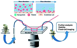
On-chip evaluation of platelet adhesion and aggregation

The use of nanoparticles in diagnostic and therapeutic roles requires that we understand their interactions and behaviour within complex biological systems. Where clinical applications would involve intravenous injection of nanoparticle-based therapeutics, it will be necessary to understand the interaction between nanoparticles and native blood components.

To address this issue, Christy Haynes and coworkers from the University of Minnesota, USA, have created a microfluidic device coated with endothelial cells to mimic the walls of the vascular system. This initial in vitro approach was carried out with both activated and unactivated platelets in the presence of various (therapeutically-relevant) concentrations of fluorescently-labelled, mesophorous silica nanoparticles. The impact of nanoparticles on the critical platelet functions of adhesion and aggregation was assessed.

Microfluidic platforms are readily customised and future work would be expected to expand upon these initial conditions to advance our understanding of nanotoxicology and interaction studies.

To read more about this study and the impact of nanoparticles on platelet interactions you can access this Analyst HOT Article free until 6 January 2014:

On-chip evaluation of platelet adhesion and aggregation upon exposure to mesoporous silica nanoparticles
Donghyuk Kim, Solaire Finkenstaedt-Quinn, Katie R. Hurley, Joseph T. Buchman and Christy L. Haynes 
Analyst, 2014, Advance Article
DOI: 10.1039/C3AN01679J

Digg This
Reddit This
Stumble Now!
Share on Facebook
Bookmark this on Delicious
Share on LinkedIn
Bookmark this on Technorati
Post on Twitter
Google Buzz (aka. Google Reader)

Prizes and Awards nominations open

The Royal Society of Chemistry’s Prizes and Awards recognise achievements by individuals, teams and organisations in advancing the chemical sciences. There are over 80 Prizes and Awards available covering all areas of the chemical sciences, with prize money of up to £5000 to be won.

The main categories are:

shutterstock_93666160

Biosciences

Environment, Sustainability and Energy

Inorganic Chemistry (Dalton Division)

Materials Chemistry

Organic Chemistry

Physical Chemistry (Faraday Division)

Industry & Technology

In addition, we have a new award this year, the Industrial Analytical Science Award, to recognise and celebrate the great contribution of analytical science in industry.

To view the full list of Prizes and Awards and to make a nomination, visit www.rsc.org/awards

Nominations open until 15 January 2014

Digg This
Reddit This
Stumble Now!
Share on Facebook
Bookmark this on Delicious
Share on LinkedIn
Bookmark this on Technorati
Post on Twitter
Google Buzz (aka. Google Reader)

Enhancing transmission Raman spectroscopy (TRS) for pharmaceutical analysis

Polly-Anna Ashford is a guest web writer for Analyst. She is currently a PhD student at the University of East Anglia, UK.

Data sub-selection in transmission Raman spectroscopy

Data sub-selection in transmission Raman spectroscopy

The analysis of compound mixtures in powder and tablet form has a range of purposes, from monitoring the stability of a formulation over time and quality control of a product, to the forensic analysis of illicit substances. Transmission Raman spectroscopy is a promising candidate for this type of analysis. TRS is fast and non-destructive, it produces data that is easy to interpret, and has good penetration depth for opaque samples such as powders.

Researchers led by Jonathan Burley at the University of Nottingham (UK) have investigated ways to improve the accuracy of TRS for quantitative analysis. In this HOT Analyst paper, they report the first detailed analysis of data sub-selection for a set of transmission Raman data obtained from a model pharmaceutical formulation. Burley and co-workers also focus on the utility of low-wavenumber data, which has only become accessible in recent years. The authors anticipate that their findings may shape the future development of Raman instrumentation.

To read more about this work, please access the link below. This paper will be free to read until 6 January 2014.

Quantification of pharmaceuticals via transmission Raman spectroscopy: data sub-selection
Jonathan C. Burley, Adeyinka Aina, Pavel Matousek and Christopher Brignell
Analyst, 2014,139, 74-78
DOI: 10.1039/C3AN01293J

Digg This
Reddit This
Stumble Now!
Share on Facebook
Bookmark this on Delicious
Share on LinkedIn
Bookmark this on Technorati
Post on Twitter
Google Buzz (aka. Google Reader)

Immobilized phage proteins for detection of staphylococci

Written by Matthew J. Baker, Guest Web-writer for Analyst and Senior Lecturer in Toxicology and Analytical Chemistry at the University of Central Lancashire (UCLan), UK.

The rapid and specific detection of pathogenic bacteria, such as staphylococci, is an important worldwide concern. Staphylococci cause serious infections in humans and animals and certain species such as S. aureus and methicillin resistant S. aureus (MRSA) are developing antibiotic resistance that is quickly becoming a global crisis. The ability to detect these bacteria rapidly and in situ would allow for early treatment even in countries with poor access to healthcare.

Immobilized phage proteins for specific detection of staphylococci

Canadian researchers from McGill University and Polytechnique Montreal have recently shown the use of immobilized bacteriophage proteins to develop a biosensor for the specific detection of 8 clinical isolates of staphylococciHicham Chibli and coworkers examined several purified phage proteins from the Endolysin class of enzymes and exploited their ability to specifically bind staphylococci. Indeed these immobilized phage proteins did not bind to other closely related bacteria.

Bacteriophages have been used as recognition elements for biosensors before, but this research shows the use of specific phage proteins instead of whole bacteriophages. “Compared with whole phages, these single proteins are smaller, easier to purify, and easier to assemble in a reproducible manner of a surface” – says Jay Nadeau of McGill University – “The small size is key for development of some types of biosensors where sensitivity is dependent upon thickness of the functionalizing layer, for example, microresonators” – he adds. The ability to develop microresonators with specific detection can provide small, portable and specific biosensors for in field use.

To know more about this study, please access the link below. This paper will be free to read for the next three weeks.

Immobilized phage proteins for specific detection of staphylococci
Hicham Chibli, Hala Ghali, Soonhyang Park, Yves-Alain Peter and Jay L. Nadeau  
Analyst, 2014, 139, 179-186
DOI: 10.1039/C3AN01608K

Digg This
Reddit This
Stumble Now!
Share on Facebook
Bookmark this on Delicious
Share on LinkedIn
Bookmark this on Technorati
Post on Twitter
Google Buzz (aka. Google Reader)

HOT Articles in Analyst

Take a look at our new HOT articles just published in Analyst and free for you for the next couple of weeks:

Development of coated-wire silver ion selective electrodes on paper using conductive films of silver nanoparticles
Wanwisa Janrungroatsakul, Chutiparn Lertvachirapaiboon, Wittaya Ngeontae, Wanlapa Aeungmaitrepirom, Orawon Chailapakul, Sanong Ekgasitb and Thawatchai Tuntulani
Analyst, 2013,138, 6786-6792
DOI: 10.1039/C3AN01385E

A visible light photoelectrochemical sensor for tumor marker detection using tin dioxide quantum dot–graphene as labels
Yanhu Wang, Meng Li, Yuanna Zhu, Shenguang Ge, Jinghua Yu, Mei Yan and Xianrang Song
Analyst, 2013,138, 7112-7118
DOI: 10.1039/C3AN01410J

Retention in continuous two-dimensional thermal field-flow fractionation: comparison of experimental results with theory
Pertti Vastamäki, P. Stephen Williams, Matti Jussila, Michel Martin and Marja-Liisa Riekkola
Analyst, 2014, Advance Article
DOI: 10.1039/C3AN01047C

Cascade signal amplification for ultrasensitive electrochemical DNA detection
Jin Xu, Qiong Wang, Yun Xiang, Ruo Yuan and Yaqin Chai
Analyst, 2014, Advance Article
DOI: 10.1039/C3AN01673K

Detection and characterization of silver nanoparticles and dissolved species of silver in culture medium and cells by AsFlFFF-UV-Vis-ICPMS: application to nanotoxicity tests
E. Bolea, J. Jiménez-Lamana, F. Laborda, I. Abad-Álvaro, C. Bladé, L. Arola and J. R. Castillo
Analyst, 2014, Advance Article
DOI: 10.1039/C3AN01443F

MALDI mechanisms: wavelength and matrix dependence of the coupled photophysical and chemical dynamics model
Richard Knochenmuss
Analyst, 2014, Advance Article
DOI: 10.1039/C3AN01446K

Target-induced quenching for highly sensitive detection of nucleic acids based on label-free luminescent supersandwich DNA/silver nanoclusters
Guangfeng Wang, Yanhong Zhu, Ling Chen, Lun Wang and Xiaojun Zhang
Analyst, 2014, Advance Article
DOI: 10.1039/C3AN01702H

LED-based interferometric reflectance imaging sensor for the detection of amyloid-β aggregation
Xin R. Cheng, George G. Daaboul, M. Selim Ünlü and Kagan Kerman
Analyst, 2014, Advance Article
DOI: 10.1039/C3AN01307C

Improved accuracy for label-free absolute quantification of proteome by combining the absolute protein expression profiling algorithm and summed tandem mass spectrometric total ion current
Qi Wu, Yichu Shan, Yanyan Qu, Hao Jiang, Huiming Yuan, Jianxi Liu, Shen Zhang, Zhen Liang, Lihua Zhang and Yukui Zhang
Analyst, 2014, Advance Article
DOI: 10.1039/C3AN01738A

Focussed ion beam serial sectioning and imaging of monolithic materials for 3D reconstruction and morphological parameter evaluation
Mercedes Vázquez, David Moore, Xiaoyun He, Aymen Ben Azouz, Ekaterina Nesterenko, Pavel Nesterenko, Brett Paull and Dermot Brabazon
Analyst, 2014, Advance Article
DOI: 10.1039/C3AN01827J

Digg This
Reddit This
Stumble Now!
Share on Facebook
Bookmark this on Delicious
Share on LinkedIn
Bookmark this on Technorati
Post on Twitter
Google Buzz (aka. Google Reader)

Therapeutic screening for Alzheimer’s disease

Scientists in Canada and the US hope a system they have developed for monitoring amyloid-β aggregation on a chip could be used to find new treatments for Alzheimer’s disease.

Alzheimer’s disease is a complex neurodegenerative condition and the most common cause of dementia. In the US, more than 5 million people are estimated to have the condition. There is currently no known cure. Research has uncovered that the self-aggregation of the amyloid-β (Aβ) peptide plays a vital role in the development of Alzheimer’s disease. Methods that study the interaction of Aβ with potential new drugs are therefore extremely important.

Now, Kagan Kerman at the University of Toronto at Scarborough, and colleagues, have created a sensing platform to aid the drug screening process. ‘We have successfully demonstrated a novel method for high throughput screening of small molecules modulating Aβ growth and it provides a promising platform to facilitate therapeutics discovery for Alzheimer’s disease,’ says Kerman.

To read the full article, please visit Chemistry World.

LED-based interferometric reflectance imaging sensor for the detection of amyloid-β aggregation
Xin R. Cheng, George G. Daaboul, M. Selim Ünlü and Kagan Kerman
Analyst, 2014, Advance Article
DOI: 10.1039/C3AN01307C, Paper

Digg This
Reddit This
Stumble Now!
Share on Facebook
Bookmark this on Delicious
Share on LinkedIn
Bookmark this on Technorati
Post on Twitter
Google Buzz (aka. Google Reader)