Marblar Micromixer Challenge!

If you haven’t already heard of Marblar, the crowd-sourcing website that enables many minds to work together to realise the potential of new technologies, then now is the time to find out more, as the Royal Society of Chemistry is sponsoring a Marblar challenge to find  applications for a new microfluidic mixing device developed by Huanming Zia and co-workers based on their 2012 communication in Lab on a Chip!

Converting steady laminar flow to oscillatory flow through a hydroelasticity approach at microscales
H. M. Xia, Z. P. Wang, W. Fan, A. Wijaya, W. Wanga and Z. F. Wanga  
DOI: 10.1039/C1LC20667B

The microfluidic mixer is a non-powered device that can be added in-line to convert laminar flow to oscillatory flow, achieving effective mixing in milliseconds – ordinarily the laminar flow behaviour at these scales means fluid streams only mix very slowly via diffusion, which has been a big limitation for development of microfluidic products.

If you might have an idea or think you could improve on someone else’s, then join in the discussion and maybe even submit an idea or two to win points and marbles!

Digg This
Reddit This
Stumble Now!
Share on Facebook
Bookmark this on Delicious
Share on LinkedIn
Bookmark this on Technorati
Post on Twitter
Google Buzz (aka. Google Reader)

On-chip investigation: How do cells feel the force?

Posted on behalf of Alphonsus Ng, Lab on a Chip web writer

Researchers at the City University of Hong Kong and Jinan University, China, have developed a rapid approach to study how cells “feel” the forces around them.

Cells sense and react to their physical surroundings through mechanotransduction—the translation of mechanical stimulation into biochemical signals. These signals, in turn, regulate many important physiological processes such as those in the immune system, bone metabolism and blood circulation. Since defects in mechanotransduction are associated with various human diseases,1 understanding their molecular basis may aid in the development of new therapeutics. Unfortunately, most techniques for studying mechanotransduction are slow and labour-intensive, requiring meticulous manipulation of single cells for mechanical stimulation and analysis.

To address this challenge, a research team led by Prof. Mengsu Yang has developed a new microfluidic chip that can isolate individual cells and apply precise whole-cell mechanical stimulation.

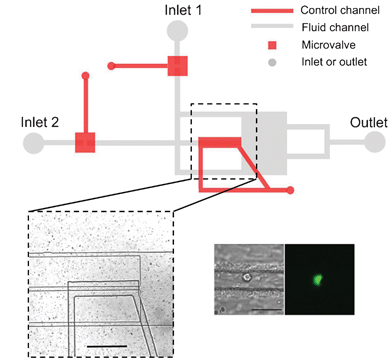
The microfluidic chip is assembled by sandwiching a deflectable polymer membrane between a fluid layer and a control layer. The chip comprises three parallel channels, two microvalves and one compressive component in the middle channel (see picture). By modulating the pressure in the control channels, the height of the middle channel can be adjusted to trap or compress an individual cell from a suspension. The microvalves can be opened or closed to exchange the reagent environment surrounding the trapped cell.

Using this chip, the research team studied the intracellular calcium signaling in HL60 cells (leukemic cells) triggered by whole-cell compression. They demonstrate that mechanical compression can activate ion channels in the cell membrane, causing extracellular calcium ions to transport inside the cell. Interestingly, they show that an intact cytoskeleton was not required for the activation of mechanosensitive ion channels in HL60 cells—the involvement of the cytoskeleton in mechnotranduction remains a much-debated topic in the field.

Taken together, the research team successfully demonstrated that this microfluidic chip is a fast, useful platform for the study of mechanotransduction in individual suspension cells. In the future, the throughput of this chip may be extended by parallelizing several compressive components on one chip.

1.            Jaalouk, D. E.; Lammerding, J. Nature Reviews Molecular Cell Biology 2009, 10, 63-73.

Microfluidics study of intracellular calcium response to mechanical stimulation on single suspension cells
Tao Xu, Wanqing Yue, Cheuk-Wing Li,  Xinsheng Yao and Mengsu Yang
DOI: 10.1039/C3LC40880A

Alphonsus Ng is a PhD student in the Wheeler Microfluidics Laboratory, University of Toronto, Canada

Digg This
Reddit This
Stumble Now!
Share on Facebook
Bookmark this on Delicious
Share on LinkedIn
Bookmark this on Technorati
Post on Twitter
Google Buzz (aka. Google Reader)

Awards for LOC Research Highlights writer Ali Khademhosseini

Congratulations to Ali Khademhosseini, writer of the regular Lab on a Chip Research Highlights articles, who has this year received three prestigious research awards!

In June, he received the 2013 Owens Corning Early Career Award of the American Institute for Chemical Engineers (AIChE) for outstanding contributions in applying micro/nanoscale technologies to engineer functional biomaterials for regenerative medicine. This award recognizes outstanding independent contributions to the scientific, technological, or educational areas of materials science and engineering by a member of the Materials Engineering and Sciences Division of the AIChE under 40. The award will be presented at November’s annual meeting of AIChE in San Francisco.

Professor Khademhosseini was also recently awarded the 2013 IEEE Engineering in Medicine and Biology Society’s Technical Achievement Award. The award is given to recognize outstanding achievement, contribution and/or innovation in a technical area of biomedical engineering. He received the award at the annual EMBS meeting in Japan last week.

In April, we blogged to congratulate Ali on winning the Controlled Release Society Young Investigator award, which will be presented at the end of this month in Hawaii!

View some of Ali’s recent Research Highlights articles here:

Research highlights
Imee G. Arcibal, Donald M. Cropek, Mehmet R. Dokmeci and Ali Khademhosseini 
DOI: 10.1039/C3LC90037A

Research highlights
João Ribas, Mark W. Tibbitt, Mehmet R. Dokmeci and Ali Khademhosseini
DOI: 10.1039/C3LC90032K

Research highlights
Šeila Selimović, Mehmet R. Dokmeci and Ali Khademhosseini
DOI: 10.1039/C3LC90025H

Research highlights
Šeila Selimović, Mark W. Tibbitt, Mehmet R. Dokmeci and Ali Khademhosseini
DOI: 10.1039/C3LC90018E

Digg This
Reddit This
Stumble Now!
Share on Facebook
Bookmark this on Delicious
Share on LinkedIn
Bookmark this on Technorati
Post on Twitter
Google Buzz (aka. Google Reader)

Worm on the run: measuring forces exerted by a nematode in motion

Posted on behalf of Aleksandra (Sasha) Denisin, Lab on a Chip web writer.

Researchers at the University of Canterbury in New Zealand have created a platform measuring mechancial forces exerted by the microscopic worm C. elegans in locomotion. The platform can measure all of the important worm locomotion parameters in a single assay. The team has noted that the worm’s crawling behaviors and thrust forces correlate to the structure of their microenvironment as the worm adjusts its behavior via mechanical sensing of its surroundings.

C. elegans are soil-dwelling worms made up of just 959 cells (adult hermaphrodite). The worms are 1 mm long and 80 μm in body diameter. In labs studying C. elegans, the worms crawl on agar plates in search of food (usually E. coli) by twisting their body in sinusoidal waves.1 The sense of touch is crucial to these nematodes: 6 touch receptor neurons (mechanoreceptor neurons) allow the animal to detect external mechanical feedback with the environment as well as internal forces.2

The group led by Maan M. Alkaisi of The MacDiarmid Institute, New Zealand, and Wenhui Wang now at Tsinghua University, China, developed micropillar arrays of various arrangements and dimensions to modify the worm’s microenvironment and measure forces exerted by the worm as it crawls on the substrate. The researchers were also able to quantify locomotion parameters such as speed, amplitude of sine wave, and wavelength. Their unique assay combines 3 key elements: variable substrate topology, worm thrust force measurements, and locomotive metrics. One version of the device is also compatible with different substrates by mounting pillars on top of migrating worms instead of allowing worms to migrate directly on the microstructures.

The group found that reduced pillar spacing caused wild-type worms to exert twice the force to crawl through the substrate, especially in a honeycomb pillar arrangement. The worms were also much slower at moving in the narrow pillar structures and the frequency of sine wave propagations correlated to resistivity of the physical environment. The platform presented in this study enables many exciting studies in C. elegans locomotion and touch response, especially as  many genetic mutant strains are available with changes to the number of mechanoreceptor neurons and muscle arms. 3

References:

1. S. Berri, J. H. Boyle, M. Tassieri, I. A. Hope and N. Cohen, HFSP journal, 2009, 3, 186-193.
2. M. B. Goodman, in WormBook: the online review of C. elegans biology, NIH Public Access, 2006, available from
http://www.ncbi.nlm.nih.gov/books/NBK20187/.http://www.wormbook.org/chapters/www_mechanosensation/mechanosensation.html.
3. C. I. Bargmann and I. Mori, in C. elegans II. 2nd edition., ed. D. L. Riddle, T. Blumenthal, B. J. Meyer and J. R. Priess, Cold Spring Harbor Laboratory Press, NY, 1997, available from http://www.ncbi.nlm.nih.gov/books/NBK20187/.

On-chip analysis of C. elegans muscular forces and locomotion patterns in microstructured environments
Shazlina Johari, Volker Nock, Maan M. Alkaisi, and Wenhui Wang. Lab Chip, 2013, 13, 1699-1707.
DOI: 10.1039/c3lc41403e

Sasha is a PhD student at Stanford University working with Professor Beth Pruitt’s Microsystems Lab. Her research interests focus on designing microscale devices for studying cell mechanobiology and the biophysical underpinnings of cell-cell and cell-substrate interactions.

Digg This
Reddit This
Stumble Now!
Share on Facebook
Bookmark this on Delicious
Share on LinkedIn
Bookmark this on Technorati
Post on Twitter
Google Buzz (aka. Google Reader)

Free to access HOT articles!

 These articles are HOT as recommended by the referees. And we’ve made them free to access for 4 weeks*

A Microfluidic Platform for Evaporation-based Salt Screening of Pharmaceutical Parent compounds
Sachit Goyal, Michael R. Thorson, Cassandra L. Schneider, Geoff G. Z. Zhang, Yuchuan Gong and Paul J. A. Kenis  
DOI: 10.1039/C3LC41271G


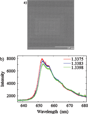
Atomically flat symmetric elliptical nanohole arrays in a gold film for ultrasensitive refractive index sensing
Gabriela Andrea Cervantes Tellez, Sa’ad Hassan, R. Niall Tait, Pierre Berini and Reuven Gordon  
DOI: 10.1039/C3LC41411F


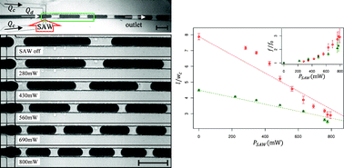
SAW-controlled drop size for flow focusing
Lothar Schmid and Thomas Franke
DOI: 10.1039/C3LC41233D


On-chip analysis of C. elegans muscular forces and locomotion patterns in microstructured environments
Shazlina Johari, Volker Nock, Maan M. Alkaisi and Wenhui Wang
DOI: 10.1039/C3LC41403E


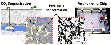
Aquifer-on-a-Chip: understanding pore-scale salt precipitation dynamics during CO2 sequestration
Myeongsub Kim, Andrew Sell and David Sinton 
DOI: 10.1039/C3LC00031A

This article is featured in the collection Lab on a Chip Top 10%


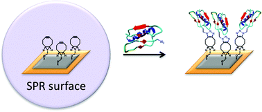
Site-specific peptide and protein immobilization on surface plasmon resonance chips via strain-promoted cycloaddition
Angelique E. M. Wammes, Marcel J. E. Fischer, Nico J. de Mol, Mark B. van Eldijk, Floris P. J. T. Rutjes, Jan C. M. van Hest and Floris L. van Delft   Shazlina Johari, Volker Nock, Maan M. Alkaisi and Wenhui Wang
DOI: 10.1039/C3LC41338A


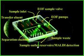
Microfluidic LC device with orthogonal sample extraction for on-chip MALDI-MS detection
Iulia M. Lazar and Jarod L. Kabulski
DOI: 10.1039/C3LC50190F


Digital microfluidics-enabled single-molecule detection by printing and sealing single magnetic beads in femtoliter droplets
Daan Witters, Karel Knez, Frederik Ceyssens, Robert Puers and Jeroen Lammertyn  
DOI: 10.1039/C3LC50119A


Control of neural network patterning using collagen gel photothermal etching
Aoi Odawara, Masao Gotoh and Ikuro Suzuki
DOI: 10.1039/C3LC00036B


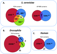
Microfluidic large scale integration of viral–host interaction analysis
Ya’ara Ben-Ari, Yair Glick, Sarit Kipper, Nika Schwartz, Dorit Avrahami, Efrat Barbiro-Michaely and Doron Gerber  
DOI: 10.1039/C3LC00034F

*Free access to individuals is provided through an RSC Publishing personal account. Registration is quick, free and simple

Digg This
Reddit This
Stumble Now!
Share on Facebook
Bookmark this on Delicious
Share on LinkedIn
Bookmark this on Technorati
Post on Twitter
Google Buzz (aka. Google Reader)

SERS-active nanostructures synthesized within a microfluidic channel

Posted on behalf of Kathryn Mayer, Lab on a Chip web writer

A new method for the direct, bottom-up growth of a SERS substrate within a microfluidic channel, developed at the University of Connecticut, will enable the inexpensive fabrication of SERS-integrated devices.

Surface enhanced Raman scattering (SERS) spectroscopy is a powerful method for analyte detection and identification.  SERS relies on the electric field enhancement provided by nanostructured metal surfaces to amplify the Raman scattering “fingerprint” of adsorbed molecules, enabling specific detection down to the single molecule level1.  Previously, SERS integration into practical devices has been limited, partly due to the cost and difficultly of fabricating SERS substrates and microfluidic channels separately, then aligning and bonding them together.  In this HOT article, researchers at the University of Connecticut led by Prof. Yu Lei have devised a method to fabricate a novel nanostructured SERS substrate directly within existing microfluidic channels, greatly simplifying the construction of such devices2

Previous SERS-integrated microfluidic devices have utilized silver plates fabricated via laser writing3 and silver nanoparticles created using physical vapor deposition4 as SERS substrates. These devices provide high detection sensitivity, but are costly to produce due to the top-down fabrication methods used.  The advantage of the new method by Parisi et al. is the ability to synthesize a SERS substrate bottom-up and in situ, reducing the complexity and cost of device fabrication.  SERS experiments can then be carried out immediately.

In situ electrodeposition and galvanic displacement are used to fabricate a nanostructured SERS substrate (right) within a microfluidic device (left)

In the new technique, copper nanowalls coated with carbon are synthesized inside a microfluidic channel via electrodeposition.  This is accomplished by applying a voltage across part of the channel while copper acetate flows through.  Next, silver nitrate flows through the channel, and a galvanic replacement reaction results in silver nanoparticles coating the nanowalls.  Then, an analyte solution flows through the channel and adsorbs onto the substrate, allowing the user to measure SERS.  The authors used crystal violet as an example analyte to demonstrate the sensitive in-channel detection capability, measuring the SERS of analyte concentrations down to 50 pM.
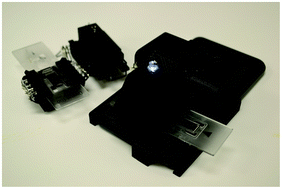
The researchers hope that their fabrication technique will ultimately allow SERS to be integrated into various microfluidics-based biological and chemical sensing platforms, increasing the power and flexibility of these devices. 

References

1. J. Kneipp et al., Chem. Soc. Rev., 37, 1052–1060, 2008. 
2. J. Parisi et al., Lab on a Chip, 13, 1501–1508, 2013.
3. B.-B. Xu et al., Lab on a Chip, 11, 3347–3351, 2011.
4. Z. Geng et al., Sensors and Actuators A, 169 (1), 37–42, 2011.

Read this HOT article in Lab on a Chip today:

In situ synthesis of silver nanoparticle decorated vertical nanowalls in a microfluidic device for ultrasensitive in-channel SERS sensing
Joseph Parisi, Liang Su and Yu Lei
DOI: 10.1039/C3LC41249K

 This article is featured in the web collection Lab on a Chip Top 10%


Katie Mayer is a post-doctoral researcher in the Walt Laboratory at Tufts University, USA

Digg This
Reddit This
Stumble Now!
Share on Facebook
Bookmark this on Delicious
Share on LinkedIn
Bookmark this on Technorati
Post on Twitter
Google Buzz (aka. Google Reader)

Pioneers of Miniaturisation Lecture 2013 – deadline extended to 22nd July!

Send your nominations for the Pioneers of Miniaturisation Lectureship to Lab on a Chip Managing Editor Harp Minhas by 22nd July!

Miniaturisation plays a significant role in our daily lives as all our handheld devices become smaller and other larger devices become handheld or desktop based and this trend is set to continue. The use of micro and nano fluidic technologies will impact on a diverse range of industries ranging from their use in motor cars, through health improvement applications to their use in protecting national and environmental security needs.

At Lab on a Chip, we strongly believe in this technology and have been willing to show the necessary commitment and financial support to back the development of this research community. It is in this vein we present this award to honour and support the up and coming, next generation pioneers in this field of endeavour. So the Lab on a Chip Journal will again join forces with Corning Incorporated to award the eighth ‘Pioneers of Miniaturisation’ Lectureship at µTAS, including a certificate of recognition and a prize of $5000. The lectureship will be presented at the µTAS 2013 Conference in Freiburg, Germany.

Who should you nominate?

The award is for early to mid-career scientists (nominees must be no older than 45 by the closing date for nominations).
The award is for extraordinary or outstanding contributions to the understanding or development of miniaturised systems. This will be judged mainly through their top 1-3 papers and/or an invention documented by patents/or a commercial product. Awards and honorary memberships may also be considered.The awardee is required to give a short lecture at the µTAS Conference in the same year.

The 2012 Pioneers of Miniaturisation Lectureship was awarded to Professor Andrew deMello, ETH Zurich, Switzerland. 

See here for further information, including past winners.

Nominations to Lab on a Chip Managing Editor Harp Minhas by 22nd July

Digg This
Reddit This
Stumble Now!
Share on Facebook
Bookmark this on Delicious
Share on LinkedIn
Bookmark this on Technorati
Post on Twitter
Google Buzz (aka. Google Reader)

Realistic networks of blood vessels in microfluidic devices

Posted on behalf of Andries van der Meer, Lab on a Chip web writer

Professor Noo Li Jeon and co-workers from Seoul National University have published an important paper in Lab on a Chip about creating vascular networks in microfluidic devices. The networks are so well-developed that they can even be flushed with fluids and cells.

In the paper, the researchers describe a new method to culture endothelial cells in a microfluidic, three-dimensional environment. The endothelial cells form a network of microvessels within a few days. The researchers show that by adding other cell types to the culture, the network can be used to study specific interactions of vessels with connective tissue, tumor tissue and blood. The endothelial network is so mature that it can be flushed with medium without leakage and that the endothelial cells respond to the mechanical forces that result from the fluid flowing over their surface.

The perfusion is nicely illustrated in this movie from the paper, showing microbeads flowing from one side of the network (top) to the other (bottom).

Additionally, the devices give a lot of structural information about the microvessels, as can be seen in the above picture from the article. It shows an endothelial network in a microfluidic device, with fluorescent labels for cell nuclei, cytoskeletal actin fibers and the cell-cell adhesion molecule CD31. The network displays a branching morphology with clearly delineated vessels and high integrity.

Taken together, this is probably the most realistic laboratory model of the microvasculature to date. The versatility of the model is demonstrated by the extensive characterization of cell-cell interactions and the functional responses of endothelial cells to flow. The realism of the model, together with a high degree of control over many important cell culture parameters makes this model superior to many current laboratory models of microvascular networks.

In the future, these types of models will be useful for studying pathological blood vessel formation, for example by systematically changing the cell culture environment or by using patient-derived cells.

Engineering of functional, perfusable 3D microvascular networks on a chip
Sudong Kim, Hyunjae Lee, Minhwan Chunga and Noo Li Jeon
DOI: 10.1039/C3LC41320A

This article is from the themed collection Lab on a Chip Top 10%

Andries van der Meer, PhD, is a post-doctoral fellow at the Wyss Institute for Biologically Inspired Engineering, Harvard University, USA.

Digg This
Reddit This
Stumble Now!
Share on Facebook
Bookmark this on Delicious
Share on LinkedIn
Bookmark this on Technorati
Post on Twitter
Google Buzz (aka. Google Reader)

Chang Lu awarded NIH funding to continue Lab on a Chip work

Congratulations go to Chang Lu at the School of Biomedical Engineering and Sciences at Virginia Tech has been awarded a new National Institutes of Health project to continue his work with Albert Baldwin at The University of North Carolina on the molecular mechanisms of cancer.

Preliminary work by Chang Lu and co-workers published in Lab on a Chip in 2011 focused on histone modification analysis using a microfluidic platform. Their microfluidics device enables traditional chromatin immunoprecipitation assays (ChIP) using much lower numbers of cells in a much quicker time. All steps are conducted on-chip. The assay is coupled with RT-PCR to give a speedier method for analysis of scarce biological samples.

This new grant from the NIH is to continue this work in designing more advanced, more sensitive on-chip ChIP assays as part of the institutes’ National Cancer Initiative.

Read the first paper in Lab on a Chip today:

Histone modification analysis by chromatin immunoprecipitation from a low number of cells on a microfluidic platform
Tao Geng, Ning Bao,   Michael D. Litt, Trevor G. Glaros, Liwu Lid and Chang Lu  
DOI: 10.1039/C1LC20253G

Digg This
Reddit This
Stumble Now!
Share on Facebook
Bookmark this on Delicious
Share on LinkedIn
Bookmark this on Technorati
Post on Twitter
Google Buzz (aka. Google Reader)

Taming shape-shifters: analyzing dynamic protein structures on-chip

Posted on behalf of Aleksandra (Sasha) Denisin, Lab on a Chip web writer.

Researchers at York University in Toronto have developed an integrated microfluidic device to facilitate analysis of modified protein structures after biochemical reactions.  

The platform guides a protein right through from a controlled reaction, functional labeling, and fragmentation zones to a coupled mass spectrometer for determination of the protein’s secondary structure. Using this device, the group conducts the first dynamic structural analysis of the β-lactamase enzyme TEM-1 to identify key regions of the enzyme that affect catalytic activity.  TEM-1 is the enzyme that provides bacterial resistance to β-lactam antibiotics.

Protein binding events crucial to protein function can change protein shape directly at the binding site or through another site (an allosteric mechanism). One example of allostery is when the binding of oxygen to one heme site in hemoglobin increases oxygen affinity at other heme sites due to structural changes. For hemoglobin, functional labeling during binding events and mass spectrometry (MS) protein structure analysis was used to find the 3 sets of residues responsible for the allosteric mechanism at each heme site.1 The integrated microfluidic platform described here by Derek J. Wilson’s group enables similar experiments with faster sample transfer to electrospray ionization for MS analysis.

This integrated microfluidic platform first incubates the protein of interest with a reactant for a controlled time. Then, the amide protons of the backbone are functionally labeled with deuterium and the labeled protein is digested in a pepsin-functionalized region. Finally, the fragments are ionized and transferred into a time-of-flight mass spectrometer (Q-TOF MS). Labeled peptides are quickly delivered to the spectrometer resulting in high retention of the proton label.

Evaluating TEM-1 after incubation with its inhibitor clavulanate, Wilson’s group notes that regions of the protein which undergo slow changes in deuterium uptake correspond to allosteric transitions and occur on the protein periphery. Rapid changes occur to reposition specific residues, “loosening” some structures and “locking up” others near the active site.

These measurements are the first to analyze TEM-1 inhibition dynamics and demonstrate the great utility of this microfluidic platform to facilitate exploration of allosteric mechanisms and other dynamic structural changes affecting protein function.

1. I. A. Kaltashov and S. J. Eyles, in Mass Spectrometry on the Frontiers of Molecular Biophysics and Structural Biology: Perspectives and Challenges, Wiley Online Library, 2012, pp.186-208.

An electrospray MS-coupled microfluidic device for sub-second hydrogen/deuterium exchange pulse-labelling reveals allosteric effects in enzyme inhibition
Tamanna Rob, Preet Kamal Gill, Dasantila Golemi-Kotra, and Derek J. Wilson
DOI: 10.1039/C3LC00007A

Sasha is a PhD student at Stanford University working with Professor Beth Pruitt’s Microsystems Lab. Her research interests focus on designing microscale devices for studying cell mechanobiology and the biophysical underpinnings of cell-cell and cell-substrate interactions.

Digg This
Reddit This
Stumble Now!
Share on Facebook
Bookmark this on Delicious
Share on LinkedIn
Bookmark this on Technorati
Post on Twitter
Google Buzz (aka. Google Reader)