A versatile valving toolkit for automating fluidic operations in paper microfluidic devices
New YouTube Videos
January’s Free HOT Articles
These HOT articles, published in January 2015 were recommended by our referees and are free* to access for 4 weeks
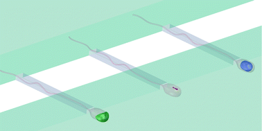
Microfluidic single sperm entrapment and analysis
B. de Wagenaar, J. T. W. Berendsen, J. G. Bomer, W. Olthuis, A. van den Berg and L. I. Segerink
Lab Chip, 2015, Advance Article
DOI: 10.1039/C4LC01425A, Paper
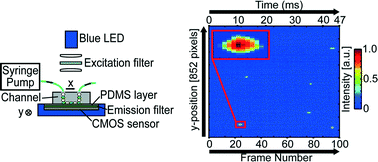
Optofluidic ultrahigh-throughput detection of fluorescent drops
Minkyu Kim, Ming Pan, Ya Gai, Shuo Pang, Chao Han, Changhuei Yang and Sindy K. Y. Tang
Lab Chip, 2015, Advance Article
DOI: 10.1039/C4LC01465K, Paper
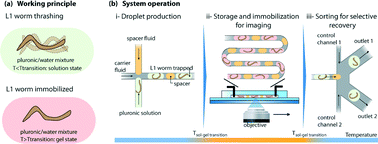
Hydrogel-droplet microfluidic platform for high-resolution imaging and sorting of early larval Caenorhabditis elegans
Guillaume Aubry, Mei Zhan and Hang Lu
Lab Chip, 2015, Advance Article
DOI: 10.1039/C4LC01384K, Paper
Take a look at our Lab on a Chip 2015 HOT Articles Collection!
Culturing Cells on Druggable Bubbles
When testing the effects of drugs on cells and tissues, laboratory scientists generally use a pretty crude approach. They simply mix the desired concentration of the drug with the culture medium and then add it to separate plastic wells in which cells or tissues have been cultured. Of course, this approach has its limitations: when testing many different concentrations and mixtures of drugs, the amount of wells needed for an experiment grows very quickly to overwhelming numbers. Moreover, all of the drug is generally added at once, not taking into account the gradual pharmacokinetic profiles that you would normally find in the human body.
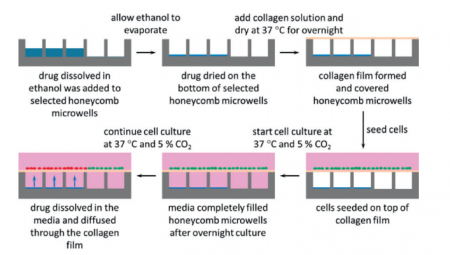
In a paper in Lab on a Chip, scientists from Corning, Inc. and Massachusetts General Hospital demonstrate a new, microengineered approach for treating cultured cells with drugs. Instead of growing the cells on flat surfaces, cells are grown on micro-modified wells plates that contain regular patterns of tiny holes. The microholes can be filled with dried-up drug and then sealed off with a semi-permeable layer of collagen. When cells are grown on the collagen, culture medium starts seeping into the air-filled microholes. The dried-up drug then dissolves and diffuses through the collagen layer, exposing the microscopic patch of cells around the microhole to the drug (see also the figure below).
The authors only show the results of a simple proof-of-principle experiment with Nefazodone-filled holes leading to local toxicity for cultured liver cells. The technique looks promising however, and it will be interesting to see further development. How easy will it be to load holes with different concentrations and mixtures of drugs? Can the process of drugs diffusing into the medium be controlled? Can we control time-release profiles by changing the shape of the holes, by changing the surface properties of the holes, or maybe by changing the gas content of the medium?
It will also be interesting to see whether the technique can be combined with high-throughput imaging, like fluorescence microscopy or scanning electrochemical microscopy as was recently shown for cells in microscopic wells by Sridhar, et al.
Make sure to check out the paper by Goral, et al. in which they outline their technique while it is still free* to access.
New You Tube Videos
Chip-on-a-cell
Article written by Jennifer Newton
Scientists in Spain have flipped the cell-on-a-chip concept to bring us a chip-on-a-cell
Field emission scanning electron microscopy images of a barcode attached to the zona pellucida of a mouse embryo
Jose Antonio Plaza of the Institute of Microelectronics Barcelona and colleagues affixed polysilicon chips, which act as barcodes, onto the outer surface of the zona pellucida, a membrane that surrounds immature egg cells and embryos. Although silicon nanowires penetrated the membrane to attach the chip, they did not interfere with embryo development in tests on mouse embryos.
To read the full article visit Chemistry World.
Silicon-nanowire based attachment of silicon chips for mouse embryo labelling
S. Durán, S. Novo, M. Duch, R. Gómez-Martínez, M. Fernández-Regúlez, A. San Paulo, C. Nogués, J. Esteve, E. Ibañez and J. A. Plaza
Lab Chip, 2015, Advance Article
DOI: 10.1039/C4LC01299B, Paper
New YouTube Videos
New You Tube videos
December’s Free HOT Articles
These HOT articles, published in December 2014 were recommended by our referees and are free* to access for 4 weeks
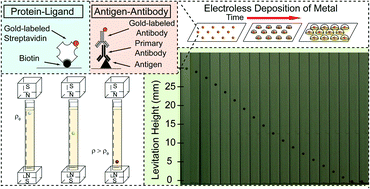
Metal-Amplified Density Assays, (MADAs), including a Density-Linked Immunosorbent Assay (DeLISA)
Anand Bala Subramaniam, Mathieu Gonidec, Nathan D. Shapiro, Kayleigh M. Kresse and George M. Whitesides
Lab Chip, 2015, Advance Article
DOI: 10.1039/C4LC01161A, Paper
High process yield rates of thermoplastic nanofluidic devices using a hybrid thermal assembly technique
Franklin I. Uba, Bo Hu, Kumuditha Weerakoon-Ratnayake, Nyote Oliver-Calixte and Steven A. Soper
Lab Chip, 2015, Advance Article
DOI: 10.1039/C4LC01254B, Paper
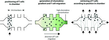
Real-time tracking, retrieval and gene expression analysis of migrating human T cells
Matthias Mehling, Tino Frank, Cem Albayrak and Savaş Tay
Lab Chip, 2015, Advance Article
DOI: 10.1039/C4LC01038H, Paper
Take a look at our Lab on a Chip 2014 HOT Articles Collection!
Saving Stripes: using gratings to prevent destructive air-water interfaces
Researchers at National Taiwan University design grating structures to prevent air-water interfaces from destroying lipid bilayers, enabling robust bioassays of synthetic membranes.
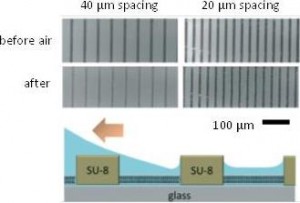
Supported lipid bilayers (SLBs) are useful as platforms to simulate cell membranes for evaluating transport of toxins and viral particles1 and screening new pharmaceutical reagents. Yet a significant challenge is maintaining the integrity of SLBs throughout an experiment. Air-water interfaces, commonly formed during reagent changes and rinses, peel apart SLBs and delaminate them from the substrate. Strategies to preserve SLB integrity involve coating SLBs with polymers to increase their rigidity or adding proteins and sugars to form protective layers with a high bending modulus above the membrane. These methods modify the chemical structure and environment of SLBs, preventing analysis of membrane properties and specific assays of membrane-tethered species. Thus, Chung-Ta Han and Ling Chao developed a substrate with patterned gratings to prevent air-water interfaces from directly contacting SLBs when an air bubble is introduced into a microchannel with SLBs.
The grating structures, fabricated by standard photolithography, are perpendicular to fluid flow in the microchannel and act as obstacles to air-water interfaces contacting SLBs directly by a ‘tenting’ mechanism (see figure at right). Holding the obstacle height constant at 2 μm, Han and Chao evaluated obstacle spacing at different flow rates influenced SLB stability after treatment with an air bubble. 40 μm spacing was found to efficiently preserve SLBs from air-water interfaces at a practical range of flow rates: 60 – 6000 mm/min. The authors also confirmed the integrity of the membranes by comparable diffusivity measurements within the SLBs before and after air-bubble treatment. Finally, the authors demonstrated that air bubbles did not affect receptor-ligand interactions between species embedded in the SLBs and surrounding buffer when SLBs were protected using the microfabricated obstacles.
This platform uses integrated barriers to protect SLBs from air-water interfaces, creating SLBs with native properties to study biomolecule behavior within membranes and perform high throughput analytical assays utilizing synthetic membranes.
Download the full article now – free* access for a limited time only!
Using a patterned grating structure to create lipid bilayer platforms insensitive to air bubbles
Chung-Ta Han and Ling Chao. Lab Chip, 2015, 15, 86 – 93.
DOI: 10.1039/c4lc00928b
[1] I. Kusters, A. M. Van Oijen and A. J. Driessen, ACS Nano, 2014, 8, 3380-3392.
*Access is free until 06.02.15 through a registered RSC Publishing account.