When testing the effects of drugs on cells and tissues, laboratory scientists generally use a pretty crude approach. They simply mix the desired concentration of the drug with the culture medium and then add it to separate plastic wells in which cells or tissues have been cultured. Of course, this approach has its limitations: when testing many different concentrations and mixtures of drugs, the amount of wells needed for an experiment grows very quickly to overwhelming numbers. Moreover, all of the drug is generally added at once, not taking into account the gradual pharmacokinetic profiles that you would normally find in the human body.
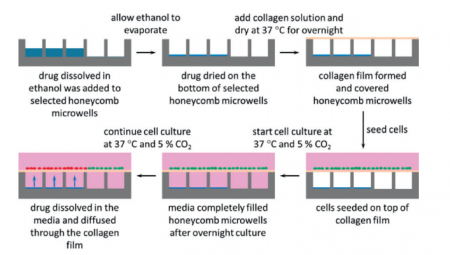
In a paper in Lab on a Chip, scientists from Corning, Inc. and Massachusetts General Hospital demonstrate a new, microengineered approach for treating cultured cells with drugs. Instead of growing the cells on flat surfaces, cells are grown on micro-modified wells plates that contain regular patterns of tiny holes. The microholes can be filled with dried-up drug and then sealed off with a semi-permeable layer of collagen. When cells are grown on the collagen, culture medium starts seeping into the air-filled microholes. The dried-up drug then dissolves and diffuses through the collagen layer, exposing the microscopic patch of cells around the microhole to the drug (see also the figure below).
The authors only show the results of a simple proof-of-principle experiment with Nefazodone-filled holes leading to local toxicity for cultured liver cells. The technique looks promising however, and it will be interesting to see further development. How easy will it be to load holes with different concentrations and mixtures of drugs? Can the process of drugs diffusing into the medium be controlled? Can we control time-release profiles by changing the shape of the holes, by changing the surface properties of the holes, or maybe by changing the gas content of the medium?
It will also be interesting to see whether the technique can be combined with high-throughput imaging, like fluorescence microscopy or scanning electrochemical microscopy as was recently shown for cells in microscopic wells by Sridhar, et al.
Make sure to check out the paper by Goral, et al. in which they outline their technique while it is still free* to access.


