New YouTube Videos
Scientists can quickly detect waterborne pathogens using a smartphone!
Web writer Claire Weston, Imperial College London, writes more…
More than half a billion people have to survive with unimproved water, as providing safe drinking water is still a problem in many parts of the developing world. Of the waterborne pathogens, Giardia lambia (G. lambia) is one of the most common intestinal parasites that are difficult to remove using traditional water purification methods. Current methods for their detection take up to two days and require analysis laboratories with trained specialists and expensive equipment. Because of this there is an ongoing effort to design low-cost and field-portable methods that can rapidly analyse large volumes of water.
Ozcan and co-workers at UCLA have developed a method for the detection of G. lambia cysts in water using a light weight attachment to a smartphone. The attachment consists of a fluorescence microscope, aligned to the smartphone camera, and a disposable water sample cassette that can hold 20 mL of water. The whole test can be carried out in just 1 hour, from taking the sample from the source, to receiving the total number of cysts detected in the sample.
The process is relatively simple, with the test sample first being fluorescently labelled and then filtered through a membrane that traps the G. lambia cysts. A fluorescence image is taken and wirelessly transmitted to servers using an app designed by the group. Digital analysis is carried out using a machine learning algorithm that can specifically recognise the cysts over other fluorescent micro-objects. The results of this analysis are then transmitted back to the phone and displayed on the app.

The group were able to achieve an impressive limit of detection of 12 cysts per 10 mL of sample, citing several factors that led to this limit. They have put forward a number of suggestions for how they hope to further improve their system, so it will be interesting to hear more from this group.
To download the full article for free* click the link below:
Rapid imaging, detection and quantification of Giardia lamblia cysts using mobile-phone based fluorescent microscopy and machine learning
Hatice Ceylan Koydemir, Zoltan Gorocs, Derek Tseng, Bingen Cortazar, Steve Feng, Raymond Yan Lok Chan, Jordi Burbano, Euan McLeod and Aydogan Ozcan
DOI: 10.1039/C4LC01358A
About the web writer
Claire Weston is currently studying for a PhD at Imperial College London, focussing on developing novel photoswitches and photoswitchable inhibitors.

New You Tube videos
Hooked on a Feeling: measuring cell-substrate adhesion with ISFET devices
By developing an ion-sensitive field-effect transistor with small gate dimensions, scientists at the University of Applied Sciences Kaiserslautern in Germany were able to measure cell-substrate adhesion on the single cell scale.
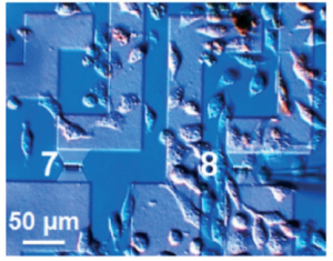
To survive, most mammalian cells attach to other cells and the extracellular environment in order to regulate their growth, proliferation, and migration. Electrical impedance spectroscopy is one way to quantitatively monitor cell-substrate interactions. The strength of cellular adhesion to a substrate with integrated electrodes can be measured by comparing the ratio of the readout voltage to the applied alternating current. Yet this method is limited groups of many cells as the size of the microelectrode must be larger than 100 μm in diameter. Smaller features are subject to greater interface impedance between the electrode and liquid media and this background impedance overwhelms the desired cell-substrate measurements. Suslorapova and colleagues thus used an ion-sensitive field-effect transistor (ISFET) with small gate dimensions to overcome this limitation. The group was able measure the effects of enzymatic digestion with trypsin and an apoptosis-inducing drug on single cell detachment using the ISFET devices with a 16 by 2 square micron gate.
The authors create an equivalent circuit model to interpret recorded impedance spectra from their single cell and small cell groups grown in contact with the field-effect transistor devices. The seal resistance and membrane capacitance parameters which can be extracted from the measured transistor transfer function (TTF) provide measures of cell shape and adhesion to the substrate. Changes in TTF correspond to adhesion of individual cells on top of the ISFET gates. This platform and the model developed to interpret TTF signal opens exciting avenues to monitoring cell adhesion in high throughput yet still at single cell resolution.
Download the full research paper paper for free* for a limited time only!
Electrical cell-substrate impedance sensing with field-effect transistors is able to unravel cellular adhesion and detachment processes on a single cell level
A. Susloparova , D. Koppenhöfer , J. K. Y. Law , X. T. Vu and S. Ingebrandt. Lab Chip, 2015, 15, 668-679. DOI: 10.1039/C4LC00593G
Google Glass to monitor plant health

‘Okay Glass, image a leaf’
Scientists in the US have developed their very own pair of rose-tinted spectacles by adapting Google Glass to measure the chlorophyll concentration of leaves.
Aydogan Ozcan and his research group at the University of California are passionate about creating new technologies through innovative, photonic methods and are well acquainted with the possibilities of wearable technology in scientific research. Chlorophyll concentration is a handy metric for monitoring plant health and the system devised by Ozcan’s team combines Google Glass with a custom made leaf holder and bespoke software to determine just that.
To read the full article visit Chemistry World.
Quantification of plant chlorophyll content using Google Glass
Bingen Cortazar, Hatice Ceylan Koydemir, Derek Tseng, Steve Feng and Aydogan Ozcan
Lab Chip, 2015, Advance Article
DOI: 10.1039/C4LC01279H, Paper
New YouTube Videos
Silver lining for paper Ebola test
Article written by Vicki Davison

Ebola, yellow fever and dengue can be tested for in one go
Researchers in the US have developed a silver nanoparticle-based paper test to simultaneously detect dengue, yellow fever and Ebola. This could provide a cheap and reliable diagnosis for all three diseases, that’s as quick as a home pregnancy test.
The Ebola epidemic in West Africa underscores an urgent need for rapid diagnostics; quick identification and patient isolation can benefit the sick and the healthy. However, dengue, yellow fever and Ebola all initially manifest as a fever and headache, so are easily mixed up.
To read the full article please visit Chemistry World.
Multicolored silver nanoparticles for multiplexed disease diagnostics: distinguishing dengue, yellow fever, and Ebola viruses
Chun-Wan Yen, Helena de Puig, Justina O. Tam, José Gómez-Márquez, Irene Bosch, Kimberly Hamad-Schifferli and Lee Gehrke
Lab Chip, 2015, Advance Article
DOI: 10.1039/C5LC00055F, Communication


