EMBL Microfluidics 2018 Conference

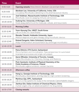
EMBL Microfuidics 2018 Conference will be held at EMBL Heidelberg, Germany between 15th-17th July 2018

“The EMBL Microfluidics Conference 2018 aims to bring together top researchers in the field and to spark scientific exchange, also across different disciplines. The latest Lab-on-a-Chip technologies and applications will be presented, which should be of major interest for experts as well as scientists looking for a first glance at this exciting new technology.”

Over the past years microfluidic approaches have been used for a variety of applications, including nucleotide sequencing, functional genomics, single-cell/single-molecule studies and diagnostics. Many of these applications, including next-generation sequencing devices, have been revolutionised by miniaturisation, paving the way for global gene analysis and hence transforming biology. Small objects such as cells, or even discrete parts thereof, can be exposed to unique conditions, facilitating entirely novel approaches in modern biology and chemistry.

Confirmed speakers include Lab on a Chip Associate Editors Petra Dittrich (ETH Zurich) and Hang Lu (Georgia Institute of Technology), Lab on a Chip Editorial Board member Piotr Garstecki (Polish Academy of Sciences) and Lab on a Chip Advisory Board members Amy E. Herr (UC Berkeley) and Dave Weitz (Harvard University).

LOCATION & DATES 

EMBL Heidelberg, Germany 15 – 17 Jul 2018

DEADLINES:

 Registration – 4 Jun 2018 

Abstract – 23 Apr 2018

Lab on a Chip  Editor-in-Chief, Abe Lee will be chairing a session during the conference and Deputy Editor, Maria Southall will also be attending the conference.

For further information on the conference, please visit the main website. To register, please click here.

Digg This
Reddit This
Stumble Now!
Share on Facebook
Bookmark this on Delicious
Share on LinkedIn
Bookmark this on Technorati
Post on Twitter
Google Buzz (aka. Google Reader)

Why should we use optofluidics for monitoring marine environment?

Phosphorus is found in natural waters and exerts a major influence on the composition and structure of aquatic ecosystems. It is a crucial nutrient for planktons and algae, which feed fish and other marine organisms. However, human activities may result in excess amount of phosphorus, which, in turn, causes harmful algae to bloom in natural waters. The bloom creates a hostile environment to other forms of marine life by consuming the available oxygen in the sea, and producing toxins. Sea organisms such as fish swim away from the blooms, but the ones that cannot swim, such as shellfish, unfortunately die. We do care about this occurrence as it negatively affects natural life and the economy. There is only one way to interpret the effect of continuously changing phosphorus levels on the strength of the biological pump: real-time monitoring of phosphate levels in the marine environment!

Figure 1. The design of the Fabry-Pérot microcavity, consisting of two parallel mirrors (reflectors) fabricated by coating the surface of the optical fibers with a gold layer. The light is reflected by the mirrors multiple times to enhance the signal. Adapted from Zhu et al., 2017.

Conventional vs. optofluidic monitoring instruments

Conventional phosphate monitoring instruments are mostly used for on-site sampling, then the fresh sample is transported to a laboratory for determining the phosphate level. Laboratories complete one round of analysis in 20 min, often using spectrophotometrical measurement tools. Given the conditions, real-time phosphate monitoring easily becomes laborious, time consuming, and costly. To address this challenge, researchers in Chinese Academy of Sciences, Wuhan University, and The First Institute of Oceanography in China collaborated to develop a portable optofluidic phosphate monitoring tool. However, prototyping an optofluidic marine phosphate detection tool is not straightforward because an absorption cell—a component core to the measurement unit—is simply too big to fit in a microchip. Instead of using a bulky absorption cell, researchers considered integrating a Fabry-Pérot cavity in the microsystem. The Fabry-Pérot cavity consists of two parallel optical fibers with a spacing in between. The cross-sectional surface of each optical fiber is coated with a thin layer of gold to create reflector surfaces (Figure 1) in order to enhance the absorption of phosphate. Shortening the spacing between the reflectors decreases the analysis time from minutes to seconds.


How it works?

In the microchip, filtered water sample and a chromogenic reagent are injected into a curved microchannel. After the chromogenic reaction, the water-soluble components are transported into the optical section (Figure 2). The probe light is sent into the Fabry-Pérot cavity via one of the fibers, bounces between the reflectors multiple times to increase the optical feedback and then analyzed by the detector. The obtained absorbance value, therefore, increases linearly with increasing phosphate concentration. In this microsystem, phosphate detection range is 0.1-100 µmol per liter (400 times greater than the range of a conventional instrument) and detection time is 4 seconds (300 times shorter than detection time of a conventional instrument). The authors of the paper think that this technology can be applied to detect other nutrient levels as well as pH changes in marine environment.


optofluidic phosphate monitoring

Figure 2. A schematic of the optofluidic microchip consisting of two parts: the microfluidics circuit forming the microreactor in the microchannel, and the optical part to provide optical feedback for enhanced absorption analysis. Adapted from Zhu et al., 2017.

To download the full article for free* click the link below:


Optofluidic marine phosphate detection with enhanced absorption using a Fabry–Pérot resonator

M. Zhu, Y. Shi, X. Q. Zhu, Y. Yang, F. H. Jiang, C. J. Sun, W. H. Zhaoc, and X. T. Hanc

Lab Chip, 2017, Lab on a Chip Recent Hot Articles

DOI: 10.1039/C7LC01016H


About the Webwriter

Burcu Gumuscu is a postdoctoral fellow in Herr Lab at UC Berkeley in the United States. Her research interests include development of microfluidic devices for quantitative analysis of proteins from single-cells, next generation sequencing, compartmentalized organ-on-chip studies, and desalination of water on the microscale.

*until 16th February 2018

Digg This
Reddit This
Stumble Now!
Share on Facebook
Bookmark this on Delicious
Share on LinkedIn
Bookmark this on Technorati
Post on Twitter
Google Buzz (aka. Google Reader)

Lab on a chip – from molecular assays to organs on a chip: a symposium by the Royal Society of Chemistry and ETH Zurich

The Lab on a chip – from molecular assays to organs on a chip symposium will be held on 10th April 2018, Basel, Switzerland. 

The Royal Society of Chemistry journal Lab on a Chip and ETH Zurich are delighted to present this symposium, which will showcase the high impact research from the groups of the Lab on a Chip Editorial Board members. The research presented will be on a wide variety of cutting-edge topics in line with the ethos and scope of the journal.

Editor-in-Chief, Abe Lee, Associate Editor, Petra Dittrich and Executive Editor, Sam Keltie warmly invite you to take part in this event and look forward to welcoming you in Basel.

Final programme

Aims

The symposium will bring together exceptional researchers – all leading names in their field – for an outstanding plenary programme, together with an open lunch for all attendees that will provide many networking opportunities.

Registration

Registration for the event is required, as we have limited spaces at the venue. Registration costs are students & graduate students 20 CHF, postdocs & group leaders: 40 CHF, industry 80 CHF.  Book now

Lunch and coffee

Lunch and coffee are provided and are included with registration.

Venue

Hotel Bildungszentrum 21, Missionsstr. 21, CH – 4055 Basel, Switzerland


Contact Information

Dr Sam Keltie

Executive Editor, Lab on a Chip

Royal Society of Chemistry

loc-rsc@rsc.org

Digg This
Reddit This
Stumble Now!
Share on Facebook
Bookmark this on Delicious
Share on LinkedIn
Bookmark this on Technorati
Post on Twitter
Google Buzz (aka. Google Reader)

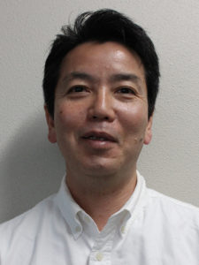
Manabu Tokeshi – Our new Associate Editor

We are delighted to announce our new Associate Editor – Manabu Tokeshi!

“I am excited to join the editorial team of Lab on a Chip, my favorite Journal ever since its inception.  I am looking forward to seeing your excellent research in this Journal.”

Manabu Tokeshi is a Professor at the Division of Applied Chemistry at Hokkaido University, Japan and a visiting Professor at the ImPACT Research Center for Advanced Nanobiodevice, Innovative Research Center for Preventive Medical Engineering, and Institute of Innovation for Future Society at Nagoya University.

He received his PhD degree from Kyushu University, Japan. After a research fellowship of the Japan Society of Promotion of Science at The University of Tokyo, he worked at Kanagawa Academy of Science and Technology as a researcher, group subleader and group leader. Before joining Hokkaido University as Professor in 2011, Manabu worked at the Institute of Microchemistry Technology Co. Ltd. as President and at Nagoya University as an Associate Professor.

Professor Tokeshi is a board member of the Chemical & Biological Microsystem Society (CBMS) which oversees the International Conference on Miniaturized Systems for Chemical and Life Sciences (mTAS). He has received various awards for his work, including the Outstanding Researcher Award on Chemistry and Micro-Nano Systems from the Society for Chemistry and Micro-Nano Systems (2007), the Lab on a Chip/Corning Inc Pioneers in Miniaturization Lectureship (2007) and the Masao Horiba Award from HORIBA, Ltd. (2011).

His research interests are in the development of micro- and nano-systems for chemical, biochemical, and clinical applications. You can find out more about Manabu’s research on his homepage.

Manabu will be handling papers from 1st January 2017, so submit your best work to him!

Digg This
Reddit This
Stumble Now!
Share on Facebook
Bookmark this on Delicious
Share on LinkedIn
Bookmark this on Technorati
Post on Twitter
Google Buzz (aka. Google Reader)

SLAS 2018


SLAS will host SLAS2018, the seventh Annual International SLAS International Conference and Exhibition, in San Diego, California, from Feb. 3-7, 2018.


Through a unique combination of education, access to innovative technologies and intelligent peer networking, SLAS2018 delivers unmatched value for professionals and students looking to discover the latest life sciences technologies and how they can be applied to drive research objectives. SLAS 2018 invites research scientists, engineers, academics and business leaders to submit abstracts for presentation.

SLAS is a global community of more than 20,000 life sciences professionals—from academia, government and industry—collectively focused on leveraging the power of technology to achieve scientific objectives. Showcase your research on this global stage by presenting at SLAS2018


Key deadlines:

18th December: Early-Bird Registration Discount

Monday, January 22, 2018 (Final Poster Abstract Submission Deadline)

Digg This
Reddit This
Stumble Now!
Share on Facebook
Bookmark this on Delicious
Share on LinkedIn
Bookmark this on Technorati
Post on Twitter
Google Buzz (aka. Google Reader)

Emerging Investigator Series – Ashleigh Theberge

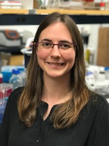
We are delighted to introduce our latest Lab on a Chip Emerging Investigator, Ashleigh Theberge!

Ashleigh Theberge is an Assistant Professor of Chemistry at the University of Washington and Adjunct Assistant Professor of Urology at the University of Washington School of Medicine. She received her BA in Chemistry at Williams College and her PhD in Chemistry at the University of Cambridge, UK with Wilhelm Huck. During her graduate work, she was a Visiting Scientist with Andrew Griffiths at the Université de Strasbourg, France. Her graduate research focused on droplet-based microfluidics for chemical synthesis and analysis. She completed her postdoctoral fellowship in Biomedical Engineering and Urology with David Beebe, William Ricke, and Wade Bushman at the University of Wisconsin-Madison. In 2014, she was awarded an NIH K Career Development Award from the NIDDK in Urology. She joined the faculty at the University of Washington in 2016. Her group develops new microscale culture and analysis methods to study cell-cell, cell-extracellular matrix, and host-microbe interactions, with a focus on inflammation in urologic diseases and asthma.

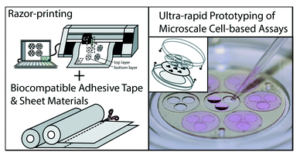
Read Ashleigh’s Emerging Investigator series paper “Upgrading well plates using open microfluidic patterning” and find out more about her in the interview below:

Your recent Emerging Investigator Series paper focuses on upgrading well plates using microfluidic patterning. How has your research evolved from your first article to this most recent article? 

My research has changed dramatically since my first article, which focused on chemical synthesis in droplet-based microfluidics. The common thread in both articles is manipulation of fluids on the microscale, but almost everything else is different! While my graduate work focused on chemical applications of microfluidics in conventional closed microfluidic channels, I now work on methods for studying cell signaling in biological systems using primarily open microfluidic channels. For example in this most recent article, the “walls” of the “channel” are comprised of the floor of the well plate, a plastic insert, and two free air-liquid interfaces.

What aspect of your work are you most excited about at the moment?

I am excited about our group’s fundamental work advancing fluid manipulation in open microfluidics using capillary flow. On the applications side, I am excited to be exploring signaling in ever-more complex cellular systems, including “multikingdom” systems where we study the chemical signals exchanged between host and microbe. I am also happy to work with a fantastic group of students who are taking our group in new directions both biologically and technologically.

In your opinion, what is the biggest advantage of the presented patterning device over the current systems?

Our method enables biologists to easily “upgrade” any existing assay developed in conventional well plates to a co- or multiculture assay. Well plates are the most common cultureware used for cell-based assays, and our device is a simple plastic insert that fits within the well and enables the user to pipette hydrogels (such as collagen, Matrigel, or synthetic gels) to make biocompatible partitions that enable segregated cell culture and maintain soluble factor signaling through the gel wall. Our device enables biologists to use a familiar off-the-shelf platform (well plates) and culture cells on surfaces that have been optimized by decades of industry experience with tissue culture treated surfaces. Since our device uses “open microfluidics,” it does not require bonding of multiple layers and can be fabricated in a single step via injection molding to scale up production.

What do you find most challenging about your research?

A challenge is the interdisciplinary of our work; but this is also what I love about it. Much of our work involves new methods to isolate and study chemical signals from complex cultures – falling under the category of analytical chemistry or bioengineering. But we also focus heavily on disease mechanisms, delving deeply into biology, and our work in the fundamentals of open microfluidics is at the interface of physics and chemistry. Our work also touches on applied aspects of microscale device fabrication, traditionally falling under mechanical engineering, such as our recent paper in press at Lab on a Chip entitled “Fundamentals of rapid injection molding for microfluidic cell-based assays.”

In which upcoming conferences or events may our readers meet you?

I regularly attend microTAS and the Gordon Research Conference on Microfluidics. I also plan to attend the IEEE EMBS Micro and Nanoengineering in Medicine Conference and biological conferences related to our group’s focus areas (microbiome, urologic research, etc.).

How do you spend your spare time?

I try to spend spare time outdoors and enjoy sailing, open water swimming, and climbing. I also like to travel – both to explore different cultures and also wilderness such as the Yukon Territory, Patagonia, and national forest land nearby in Washington.

Which profession would you choose if you were not a scientist?

I would enjoy having a leadership role in a company or organization. I like brainstorming ideas to develop a vision for large projects and working in teams toward a common goal – be it a product, a discovery, or a process. I decided to be a scientist because the scientific method provides an opportunity to rigorously explore questions, but there are many complex problems outside of science that require exciting new strategies and the cohesive effort of many minds to solve.

Can you share one piece of career-related advice or wisdom with other early career scientists?

One strategy that has been helpful for me is to be persistent in looking for opportunities to support projects that I care about. As an undergraduate researcher, I designed my own project that required augmenting our department’s gas chromatograph. I was able to secure the funds by talking with different offices on campus (dean’s office initiatives, student project grants, etc.); I was turned down many times but even those experiences helped me to refine my explanation of the project to have a more compelling conversation with the next person I approached. As an undergraduate summer intern working for Merck, my department did not have funds to send me to a conference to present my work, so I asked the human resources department to fund my trip as a way to promote the intern program as well as present my science; they agreed. In graduate school, I set up an international collaboration in order to develop the expertise required to pursue a multidisciplinary project that I had designed. So for early career scientists, I think it is key to remember that there are many opportunities (even if they are not always obvious at first), and facing road blocks or being turned down several times doesn’t preclude a successful outcome.

Digg This
Reddit This
Stumble Now!
Share on Facebook
Bookmark this on Delicious
Share on LinkedIn
Bookmark this on Technorati
Post on Twitter
Google Buzz (aka. Google Reader)

MMB 2018

The 9th International Conference on Microtechnologies in Medicine and Biology (MMB 2018) is being held in California, USA on March 26-28, 2018

The primary purpose of the conference is to foster interactions between biologists and medical researchers; chemists, physicists and engineers to enhance and strengthen the potential of microtechnologies in revolutionizing the fields of medicine and biological sciences through the development of new research tools and technologies.

The conference is set to have a great talks, with Keynote lectures from Seok “Sid” Chung, Korea University; Jianping Fu, University of Michigan; Amy Herr, University of California, Berkeley; Henry Hess, Columbia University; Marianna Kruithof-de Julio, University of Bern; and Milica Radisic, University of Toronto.

Key Dates:

Late News deadline: 6th February 2018

Early Bird Registration: 13th February 2018

Regular Registration: 21st March 2018


Digg This
Reddit This
Stumble Now!
Share on Facebook
Bookmark this on Delicious
Share on LinkedIn
Bookmark this on Technorati
Post on Twitter
Google Buzz (aka. Google Reader)

Spotting cancer: one in a billion cells

The human body consists of tens of trillions cells, all of which theoretically should have the same genome. Depending on genetic and environmental factors, some of these cells experience point mutations. Although most of those mutations are cleaned up by DNA repair enzymes, about 0.01% of them stay. A low percentage of the persistent mutations turn out to be ‘cancer’ while others stay recessive. Several genes, including the ones responsible for cell growth cycle, cause the persistent mutations. Uncontrolled growth of cells leads to formation of tumor, which is now prone to experience more mutations due to continuous proliferation. These mutations create heterogeneity among the cell population of a tumor, and eventually some cells leave their original tumor and start a new one in another organ of the body. When a cell leaves its parent tumor, it starts circulating in blood vessels before settling in. Those cells are called circulating tumor cells (CTCs), and around 1-10 CTC can be found in 1 mL of blood (which contains about 1 billion red blood cells, and 1 million white blood cells). Capturing ultra-rare CTCs has enormous implications in early cancer diagnosis. Often times, analysis of at least 10 mL of blood is necessary to capture sufficient CTCs to confirm their presence. The available technology can achieve CTC capturing in about 10 hours leading to the loss of target cells and decay of detection biomarkers.

Microfluidic devices are well known for precise sorting of microscale materials. Parallelizing microscale sorting in microfluidic devices enables high-throughput sample processing. In the light of this principle, David Issadore, Jina Ko, and their fellow researchers at University of Pennsylvania coined CaTCh FISH, a circulating tumor cell fluorescence in situ hybridization platform for rapid detection of CTCs. David Issadore kindly accepted to talk about this exciting lab-on-a-chip device. According to David, “the main strength of the CaTCh FISH is that it preserves the sensitivity and specificity of microfluidic cell sorting and RNA FISH, but through clever engineering allows these normally very slow laboratory-based operations to be performed rapidly and automatically on a chip.”

RNA FISH, high-throughput cell handling

Figure 1. Overview of the CaTCh FISH platform. Whole blood sample is processed in TEMPO step, where magnetic nano particle based cell separation is followed by single-cell RNA analysis (modified from Ko et al., 2017).

Processing a whole blood sample using CaTCh FISH involves three steps (Figure 1). First, white blood cells are labelled with magnetic nanoparticles. Second, whole blood is passed through a magnetic micropore filter to selectively trap magnetically labelled cells. “Our magnetic micropore device rapidly and precisely removes all of the cells that we know are not CTCs”, says David. The operating principle of magnetic micropore filter is based on strong and highly localized microscale field gradients formed at the edge of micropores to enable application of high flow rates. Third, single cell RNA analysis is performed on isolated cells using rapid in-situ hybridization strategy so that CTCs can be identified within the isolated cell population. In this way, targeted CTCs can be isolated from the rest of the cell population regardless of their physical and molecular properties. Analysis of a 10 mL blood sample takes less than an hour.

As a key novelty, the researchers maintained high-throughput processing and high sensitivity at the same time by integrating the FISH technique (hybridization of 20-50 fluorescently-labelled oligonucleotide probes to the target RNA, and subsequent fluorescence-signal based detection to enhance signal-to-noise ratio) in a microfluidic chip. CaTCh FISH has also been tested in patients with pancreatic cancer and detected CTCs in the real patient samples.

“The CaTCH FISH technology can be easily modified to measure other rare cells, for the diagnosis of other cancers or for stem cell research for example, by modifying the RNA FISH probes”, says David. He considers converting the platform to a high-precision hospital-based diagnostic tool and he collaborates with a company in the Bay area for this process. The CaTCh FISH device is poised to have a big impact on the way cancer is diagnosed.

To download the full article for free* click the link below:

A magnetic micropore chip for rapid (<1 hour) unbiased circulating tumor cell isolation and in situ RNA analysis

Jina Ko, Neha Bhagwat, Stephanie S. Yee, Taylor Black, Colleen Redlinger, Janae Romeo, Mark O’Hara, Arjun Raj, Erica L. Carpenter, Ben Z. Stanger and David Issadore

Lab Chip, 2017, Paper

DOI: 10.1039/C7LC00703E


About the Webwriter

Burcu Gumuscu is a postdoctoral fellow in Herr Lab at UC Berkeley in the United States. Her research interests include development of microfluidic devices for quantitative analysis of proteins from single-cells, next generation sequencing, compartmentalized organ-on-chip studies, and desalination of water on the microscale.

*until 5th January 2018

Digg This
Reddit This
Stumble Now!
Share on Facebook
Bookmark this on Delicious
Share on LinkedIn
Bookmark this on Technorati
Post on Twitter
Google Buzz (aka. Google Reader)

1st UK Microfluidics for Analytical Chemistry Conference

1st UK Microfluidics for Analytical Chemistry Conference will be held on Thursday 1 February 2018 at the National Oceanography Centre in Southampton, UK.


This one day conference aims to bring together UK researchers developing and applying microfluidic systems for chemical and biochemical analysis. The meeting will cover all types of devices (lab-on-chip, digital microfluidics, paper microfluidics, total analytical systems etc.) and concern application to all forms of molecular analysis (biomedical, environmental, forensic, quality control etc.)

It will provide an opportunity to discuss recent developments in the field and develop future research opportunities as part of an overall aim to nurture and promote the UK microfluidic and analytical communities. This will also offer PhD students and early career researchers a chance to present their work.

Sessions will feature presentations by invited speakers, including keynote presentations from Nicole Pamme (Advisory Board Member for Lab on a Chip) and Joshua Edel (Advisory Board Member for Analyst), in addition to presentations selected from submission of abstracts. There will also be a chance to view the posters and exhibition, with additional networking possible during a wine reception at the end of the event.

Key deadlines:

Abstract deadline – 3rd December, 2018

Earlybird registration deadline -17th December, 2018

Standard Registration deadline – 12th January, 2018


To register, please click here and for more information, please visit the Conference website here.

Digg This
Reddit This
Stumble Now!
Share on Facebook
Bookmark this on Delicious
Share on LinkedIn
Bookmark this on Technorati
Post on Twitter
Google Buzz (aka. Google Reader)

Art in Science Competition Winner Announced at MicroTAS 2017

Lab on a Chip and the National Institute of Standards Technology (NIST) were pleased to present the Art in Science award at the µTAS 2017 Conference on 26 October 2017. The award highlights the aesthetic value in scientific illustrations while still conveying scientific merit. Many fantastic submissions were received this year with the winner selected by Maria SouthallLab on a Chip Deputy EditorDarwin Reyes, NIST and Petra DittrichLab on a Chip Associate Editor member.

And the winner is…

Give Bubbles a Chance
Maria Cristina Letizia, EPFL, SWITZERLAND


Runners up are…

Reflections
Dorothea Helmer, Karlsruhe Institute of Technology, GERMANY

Mondrian’s Micropillars
Eloise Pariset, CEA – LETI, FRANCE

A big thank you to all the contributors this year!

Digg This
Reddit This
Stumble Now!
Share on Facebook
Bookmark this on Delicious
Share on LinkedIn
Bookmark this on Technorati
Post on Twitter
Google Buzz (aka. Google Reader)