Archive for the ‘Hot Articles’ Category

Worm on the run: measuring forces exerted by a nematode in motion

Researchers at the University of Canterbury in New Zealand have created a platform measuring mechancial forces exerted by the microscopic worm C. elegans in locomotion. The platform can measure all of the important worm locomotion parameters in a single assay. The team has noted that the worm’s crawling behaviors and thrust forces correlate to the structure of their microenvironment as the worm adjusts its behavior via mechanical sensing of its surroundings.
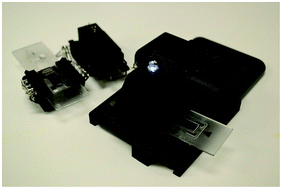
C. elegans are soil-dwelling worms made up of just 959 cells (adult hermaphrodite). The worms are 1 mm long and 80 μm in body diameter. In labs studying C. elegans, the worms crawl on agar plates in search of food (usually E. coli) by twisting their body in sinusoidal waves.1 The sense of touch is crucial to these nematodes: 6 touch receptor neurons (mechanoreceptor neurons) allow the animal to detect external mechanical feedback with the environment as well as internal forces.2

The group led by Maan M. Alkaisi of The MacDiarmid Institute, New Zealand, and Wenhui Wang now at Tsinghua University, China, developed micropillar arrays of various arrangements and dimensions to modify the worm’s microenvironment and measure forces exerted by the worm as it crawls on the substrate. The researchers were also able to quantify locomotion parameters such as speed, amplitude of sine wave, and wavelength. Their unique assay combines 3 key elements: variable substrate topology, worm thrust force measurements, and locomotive metrics. One version of the device is also compatible with different substrates by mounting pillars on top of migrating worms instead of allowing worms to migrate directly on the microstructures.

The group found that reduced pillar spacing caused wild-type worms to exert twice the force to crawl through the substrate, especially in a honeycomb pillar arrangement. The worms were also much slower at moving in the narrow pillar structures and the frequency of sine wave propagations correlated to resistivity of the physical environment. The platform presented in this study enables many exciting studies in C. elegans locomotion and touch response, especially as  many genetic mutant strains are available with changes to the number of mechanoreceptor neurons and muscle arms. 3

References:

1. S. Berri, J. H. Boyle, M. Tassieri, I. A. Hope and N. Cohen, HFSP journal, 2009, 3, 186-193.
2. M. B. Goodman, in WormBook: the online review of C. elegans biology, NIH Public Access, 2006, available from
http://www.ncbi.nlm.nih.gov/books/NBK20187/.http://www.wormbook.org/chapters/www_mechanosensation/mechanosensation.html.
3. C. I. Bargmann and I. Mori, in C. elegans II. 2nd edition., ed. D. L. Riddle, T. Blumenthal, B. J. Meyer and J. R. Priess, Cold Spring Harbor Laboratory Press, NY, 1997, available from http://www.ncbi.nlm.nih.gov/books/NBK20187/.

On-chip analysis of C. elegans muscular forces and locomotion patterns in microstructured environments
Shazlina Johari, Volker Nock, Maan M. Alkaisi, and Wenhui Wang. Lab Chip, 2013, 13, 1699-1707.
DOI: 10.1039/c3lc41403e

Sasha is a PhD student at Stanford University working with Professor Beth Pruitt’s Microsystems Lab. Her research interests focus on designing microscale devices for studying cell mechanobiology and the biophysical underpinnings of cell-cell and cell-substrate interactions.

Digg This
Reddit This
Stumble Now!
Share on Facebook
Bookmark this on Delicious
Share on LinkedIn
Bookmark this on Technorati
Post on Twitter
Google Buzz (aka. Google Reader)

Free to access HOT articles!

 These articles are HOT as recommended by the referees. And we’ve made them free to access for 4 weeks*

A Microfluidic Platform for Evaporation-based Salt Screening of Pharmaceutical Parent compounds
Sachit Goyal, Michael R. Thorson, Cassandra L. Schneider, Geoff G. Z. Zhang, Yuchuan Gong and Paul J. A. Kenis  
DOI: 10.1039/C3LC41271G


Atomically flat symmetric elliptical nanohole arrays in a gold film for ultrasensitive refractive index sensing
Gabriela Andrea Cervantes Tellez, Sa’ad Hassan, R. Niall Tait, Pierre Berini and Reuven Gordon  
DOI: 10.1039/C3LC41411F


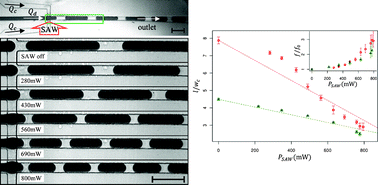
SAW-controlled drop size for flow focusing
Lothar Schmid and Thomas Franke
DOI: 10.1039/C3LC41233D


On-chip analysis of C. elegans muscular forces and locomotion patterns in microstructured environments
Shazlina Johari, Volker Nock, Maan M. Alkaisi and Wenhui Wang
DOI: 10.1039/C3LC41403E


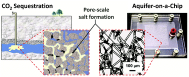
Aquifer-on-a-Chip: understanding pore-scale salt precipitation dynamics during CO2 sequestration
Myeongsub Kim, Andrew Sell and David Sinton 
DOI: 10.1039/C3LC00031A

This article is featured in the collection Lab on a Chip Top 10%


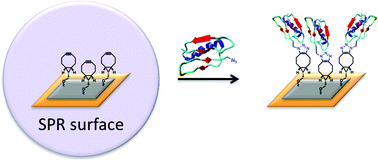
Site-specific peptide and protein immobilization on surface plasmon resonance chips via strain-promoted cycloaddition
Angelique E. M. Wammes, Marcel J. E. Fischer, Nico J. de Mol, Mark B. van Eldijk, Floris P. J. T. Rutjes, Jan C. M. van Hest and Floris L. van Delft   Shazlina Johari, Volker Nock, Maan M. Alkaisi and Wenhui Wang
DOI: 10.1039/C3LC41338A


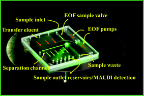
Microfluidic LC device with orthogonal sample extraction for on-chip MALDI-MS detection
Iulia M. Lazar and Jarod L. Kabulski
DOI: 10.1039/C3LC50190F


Digital microfluidics-enabled single-molecule detection by printing and sealing single magnetic beads in femtoliter droplets
Daan Witters, Karel Knez, Frederik Ceyssens, Robert Puers and Jeroen Lammertyn  
DOI: 10.1039/C3LC50119A


Control of neural network patterning using collagen gel photothermal etching
Aoi Odawara, Masao Gotoh and Ikuro Suzuki
DOI: 10.1039/C3LC00036B


Microfluidic large scale integration of viral–host interaction analysis
Ya’ara Ben-Ari, Yair Glick, Sarit Kipper, Nika Schwartz, Dorit Avrahami, Efrat Barbiro-Michaely and Doron Gerber  
DOI: 10.1039/C3LC00034F

*Free access to individuals is provided through an RSC Publishing personal account. Registration is quick, free and simple

Digg This
Reddit This
Stumble Now!
Share on Facebook
Bookmark this on Delicious
Share on LinkedIn
Bookmark this on Technorati
Post on Twitter
Google Buzz (aka. Google Reader)

Realistic networks of blood vessels in microfluidic devices

Professor Noo Li Jeon and co-workers from Seoul National University have published an important paper in Lab on a Chip about creating vascular networks in microfluidic devices. The networks are so well-developed that they can even be flushed with fluids and cells.

In the paper, the researchers describe a new method to culture endothelial cells in a microfluidic, three-dimensional environment. The endothelial cells form a network of microvessels within a few days. The researchers show that by adding other cell types to the culture, the network can be used to study specific interactions of vessels with connective tissue, tumor tissue and blood. The endothelial network is so mature that it can be flushed with medium without leakage and that the endothelial cells respond to the mechanical forces that result from the fluid flowing over their surface.

The perfusion is nicely illustrated in this movie from the paper, showing microbeads flowing from one side of the network (top) to the other (bottom).

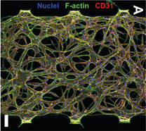
Additionally, the devices give a lot of structural information about the microvessels, as can be seen in the above picture from the article. It shows an endothelial network in a microfluidic device, with fluorescent labels for cell nuclei, cytoskeletal actin fibers and the cell-cell adhesion molecule CD31. The network displays a branching morphology with clearly delineated vessels and high integrity.

Taken together, this is probably the most realistic laboratory model of the microvasculature to date. The versatility of the model is demonstrated by the extensive characterization of cell-cell interactions and the functional responses of endothelial cells to flow. The realism of the model, together with a high degree of control over many important cell culture parameters makes this model superior to many current laboratory models of microvascular networks.

In the future, these types of models will be useful for studying pathological blood vessel formation, for example by systematically changing the cell culture environment or by using patient-derived cells.

Engineering of functional, perfusable 3D microvascular networks on a chip
Sudong Kim, Hyunjae Lee, Minhwan Chunga and Noo Li Jeon
DOI: 10.1039/C3LC41320A

This article is from the themed collection Lab on a Chip Top 10%

Andries van der Meer, PhD, is a post-doctoral fellow at the Wyss Institute for Biologically Inspired Engineering, Harvard University, USA.

Digg This
Reddit This
Stumble Now!
Share on Facebook
Bookmark this on Delicious
Share on LinkedIn
Bookmark this on Technorati
Post on Twitter
Google Buzz (aka. Google Reader)

Free to access HOT articles!

These articles are HOT as recommended by the referees. And we’ve made them free to access for 4 weeks*

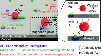
Attomolar protein detection using a magnetic bead surface coverage assay
H. Cumhur Tekin, Matteo Cornaglia and Martin A. M. Gijs
DOI: 10.1039/C3LC41285G

This article is featured in the collection Lab on a Chip Top 10%


Cost-effective and rapid blood analysis on a cell-phone
Hongying Zhu, Ikbal Sencan, Justin Wong, Stoyan Dimitrov, Derek Tseng, Keita Nagashima and Aydogan Ozcan 
DOI: 10.1039/C3LC41408F


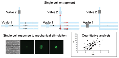
Microfluidics study of intracellular calcium response to mechanical stimulation on single suspension cells
Tao Xu, Wanqing Yue, Cheuk-Wing Li, Xinsheng Yao and Mengsu Yang 
DOI: 10.1039/C3LC40880A


Programmable parylene-C bonding layer fluorescence for storing information on microfluidic chips
Ata Tuna Ciftlik, Diego Gabriel Dupouy and Martin A. M. Gijs 
DOI: 10.1039/C3LC41280F


An electrospray ms-coupled microfluidic device for sub-second hydrogen/deuterium exchange pulse-labelling reveals allosteric effects in enzyme inhibition
Tamanna Rob, Preet Kamal Gill, Dasantila Golemi-Kotra and Derek J. Wilson
DOI: 10.1039/C3LC00007A


A simple, rapid, low-cost diagnostic test for sickle cell disease
Xiaoxi Yang, Julie Kanter, Nathaniel Z. Piety, Melody S. Benton, Seth M. Vignes and Sergey S. Shevkoplyas
DOI: 10.1039/C3LC41302K


Engineering of functional, perfusable 3D microvascular networks on a chip
Sudong Kim, Hyunjae Lee, Minhwan Chung and Noo Li Jeon 
DOI: 10.1039/C3LC41320A

This article is featured in the collection Lab on a Chip Top 10%


In situ synthesis of silver nanoparticle decorated vertical nanowalls in a microfluidic device for ultrasensitive in-channel SERS sensing
Joseph Parisi, Liang Su and Yu Lei 
DOI: 10.1039/C3LC41249K

This article is featured in the collection Lab on a Chip Top 10%


*Free access to individuals is provided through an RSC Publishing personal account. Registration is quick, free and simple


Digg This
Reddit This
Stumble Now!
Share on Facebook
Bookmark this on Delicious
Share on LinkedIn
Bookmark this on Technorati
Post on Twitter
Google Buzz (aka. Google Reader)

LOC Issue 11 online! 3D etching, digital microfluidics, lens-free microscopy

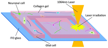
Issue 11’s significant front cover article from Ikuro Suzuki et al. at Tokyo University of Technology, Japan, describes the development of a new 3D etching method. An infrared laser allows tight control over the area of cell adhesion, selecting cell number and cell type, as a small area of the collagen gel substrate can be targeted. The researchers can guide neural network formation using this tool. 3D networks are created upon which neurons survived longer than on 2D substrates.

Control of neural network patterning using collagen gel photothermal etching
Aoi Odawara, Masao Gotoh and Ikuro Suzuki 
DOI: 10.1039/C3LC00036B

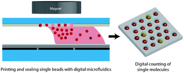
Work from Jeoren Lammertyn et al. is featured on the inside front cover. The team from University of Leuven, Belgium, use digital microfluidics to facilitate single-molecule detection for the first time. They are able to print and seal thousands of femtolitre droplets in microwells in each step. Single paramagnetic beads can be loaded into the microwells with high capacity.

Digital microfluidics-enabled single-molecule detection by printing and sealing single magnetic beads in femtoliter droplets
Daan Witters, Karel Knez, Frederik Ceyssens, Robert Puers and Jeroen Lammertyn  
DOI: 10.1039/C3LC50119A

A Frontier article from Aydogan Ozcan et al. at University of California, Los Angeles, USA, discusses progress in computational lens-free microscopy on-chip and how such technology is taking over conventional bulky optical microscopes. This article, which is also featured on the back cover, includes a discussion of the applications this new technology opens up.

Toward giga-pixel nanoscopy on a chip: a computational wide-field look at the nano-scale without the use of lenses
Euan McLeod, Wei Luo, Onur Mudanyali, Alon Greenbaum and Aydogan Ozcan
DOI: 10.1039/ c3lc50222h

For more critical reviews, HOT primary research as recommended by referees and Technical Innovations, take a look at the full issue now

Digg This
Reddit This
Stumble Now!
Share on Facebook
Bookmark this on Delicious
Share on LinkedIn
Bookmark this on Technorati
Post on Twitter
Google Buzz (aka. Google Reader)

HOT articles and technical innovations in point-of-care flow cytometry

A team from Caltech and MIT, USA, and LeukoDx, Israel, have combined a microflow cytometer and fluorescent dye to produce a portable suitcase-sized point-of-care test for leukocyte count – one of the most common clinical tests. The test can identify four different types of leukocyte using only a small blood sample.

This article was featured on the bright cover of Issue 7!

Four-part leukocyte differential count based on sheathless microflow cytometer and fluorescent dye assay
Wendian Shi, Luke Guo, Harvey Kasdan and Yu-Chong Tai 
DOI: 10.1039/C3LC41059E


A technical innovation from Oliver Hayden and Michael Helou et al. in Germany featured on the outside front cover of Issue 6 also concentrates on flow cytometry for point-of-care testing. This vastly different technique uses magnetophoresis instead of fluorescence to detect specific cancer cells in whole blood. Cell diameters are measured from time of flight information. The device integrates sample preparation for ease of point-of-care applications. The can perform cell enrichment, cell focusing and background elimination in situ.
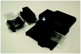
Time-of-flight magnetic flow cytometry in whole blood with integrated sample preparation
Michael Helou, Mathias Reisbeck, Sandro F. Tedde, Lukas Richter, Ludwig Bär, Jacobus J. Bosch, Roland H. Stauber, Eckhard Quandt and Oliver Hayden  
DOI: 10.1039/C3LC41310A

Remember all of our HOT articles are made free to access for 4 weeks*!

 *Free access to individuals is provided through an RSC Publishing personal account. Registration is quick, free and simple

Digg This
Reddit This
Stumble Now!
Share on Facebook
Bookmark this on Delicious
Share on LinkedIn
Bookmark this on Technorati
Post on Twitter
Google Buzz (aka. Google Reader)

Trio of HOT articles! Reversible phase transitions, super-strong adhesive and flow-switching for programmable gradients

With the Introducing series and other exciting news on the blog in the last couple of weeks, we’ve had no time for HOT articles. Here are three in brief all at once and all free to access for 4 weeks*!


 1. A team at Oak Ridge National Laboratory and The University of Tennessee, USA, led by Patrick Collier, generate femtolitre aqueous two-phase droplets in a microfluidic oil channel and demonstrate that a single droplet can be isolated, monitored and transformed reversibly. This is part of recent research efforts to mimic the phase separation that naturally occurs to create the microcompartments inside a cell’s cytoplasm. This device does not require continuous jets or high-frequency droplet formation to create the compartments of differing compositions. The researchers instead concentrated on using the tension between the oil and aqueous phases. They can reversibly generate core–shell microbeads, which could be of interest in controlled drug release.
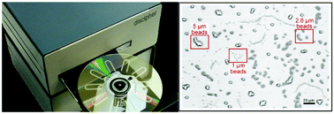
Aqueous two-phase microdroplets with reversible phase transitions
Jonathan B. Boreyko, Prahya Mruetusatorn, Scott T. Retterer and C. Patrick Collier 
DOI: 10.1039/C3LC41122B


 2. As featured on the colourful back cover of Issue 7, Sung Gap Im’s group at KAIST, South Korea, have developed a doubly cross-linked nano-adhesive system (DCNA). The aim was a super-strong adhesive system resistant to tough chemical and thermal conditions. They use initiated chemical vapour deposition (iCVD) to demonstrate their new secure sealing technique in the fabrication of microfluidic devices with flexible and rigid substrates with high strength and stability. This is doubly cross-linked due to the epoxy groups on both sides of the substrates, giving strong adhesion.

A doubly cross-linked nano-adhesive for the reliable sealing of flexible microfluidic devices
Jae Bem You, Kyoung-Ik Min, Bora Lee, Dong-Pyo Kim and Sung Gap Im  
DOI: 10.1039/C2LC41266G


 3. The third and final HOT article featured on the inside back cover of Issue 7 comes from Savaş Tay and Tino Frank at ETH Zurich, Switzerland. The cell culture platform in this HOT article is able to support a programmable diffusion-based gradient generator. Long-term experiments with 30 different gradients could be done in parallel. The microfluidic chip uses membrane valves and automation to reduce error and increase simplicity. A wide variety of cell types can be cultured in this total analysis system in flow-free conditions.

Flow-switching allows independently programmable, extremely stable, high-throughput diffusion-based gradients
Tino Frank and Savaş Tay  
DOI: 10.1039/C3LC41076E

*Free access to individuals is provided through an RSC Publishing personal account. Registration is quick, free and simple


Digg This
Reddit This
Stumble Now!
Share on Facebook
Bookmark this on Delicious
Share on LinkedIn
Bookmark this on Technorati
Post on Twitter
Google Buzz (aka. Google Reader)

Liver on a chip featured in Chemistry World!

Research from SangHoon Lee et al. on a new 3D liver on a chip that enables more detailed study of the paracrine signalling effects on liver tissue function is featured in Chemistry World today!

Read the Chemistry World article here, including independent comment from Gretchen Mahler at Binghamtom University, US. An excerpt is below:

“Researchers in South Korea have developed a three-dimensional liver model that can recreate cell signalling within the organ. The liver on a chip could cut tests on animals by providing an accurate artificial model of how the organ responds to new drugs.The liver contains two kinds of cells. 80% are hepatocytes and the remaining 20% are non-parenchymal cells, including hepatic stellate cells (HSCs). HSCs work with hepatocytes when the liver is damaged, playing a vital role in liver regeneration. Interactions between HSCs and hepatocytes are not well understood, but both direct cell–cell contact and short distance cell–cell signalling, called paracrine signalling, are known to be involved. Despite numerous artificial liver models, no study has yet looked at paracrine influence alone.”

 Design concept (top) and operation mechanism (bottom) of the chip Design concept (top) and operation mechanism (bottom) of the chip

 Spheroid-based three-dimensional liver-on-a-chip to investigate hepatocyte–hepatic stellate cell interactions and flow effects
Seung-A Lee, Da Yoon No, Edward Kang, JongIl Ju, Dong-Sik Kim and SangHoon Lee  
DOI: 10.1039/C3LC50197C

Digg This
Reddit This
Stumble Now!
Share on Facebook
Bookmark this on Delicious
Share on LinkedIn
Bookmark this on Technorati
Post on Twitter
Google Buzz (aka. Google Reader)

HOT article: Low cost, miniaturised, thermoplastic SPR biosensor

SPR biosensorAs featured on the bright inside front cover of Issue 5, this HOT article from Teodor Veres and colleagues at the National Research Council and McGill University, Canada, steps towards low cost point-of-care sensors for disease diagnosis.

The team present their all-polymeric nanoplasmonic microfluidic (NMF) transmission surface plasmon resonance (SPR) biosensor. SPR, involving light stimulated electron oscillation, is advantageous for sensing as it means label-free, real-time detection with high throughput and automation. The device is miniaturised with a view to small, point-of-care applications. The approach involves nanostructures called nanogratings for transmission SPR, which gives a more stable response. The signal can be turned by altering their characteristics and they are easily fabricated en mass.

Thermoplastic materials present an advantage over traditional PDMS for such miniaturised SPR devices as they are more mechanically robust, inert, transparent and crucially viable for large scale production and commercial applications.  The novel aspect of this work is that the nanostructured surface and the microchannels are incorporated into one substrate quickly and at low cost. Thermoplastic valves are used in large numbers for the first time for a multiplex detection scheme.

They demonstrate its application in sensing glycoprotein sCD44 at picomolar to nanomolar concentrations. Further work by the group is focused on integrating this device with a CCD spectrometer.

See the design and performance results in the full paper, now available free for 4 weeks*:

All-thermoplastic nanoplasmonic microfluidic device for transmission SPR biosensing
Lidija Malic, Keith Morton, Liviu Clime and Teodor Veres
DOI: 10.1039/C2LC41123G

Digg This
Reddit This
Stumble Now!
Share on Facebook
Bookmark this on Delicious
Share on LinkedIn
Bookmark this on Technorati
Post on Twitter
Google Buzz (aka. Google Reader)

LOC Issue 9 online! Micronanofabrication, neurotransmitters, SAW-controlled droplets and pharmaceutical screening

Abraham LeeThe packed Issue 9 begins with an editorial celebrating over three decades since the birth of microfluidics by Lab on a Chip Associate Editor Abraham Lee.

Submit your work to his editorial office today at http://mc.manuscriptcentral.com/lc!

The third decade of microfluidics
Abraham Lee
DOI: 10.1039/C3LC90031B


An urgent communication from Lothar Schmid and Thomas Franke at University of Augsburg, Germany, and Harvard University, USA, is featured on the outside front cover, in which a surface acoustic wave is applied to control droplet size in real time:

SAW-controlled drop size for flow focusing
Lothar Schmid and Thomas Franke
DOI: 10.1039/C3LC41233D

The inside front cover illustrates the work of researchers in New Zealand and Beijing led by Wenhui Wang who investigate locomotion metrics and muscular forces of C. elegans in one microfluidic assay:

On-chip analysis of C. elegans muscular forces and locomotion patterns in microstructured environments
Shazlina Johari, Volker Nock, Maan M. Alkaisi and Wenhui Wang
DOI: 10.1039/C3LC41403E


Issue 9 includes one Tutorial Review on using microfluidics to study neurotransmitters from Callie Croushore and Jonathan Sweedler at University of Illinois at Urbana-Champaign, USA, and one Critical Review discussing micronanofabrication techniques:

Microfluidic systems for studying neurotransmitters and neurotransmission
Callie A. Croushore and Jonathan V. Sweedler
DOI: 10.1039/C3LC41334A

Fabrication and multifunction integration of microfluidic chips by femtosecond laser direct writing
Bin-Bin Xu, Yong-Lai Zhang, Hong Xia, Wen-Fei Dong, Hong Ding and Hong-Bo Sun
DOI: 10.1039/C3LC50160D

The high quality research published in Issue 9 includes a HOT article from Paul Kenis et al. again at the University of Illinois at Urbana-Champaign, USA, which describes a new microfluidic platform for the screening of salt forms of pharmaceuticals:

Microfluidic Platform for Evaporation-based Salt Screening of Pharmaceutical Parent compounds
Sachit Goyal, Michael R. Thorson, Cassandra L. Schneider, Geoff G. Z. Zhang, Yuchuan Gong and Paul J. A. Kenis
DOI: 10.1039/C3LC41271G

View all of the articles in Issue 9 here

Digg This
Reddit This
Stumble Now!
Share on Facebook
Bookmark this on Delicious
Share on LinkedIn
Bookmark this on Technorati
Post on Twitter
Google Buzz (aka. Google Reader)