Archive for the ‘Chemistry World Highlights’ Category

Oil reserves put under the microscope with new lab-on-a-rock

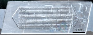
The microfluidic model is etched into a calcite crystal

Scientists in Canada have developed a new microfluidic model carved from rock, which can replicate the conditions found in underground oil reservoirs in a laboratory with more accuracy than ever before. Using it to study the processes that occur in these reservoirs could lead to greater oil yields.

David Sinton’s group, at the University of Toronto, hope that the model they’ve developed will allow them to properly study the rock structure, and see how it’s affected by oil extraction techniques. The techniques could then be optimised to make them much more efficient.

To read the full article please visit ChemistryWorld.

Chip-off-the-old-rock: the study of reservoir-relevant geological processes with real-rock micromodels*
Wen Song, Thomas W. de Haas, Hossein Fadaei and David Sinto.
Lab Chip
, 2014, Advance Article
DOI: 10.1039/C4LC00608A

*Access is free through a registered RSC account until 13 November 2014 – click here to register

Digg This
Reddit This
Stumble Now!
Share on Facebook
Bookmark this on Delicious
Share on LinkedIn
Bookmark this on Technorati
Post on Twitter
Google Buzz (aka. Google Reader)

1024 samples analysed on a single chip

Researchers in Switzerland have developed a microfluidic platform able to measure four protein biomarkers in over 1000 blood samples on a single microfluidic chip. With a dramatic reduction in reagent consumption, time and cost, this new high-throughput technology could make early disease diagnosis more affordable.

Many clinical diagnostic tests measure the amount of a specific protein in a patient’s blood. If the levels of this protein are abnormal it is often an indicator for a disease. Currently these tests are very expensive and time-consuming and are only used once a certain condition is suspected, so the patient is already showing symptoms.

Jose Garcia-Cordero and Sebastian Maerkl at the Swiss Federal Institute of Technology in Lausanne hope to change this with their new microfluidic platform capable of measuring protein biomarkers from just 5nL of human blood with comparable results to a conventional enzyme-linked immunosorbent assay (ELISA), which typically uses a 50μL sample volume. The platform uses a microspotting technique to deliver a large number of blood samples to the chip combined with a microfluidic circuit that runs four parallel immunoassays.

The microfluidic device can perform over 4000 disease biomarker assays in a single run

‘The throughput of our device is roughly 10–100 times that of current microfluidic platforms with an estimated reagent cost of US$ 0.1 per chip,’ says Maerkl. ‘By drastically reducing the cost of diagnostic tests, we hope that everyone will be able to measure a number of biomarkers on a continuous basis, allowing people to take either preventive measures or to seek early treatment for diseases such as cancer.’

Michele Zagnoni, an expert in microfluidic techniques for cancer research at the University of Strathclyde in the UK, commends the ‘outstanding increase in analytical throughput’ and adds that ‘the choice of producing a microfluidic device which can be interfaced with robotic dispensers is an appealing one for pharmaceutical companies, offering a technology that can be easily integrated with existing industrial instrumentation and procedures.’

While their current platform is based on fluorescence to quantify biomarker levels, Maerkl hopes to move to an electronic readout to make smaller and cheaper point-of-care instruments.

Read this full Chemistry World story, by Michael Parkin, here.

Digg This
Reddit This
Stumble Now!
Share on Facebook
Bookmark this on Delicious
Share on LinkedIn
Bookmark this on Technorati
Post on Twitter
Google Buzz (aka. Google Reader)

DIY cholesterol monitoring

A team of scientists from the US have developed a simple system that will allow people to test their blood cholesterol levels at home, using a smartphone.

Cholesterol is an important organic molecule that performs a crucial role in modulating the permeability and fluidity of animal cell membranes, but having too much cholesterol in the blood can be a real problem. In particular, high blood cholesterol levels are known to be a risk factor for coronary heart disease. For many people, blood cholesterol levels can be controlled through diet, by eating less saturated fat. A simple system that enables people to routinely monitor their blood cholesterol levels, using a device that many people already own, would undoubtedly save lives.

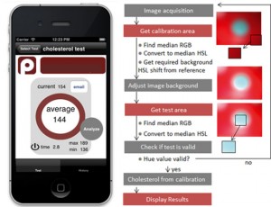
David Erickson and co-workers from Cornell University in New York may have developed just that. Their system consists of a small accessory device that attaches onto a smartphone, an app, and dry reagent test strips for measuring blood cholesterol levels that are already commercially available. A drop of blood is placed onto the test strip and an enzymatic, colorimetric reaction occurs. This strip is then placed into the accessory device and an image of the strip is generated using the camera on the phone. The app then quantifies the colour change and converts this into a blood cholesterol concentration using a calibration curve.

A screenshot of the cholesterol monitoring app (left) and the algorithm to process images of the test strips (right)

The achievement of Erickson and his colleagues should not be understated. Although what they have done may sound simple, developing a smartphone-based system that enables precise and reproducible diagnostic measurements to be taken is actually very difficult. The largest challenge comes from having to account for different lighting conditions and reaction times, differences between the cameras and camera settings in different types of phone, and the potential for misalignment of the test strip. The team overcame the lighting problem by using the accessory device to block out external light so that the test strip would be uniformly illuminated by the flash on the camera. Meanwhile, algorithms in the app account for the other potential variables.

‘This work is another excellent demonstration of cellphone-based sensing,’ says Aydogan Ozcan, a point-of-care diagnostics expert at the University of California in Los Angeles, US. Ozcan himself has developed numerous smartphone-based systems and he sees the value in this work: ‘it will provide numerous opportunities, especially for home monitoring and testing of chronic and elderly patients.’

Erickson and co-workers are now working to commercialise their system, so it may be available for the general public to purchase in the near future.

Click here to watch Erickson demonstrate the test in this great video from the Cornell Chronicle.

Or read this news article by Megan Tyler, and many more, on the Chemistry World website.

Digg This
Reddit This
Stumble Now!
Share on Facebook
Bookmark this on Delicious
Share on LinkedIn
Bookmark this on Technorati
Post on Twitter
Google Buzz (aka. Google Reader)

High-throughput chip for drug screening in 3D

A simple micro-array chip developed by scientists in China could sharpen the search for new drugs by enabling the high-throughput screening of drug candidates against cells cultured in three dimensions.

Developing new drugs for the treatment of complex diseases such as cancer is an expensive and time consuming process. Often, the first step in designing a new drug is to use high-throughput screening of large chemical libraries to identify compounds that have the desired medicinal effect.

Most cell-based drug screening technologies currently rely on cell cultures grown in two dimensions (i.e. as a flat layer of cells) and require sophisticated robotic systems to seed the cells and administer the drugs. However, Yanan Du and colleagues from Tsinghua University may have revolutionised the process by developing a micro-array chip that can be used to culture three dimensional balls of cells, called spheroids, and administer candidate drugs on a high-throughput scale.

Du’s chip consists of two plates containing arrays of micro-wells. The micro-wells of the first plate contain porous gelatin sponges. Once cells and culture medium are added, these sponges act as a rudimentary extracellular matrix enabling the cells to form spheroids. Meanwhile, the micro-wells of the second plate are filled with potential drug candidates. Pushing the two plates together brings the drug into contact with the spheroids so the effect of the drug on the cells can be observed.

The technique should be accessible to any laboratory with basic cell culture facilities

There are two key advantages to this system. Firstly, cells that are cultured in 3D more closely mimic the tissues in the body than cells grown in 2D. Consequently, the effect that the drug has on these cells is likely to be more representative of the effect that the drug would actually have in the body, making the results of the screening process more reliable. Cells cultured in 3D are also more likely to develop drug resistance so these compounds can be weeded out at an earlier stage in the drug development process. The second advantage is that the technique doesn’t require any expensive or sophisticated equipment, and so should be accessible to any laboratory with basic cell culture facilities.

‘By using microwells they can scale-down the number of cells and the amount of drug they use whilst increasing throughput by having a device compatible with standard laboratory instrumentation,’ says Tony Cass from Imperial College London, UK, who also develops devices for high throughput analysis.

Mario Cabodi, an expert in microfabrication at Boston University in Massachusetts, US, says the chip potentially has a promising future: ‘3D cell culture systems, like the one described here, might be helpful in bridging the gap between in vitro tests and in vivo results.’

Du’s team are now in the process of standardising the chip and are collaborating with drug development companies to perform cell-based tests of potential anti-cancer drugs.

To see the original Chemistry World article, and many others, click here.

Alternatively, you can download the full paper here.

Digg This
Reddit This
Stumble Now!
Share on Facebook
Bookmark this on Delicious
Share on LinkedIn
Bookmark this on Technorati
Post on Twitter
Google Buzz (aka. Google Reader)

Paper device spots antibiotic-resistant bacteria

Scientists in Canada have developed a paper-based device that checks if bacteria are resistant to certain antibiotics. The simple system could help users in remote areas pick the most appropriate treatment for bacterial infections.

Testing bacteria to see which antibiotics will be effective against them is vital for properly medicating patients. Methods exist to assess bacteria but the equipment needed is often expensive and requires highly trained operators and laboratory conditions. Now, Ratmir Derda and colleagues at the University of Alberta have designed a portable device made from paper and other low-cost materials. The team sought the help of high school students to make and test the devices to show how easy they are to construct and use.

Derda’s device consists of a paper support with a clear plastic window over an area of nutritious media permeated into a sheet of thick blotting paper. This culture media has a uniform pattern of hydrophobic spots to ensure samples disperse evenly across it. Two zones of antibiotics are added onto the media before the device is sterilised in an autoclave. After autoclaving, a cell viability dye is dropped on the culture media and the bacterial sample is then added on top of the dye. Finally, the device is sealed and incubated overnight.

In areas with no bacterial growth the dye remains blue and in areas where bacteria do grown the dye turns pink. The colours are easy to see through the device’s plastic window – there is no need to open the device. A blue area around the antibiotic zones indicates that the bacteria are susceptible to the antibiotics. The hydrophobic spots on the culture media aid quantification of the blue area to give an idea of how sensitive the bacteria are to the antibiotics.

Preparing and using the device is simple. (M: culture media; A: antibiotic zone; C: culture zone)

‘This work shows that living microorganisms can be grown and screened for antibiotic resistance using paper devices that are small and light enough to store in a person’s pocket,’ says Marya Lieberman, an expert in paper-based sensors at the University of Notre Dame in Indiana, US.

The team also found that the device could easily be stored long-term. After assembling to the point that it contained the culture media and antibiotics, it could be left for up to 70 days in a sealed bag. This storage ability along with the device’s cheap components and ease of use make it very promising for use in remote areas.

Visit Chemistry World to read this article and others by clicking here.

Or read the full paper: Antimicrobial susceptibility assays in paper-based portable culture devices, DOI: 10.1039/C3LC50887K

Digg This
Reddit This
Stumble Now!
Share on Facebook
Bookmark this on Delicious
Share on LinkedIn
Bookmark this on Technorati
Post on Twitter
Google Buzz (aka. Google Reader)

Insect-powered tweezers

Scientists in Japan have developed the first biohybrid microdevice that can function in air. The microtweezers powered by insect muscle tissue could be used to handle cells and other fragile objects as part of a microelectromechanical system (MEMS).

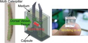
cyborg jellyfish driven by rat heart cells is part of a recent flurry of research into devices that use mammalian muscle cells to provide motor power. Higher energy efficiency and the ability to self-repair are just some of the benefits of using cells in tiny devices. However, biohybrid devices don’t typically work outside cell culture medium. Now, a team lead by Keisuke Morishima at Osaka University has used insect muscle tissue to fabricate a microtweezer device that can operate in the open air.

Morishima‘s team assembled the microtweezers from polydimethylsiloxane and muscle cells taken from moth larvae and packaged them into a capsule containing a small amount of cell medium. The capsule’s clever design exploits a surface tension effect to stop medium from spilling even if the device is flipped upside down.

‘These results indicate that it will be possible to utilise insect muscle tissue and cells as microactuators both in a wet environment, such as in a microchannel, and on a dry substrate, such as on a silicon MEMS structure,’ says Morishima. When the team layered the medium surface with paraffin to slow evaporation from the opening of the device the microtweezers were functional for up to five days.


Shuichi Takayama a microfluidics and nanotechnology specialist at the University of Michigan in the US says that ‘while a variety of muscle-based actuators have been reported, this device is interesting as it is capable of operating in harsh environments. It is a great idea to combine robust muscle tissue from insects with clever packaging.’

The group now hopes to integrate muscle tissue that functions in both wet and dry environments into devices to expand their work into the nanorobotics field.

This article was taken from Chemistry World. To read this one, and many more, click here!

Digg This
Reddit This
Stumble Now!
Share on Facebook
Bookmark this on Delicious
Share on LinkedIn
Bookmark this on Technorati
Post on Twitter
Google Buzz (aka. Google Reader)

Reprogrammable microfluidic chips

A microfluidic chip with channels that can be programmed then reset and reconfigured has been developed by scientists from France and Japan.

Water is dispensed into chip reservoirs. By selectively switching on electrodes, water is manipulated to carve out the channels

Water is dispensed into chip reservoirs. By selectively switching on electrodes, water is manipulated to carve out the channels

In recent years, scientists from across of the globe have developed a plethora of microfluidic chips to perform a variety of tasks, from PCR to cell sorting. However, a serious drawback of microfluidic technologies is that each application requires a unique arrangement of inlets, outlets and microchannels, so microfluidic chips are usually specific to one particular purpose. This, combined with the time-consuming and costly manufacturing processes required to construct microfluidic devices, makes the idea of a reprogrammable chip very attractive.

Read the full article here at Chemistry World.

Programmable and reconfigurable microfluidic chip
Raphaël Renaudot, et al.
Lab Chip, 2013, Accepted Manuscript
DOI: 10.1039/C3LC50850A, Paper

Digg This
Reddit This
Stumble Now!
Share on Facebook
Bookmark this on Delicious
Share on LinkedIn
Bookmark this on Technorati
Post on Twitter
Google Buzz (aka. Google Reader)

Personal kidney disease monitoring on your phone

Angharad Rosser-James, Publishing Editor in the Lab on a Chip editorial production team, recently wrote this fantastic article for Chemistry World. It focuses on a recent Lab on a Chip paper, and shows how the miniaturisation field can have a huge impact on our daily lives: 

A smart phone attachment and accompanying app that could be used by people in their own home to monitor the health of their kidneys have been developed by scientists in the US. The lightweight and cost-effective device contains a fluorescent assay which works with the phone’s existing camera to provide results within minutes. 

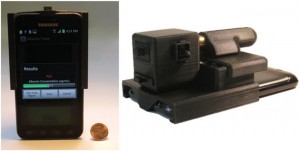
The lightweight and compact attachment is installed on the existing camera unit of a smart-phone

Millions of people die each year from chronic kidney disease with 11% of US adults thought to have some form of kidney-related problem. Early detection and treatment is the key to prevent or control kidney damage. Routine screening for kidney damage checks albumin levels in urine with high levels of the protein indicating a potential problem. These tests are currently carried out using bench-top urine analysers and require patients to make regular trips to a clinic or hospital. 

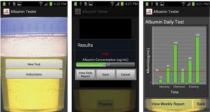
The Albumin Tester, a digital fluorescent tube reader accompanied by an android smart phone app devised by Aydogan Ozcan and colleagues at the University of California in Los Angeles could save patients from having to make so many of these trips. Weighing only 148 g, a similar weight to the smart phone itself, the whole device can be attached to the back of a smart phone. Urine is added to fluorescent assays confined within disposable test tubes and the smart phone’s camera collects images of the assays via an external plastic lens. The app converts the fluorescence signals into an albumin concentration value within 1 second. Its detection limit of 5–10 µg ml-1 is more than 3 times lower that the clinically accepted healthy threshold. 

The user-friendly app converts fluorescence signals into albumin concentrations within 1 s and can give daily or weekly reports

Ozcan envisions the device’s application in ‘the early diagnosis of kidney disease or for routine monitoring of high-risk patients, especially those suffering from chronic conditions such as diabetes, hypertension, and/or cardiovascular diseases.’ Govind Kaigala, who develops microsystems for biomolecule analysis at IBM Research in Switzerland agrees and says ‘the albumin tester is a gadget which holds the promise of a simple, rapid and low-cost test for regular use by the patient.’ 

‘This technology has the potential to make widespread impact on health care in developing as well as developed countries,’ says Olav Solgaard, an expert in optical microelectromechanical systems at Stanford University in the US. 

Ozcan anticipates that their next step is to make it possible to measure other kidney disease biomarkers, such as creatinine, using the same smart phone attachment. 

View this article on the Chemistry World website, or access the full paper: A F Coskun et alLab Chip, 2013, DOI: 10.1039/c3lc50785h

Digg This
Reddit This
Stumble Now!
Share on Facebook
Bookmark this on Delicious
Share on LinkedIn
Bookmark this on Technorati
Post on Twitter
Google Buzz (aka. Google Reader)

Circulating cancer cells spiral towards separation

Repeated biopsies of tumours can be a painful and distressing procedure for cancer patients. A new biochip developed by researchers in Singapore can isolate tumour cells from blood samples, and may one day be an alternative to more invasive methods for tracking later stage cancers. 

Operating principle of circulating tumour cell enrichment by a spiral channel with trapezoid cross-section

Operating principle of circulating tumour cell enrichment by a spiral channel with trapezoid cross-section

Deaths from cancer generally occur after the cancer has spread. Cells detach from the primary tumour and travel through the blood, subsequently forming new tumours. Being able to isolate and characterise these circulating tumour cells (CTCs) can provide information about the original tumour. However, CTCs exist in very low numbers in the blood stream and hence require enrichment and separation before analysis. 

Read the full article in Chemistry World 

Slanted spiral microfluidics for the ultra-fast, label-free isolation of circulating tumor cells
E W Majid et al, Lab Chip, 2013, Accepted manuscript, Paper
DOI: 10.1039/C3LC50617G

Digg This
Reddit This
Stumble Now!
Share on Facebook
Bookmark this on Delicious
Share on LinkedIn
Bookmark this on Technorati
Post on Twitter
Google Buzz (aka. Google Reader)

Worried about food allergens? There’s an app for that

Building on their work on a mobile phone – or cell phone – app to detect bacteria, US scientists have now adapted the technology to detect allergens in foods.

The iTube platform uses colorimetric assays and a smartphone to reveal the presence of allergens in food samples

The iTube platform uses colorimetric assays and a smartphone to reveal the presence of allergens in food samples

The device, developed by Aydogan Ozcan and his colleagues at the University of California, Los Angeles, is a compact and lightweight attachment for a mobile phone’s camera unit, which is used to image tubes containing food samples illuminated by light emitting diodes. ‘If there is an allergen in the sample, the transmitted light intensity changes,’ says Ozcan. ‘By quantifying the transmitted light intensity using a smart application on the phone, we can quantify the amount of allergen in the sample in parts per million.’

 To read the full article, visit Chemistry World.

A personalized food allergen testing platform on a cellphone
Ahmet Coskun, Justin Wong, Delaram Khodadadi, Richie Nagi, Andrew Tey and Aydogan Ozcan
Lab Chip, 2012, Accepted Manuscript
DOI: 10.1039/C2LC41152K

Digg This
Reddit This
Stumble Now!
Share on Facebook
Bookmark this on Delicious
Share on LinkedIn
Bookmark this on Technorati
Post on Twitter
Google Buzz (aka. Google Reader)