Author Archive

Corn microchips

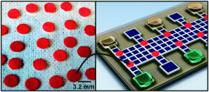
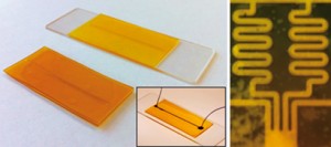
Left: macro images of zein-glass and zein-zein microfluidic devices. The inset shows a colourant-filled zein-glass microfluidic device with tubings. Right: serpentine channels for mixing two different chemical fluids

Gang Logan Liu and colleagues at the University of Illinois Urbana-Champaign, have demonstrated that by using lithography and solvent or vapour deposition bonding, thin films of zein (a protein extracted from corn by-products) can be used to construct microfluidic channels, grids and wells as a green alternative to the plastic materials currently in use.

The group tested their zein microfluidic devices using several types of fluid, including fluorescent dyes and solutions of microbeads. They found that the devices did not leak and the channels could easily be viewed through a microscope. They also extended their work to produce a concentration gradient generator, mixing dye and solvent within serpentine channels to create several different concentration mixtures.

Liu anticipates being able to use the simple bonding of the zein films to make multilayer microfluidic devices in the future.

Interested? Read Tamsin Phillips’ full Chemistry World article here or download the Lab on a Chip paper:

Green microfluidic devices made of corn proteins
Jarupat Luecha, Austin Hsiao, Serena Brodsky, Gang Logan Liu and Jozef L. Kokini
Lab Chip, 2011, Advance Article
DOI: 10.1039/C1LC20726A

Digg This
Reddit This
Stumble Now!
Share on Facebook
Bookmark this on Delicious
Share on LinkedIn
Bookmark this on Technorati
Post on Twitter
Google Buzz (aka. Google Reader)

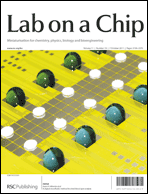
On the cover: a digital microfluidic method for dried blood spot analysis

On the front cover of Issue 19 is an article from Aaron Wheeler et al. on their new method for the analysis of dried blood spot samples. The method has the potential to offer automation of dried blood samples, which are useful for a number of clinical and pharmaceutical applications due to the small sample sizes involved and ease of storage.  The team have developed a prototype microfluidic system to quantify amino acids in which analytes are extracted, mixed with internal standards, derivatized, and reconstituted for analysis by tandem mass spectrometry.

This hot article was also recently reported on in C&EN.

A digital microfluidic method for dried blood spot analysis
Mais J. Jebrail, Hao Yang, Jared M. Mudrik, Nelson M. Lafrenière, Christine McRoberts, Osama Y. Al-Dirbashi, Lawrence Fisher, Pranesh Chakraborty and Aaron R. Wheeler
Lab Chip, 2011, 11, 3218-3224
DOI: 10.1039/C1LC20524B

Digg This
Reddit This
Stumble Now!
Share on Facebook
Bookmark this on Delicious
Share on LinkedIn
Bookmark this on Technorati
Post on Twitter
Google Buzz (aka. Google Reader)

RSC e-membership launched

This week, the RSC launched a new product, RSC e-membership, allowing anyone to access an electronic version of Chemistry World through a MyRSC account and to enjoy the benefits of electronic networking via this professional online community for £20/year.

Subscribers to this do not benefit from the professional recognition or any of the other many services and discounts available to RSC Members, but it allows chemists from around the world, many already members of another chemical society in their own country, to benefit from the highly-esteemed content in Chemistry World and the networking opportunities offered from MyRSC, which now stands at over 11,000 members. The RSC e-membership also allows subscribers to join a virtual specialist interest group on MyRSC. If you are interested in joining, please visit www.rsc.org/emembership.

Digg This
Reddit This
Stumble Now!
Share on Facebook
Bookmark this on Delicious
Share on LinkedIn
Bookmark this on Technorati
Post on Twitter
Google Buzz (aka. Google Reader)

Rumblings of the big future of small chips heard in Asia

Joong Yull Park, Sang-Hoon Lee and Shuichi Takayama

The International Symposium on Microchemistry and Microsystems 2011 (ISMM 2011), the Asian region forum on Micro Total Analysis Systems (μTAS), was held at a quiet hotel located in the southern part of Seoul, Korea, on June 2-4. Over 400 scientists and professionals arrived from diverse countries not only in Asia, but also from the U.S., the E.U., and beyond bringing with them their recent research advances and visions about the conference theme of: The Future of Miniaturized Systems.
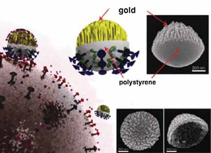
Figure 1. Nanocoral probes docking on a cancer-cell membrane. The highly roughened gold region of the nanocoral increases the molecular adsorption capacity and causes a strong surface-enhanced Raman spectroscopy signal.

The symposium opened with a plenary lecture from Professor Luke P. Lee, UC Berkely. Starting with a history of how stock market price increases paralleled commercialization of key technological breakthroughs, he predicted the future of miniaturized systems in terms of not only technology but potential financial impact as well. He emphasized the increasing need for quantitative biology and medicine and that harnessing micro/nano technology will be key. Considering that several decades were spent to move from vacuum tubes to microscale electronic components embedded in computers, he called upon both patience and hope for biochip and nanotechnology development. Specific research advances presented included Nano Satellites (Figure 1), an exciting concept for using nanoplasmonic particles to explore and visualize the inner space of living cells similar (both in concept and in terms of visual images obtained) to how macro-satellites help explore outer space.1 Combining the phenomenon of plasmon resonance, which is the collective resonant oscillation of electrons in a metal, with nanostructured particles, nanoplasmonic satellites focus and amplify light to nanometer-sized regions to shed new light on signaling pathways and cellular dynamics.
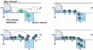
Figure 2. Size-dependent separation of spherical particles and possible rotation behaviors of spherical and nonspherical particles at the branch point.

The other plenary talks on this first day was given by Professor Minoru Seki, Chiba University, who introduced Pinched Flow Fractionation (PFF) and Hydrodynamic Filtration (HDF), a robust method to sort cells and particles by size in milliseconds (Figure 2).2 His presentation also covered calcium alginate gel fibers, anisotropic fibers, droplet-embedded fibers, and sandwich-type fibers generated in microfluidic chips. The anisotropic cross-sectional morphologies of the fibers are promising for guided growth of multiple cell types and thus for 2D/3D cell assembly which is required for tissue regeneration. His contributions are timely given the accelerating growth of cell-based therapies and associated increasing needs for cell sorting and analysis. Professor Bingcheng Lin, Chinese Academy of Sciences, proclaimed that we are ready for the future, to move microfluidics research to the next stage with increased applications, solving real-world problems, and realizing more commercialization. In the invited session, gold and silver nanoparticle-based colorimetric assay for protein and nucleic acids was presented by Professor Xiaodi Su, Institute of Materials Research and Engineering, Singarpore. The method utilizes metal nanoparticles’ unique optical properties and localized surface plasmon resonance (LSPR) for highly sensitive and lable-free detection.
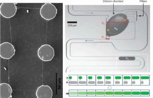
Figure 3. Left: ‘Suspended’ nanofibers connect six carbon posts. Right: Dilution module consists of input channel, dilution chamber, pillars, side channel, and output channel. Blue arrows indicate the flow direction, the dashed orange arrows the recirculatory mixing. Output droplets define a digital concentration gradient.

Plenary talks for the 2nd day were given by Professor Marc Madou, UC Irvine, and Andrew J. deMello, Imperial College London. Professor Madou introduced a way to combine photolithography, near-field electrospinning, and carbonization to pattern suspended carbon nanowire structures over incredible distances (left, Figure 3).3 These C-MEMS and C-NEMS structure were used for interdigitated electrode sensors, glucose sensors, bio-fuel cells and smart batteries. Keywords that describe Professor deMello’s lecture include ‘controlled droplet fusion’, ‘serial dilution on the microscale’, and ‘compartmentalization of single cell’. He showed a dilution module for high-throughput screening using droplet-based microfluidics (right, Figure 3), and demonstrated a homogeneous DNA-binding assay using this system. This digital concentration gradient.4 Other invited speakers included Professor Kahp-Yang Suh, Seoul National University, who introduced research opportunities in the area of ‘Body on a Chip’. One target organ he introduced was a kidney-on-a-chip. By combining microfluidics, advanced patterning technology called CFL (capillary force lithography), and multi-layer microfluidic device (MMD), a microfluidic bio-artificial kidney system was realized.5 The shear stress generated by flow in microfluidic channels is a necessary physiological stimulus for kidney cells along with hormonal stimulation. The biophysical and biochemical stimuli work synergetically to induce proper functioning of kidney cells in vitro. In other presentations, Professor Danny van Noort from the National University of Singapore, gave an aptly titled talk right before lunch about ‘Fish & Chips’. His micro fish tank array system (microaquarium) was cleverly designed to efficiently study fish embryo on a chip.

The last day of ISMM 2011 was opened by Professor Dong-Pyo Kim’s (Chungnam National University) plenary talk. He highlighted the importance of materials science and development. Despite the broad use of PDMS, particularly in academia, there is a critical need for alternate materials. For example, Professor Kim introduced various solvent resistant microreactors. He microfabricated PDMS, polyimid film and functional inorganic polymers by various lithographic techniques. The burgeoning field of microreactor technology is already starting to make a significant impact for industrial as well as benchside use, said Professor Kim.
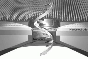
Figure 4. A single-molecule DNA translocates through a nanopore where a pair of nanoelectrodes fabricated to scan the tunnelling current across each nucleotide for label-free base sequence read-out.

Other highlights included description of the latest in single molecule DNA manipulation and sequencing technologies by Professors Kyubong Jo, Steven Soper and Tomoji Kawai. Professor Kawai, Osaka University, specifically introduced the next generation of DNA sequencing technology that utilizes gating nanopores. Two configurable nanoelectrodes enables the electrical detection of single nucleotides (Figure 4).6 Electron transport through single nucleotides occurs not by changes in the ionic current flowing parallel to the nanopore but by changes in the electric current flowing between the nanogap electrodes.

ISMM 2011 was organized in conjunction with the KBCS (Korea BioChip Society) with Professor Sang-Hoon Lee, Korea University serving as the general chair. The successful three days symposium closed on 4th June 2011. A cultural highlight of ISMM 2011 was a traditional Korean performance called Sa-mul-no-ri (Figure 5). ‘Sa-mul’ means ‘four objects’ (four traditional drums that represent thunder, wind, cloud, and rain) and ‘no-ri’ means ‘play’. The rapid and powerful drumbeat not only coordinated the dancing performers but also got the audience moving their bodies. This exciting and interactive performance of traditional music and dance symbolized well the vibrant field of miniaturized systems that we experienced at ISMM 2011 with fast-paced discoveries, cross-disciplinary interactions, and increasing collaboration with ‘audiences’ outside the micro/nanotechnology ‘performers’ including industry. It also felt like the rumblings of the exciting future where ‘play’ between the ‘four objects/subjects’ of medicine, chemistry, biology, and engineering opens unprecedented opportunities.

Figure 5. Dancing to the rhythm of Korean drums at ISMM 2011.

References
1 L. Y. Wu, B. M. Ross, S. Hong and L. P. Lee, Small, 2010, 6, 503-507.
2 S. Sugaya, M. Yamada and M. Seki, Biomicrofluidics, 2011, 5, 24103.
3 G. S. Bisht, G. Canton, A. Mirsepassi, L. Kulinsky, S. Oh, D. Dunn-Rankin and M. J. Madou, Nano Lett, 2011, 11, 1831-1837.
4 X. Niu, F. Gielen, J. B. Edel and A. J. deMello, Nat. Chem., 2011, 3, 437-442.
5 K. J. Jang and K. Y. Suh, Lab Chip, 2010, 10, 36-42.
6 M. Tsutsui, M. Taniguchi, K. Yokota and T. Kawai, Nat. Nanotechnol., 2010, 5, 286-290.
Digg This
Reddit This
Stumble Now!
Share on Facebook
Bookmark this on Delicious
Share on LinkedIn
Bookmark this on Technorati
Post on Twitter
Google Buzz (aka. Google Reader)

Lab on a Chip in C&EN!

A Lab on a Chip paper from Aaron Wheeler and coworkers at the University of Toronto has featured in the magazine Chemical & Engineering News – New Method Is Spot On.

The article describes the application of microfluidic techniques to the analysis of dried blood spot samples – read the original paper here:

A digital microfluidic method for dried blood spot analysis
Mais J. Jebrail, Hao Yang, Jared M. Mudrik, Nelson M. Lafrenière, Christine McRoberts, Osama Y. Al-Dirbashi, Lawrence Fisher, Pranesh Chakraborty and Aaron R. Wheeler
Lab Chip, 2011, Advance Article
DOI: 10.1039/C1LC20524B

Digg This
Reddit This
Stumble Now!
Share on Facebook
Bookmark this on Delicious
Share on LinkedIn
Bookmark this on Technorati
Post on Twitter
Google Buzz (aka. Google Reader)

On the cover: chemical delivery without fluid flow over cells and vapour deposition of cross-linked fluoropolymer coatings

On the cover this month are two hot articles from Nicholas A. Melosh et al. and Malancha Gupta et al, and both articles are free to download for the next 6 weeks.

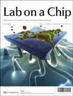
Nicholas Melosh’s article on the outside front cover depicts their method for controlled chemical delivery in microfluidic cell culture devices without fluid flow over the cells, thereby avoiding the problem of cell perturbation.

Rapid spatial and temporal controlled signal delivery over large cell culture areas
Jules J. VanDersarl, Alexander M. Xu and Nicholas A. Melosh
Lab Chip, 2011, 11, 3057-3063
DOI: 10.1039/C1LC20311H

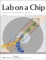
On the inside front cover, the image from Malancha Gupta highlights a vapour deposition method to line the surfaces of PDMS microfluidic devices with a cross-linked fluoropolymer barrier coating, which significantly increases the chemical compatibility of the devices.

Vapor deposition of cross-linked fluoropolymer barrier coatings onto pre-assembled microfluidic devices
Carson T. Riche, Brandon C. Marin, Noah Malmstadt and Malancha Gupta
Lab Chip, 2011, 11, 3049-3052
DOI: 10.1039/C1LC20396G

For the rest of the issue, including hot articles see here

Digg This
Reddit This
Stumble Now!
Share on Facebook
Bookmark this on Delicious
Share on LinkedIn
Bookmark this on Technorati
Post on Twitter
Google Buzz (aka. Google Reader)

HOT: on-chip single-cell lysis to detect defects in red blood cells

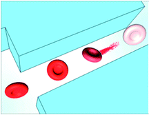
Single-cell electrical lysis has been employed by Philip S. Low, Purdue University, and Chang Lu, Virginia Tech, to rapidly detect defects in the cytosketal protein network of individual red blood cells in a large cell population.

Statistically different profiles were detected for each mutation in defective membranes, which allowed subpopulations of red blood cells to be identified and demonstrates the promise of the technique for a rapid and sensitive detection of membrane defects.

As with all our hot articles, this one’s free to access for 4 weeks – why not take a look:

Single-cell electrical lysis of erythrocytes detects deficiencies in the cytoskeletal protein network
Ning Bao, Gayani C. Kodippili, Katie M. Giger, Velia M. Fowler, Philip S. Low and Chang Lu
DOI: 10.1039/C1LC20365G

Digg This
Reddit This
Stumble Now!
Share on Facebook
Bookmark this on Delicious
Share on LinkedIn
Bookmark this on Technorati
Post on Twitter
Google Buzz (aka. Google Reader)

HOT article: a miniaturized pump for on-chip pressure separations

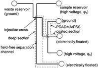
Debashis Dutta, University of Wyoming, and J. Michael Ramsey, University of North Carolina, have developed a microfluidic device with a miniaturized hydraulic pump capable of performing pressure-driven separations.

The microfluidics-based liquid chromatographic system can also inject samples and has a response time in the order of 100 ms, offering a significant advantage over those with external pumps and correspondingly large dead volumes and equilibration times.

As with all our hot articles, this one’s free to access for 4 weeks – why not take a look:

A microfluidic device for performing pressure-driven separations
Debashis Dutta and J. Michael Ramsey
DOI: 10.1039/C1LC20329K


Digg This
Reddit This
Stumble Now!
Share on Facebook
Bookmark this on Delicious
Share on LinkedIn
Bookmark this on Technorati
Post on Twitter
Google Buzz (aka. Google Reader)

HOT: cheap PDMS biochip with integrated cell positioning for imaging live cells with TIRF microscopy

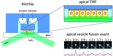
A cheap PDMS biochip has been developed by Roland Thuenauer and Alois Sonnleitner, Center for Advanced Bioanalysis, Austria, that enables the recording of individual vesicle fusion events at the apical membrane of live cells.

The cells can be grown to polarize directly on the device and then correctly placed by an integrated micro-positioning system in order to perform apical TIRF microscopy, without the need for an additional weight to force the apical membrane of the cells into the region of the evanescent wave.

As with all our hot articles, this one’s free to access for 4 weeks – why not take a look:

A PDMS-based biochip with integrated sub-micrometre position control for TIRF microscopy of the apical cell membrane
Roland Thuenauer, Kata Juhasz, Reinhard Mayr, Thomas Frühwirth, Anna-Maria Lipp, Zsolt Balogi and Alois Sonnleitner
DOI: 10.1039/C1LC20458K

Digg This
Reddit This
Stumble Now!
Share on Facebook
Bookmark this on Delicious
Share on LinkedIn
Bookmark this on Technorati
Post on Twitter
Google Buzz (aka. Google Reader)

HOT: a centrifuge on a chip and next-generation integrated microfluidics

A microfluidic chip that can mimic the functions of a centrifuge without moving parts or external forces has been designed by Dino Di Carlo and colleagues from the University of California, Los Angeles.  The Centrifuge-on-a-Chip uses fluid vortices to trap cells, and has been demonstrated to be effective in enriching rare cells from heterogeneous solutions and for performing labelling assays on-chip.

Automated cellular sample preparation using a Centrifuge-on-a-Chip
Albert J. Mach, Jae Hyun Kim, Armin Arshi, Soojung Claire Hur and Dino Di Carlo
Lab Chip, 2011, Advance Article
DOI: 10.1039/C1LC20330D


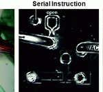
Shuichi Takayama and collaborators at the University of Michigan have provided a brief overview of current challenges associated with integrated microfluidic circuits – covering parallel, serial and embedded instruction devices as well as device architecture, and providing an outlook for the next generation of ICMs.

Next-generation integrated microfluidic circuits
Bobak Mosadegh, Tommaso Bersano-Begey, Joong Yull Park, Mark A. Burns and Shuichi Takayama
Lab Chip, 2011, Advance Article
DOI: 10.1039/C1LC20387H

Both HOT articles are free to access for 4 weeks, so why not take a look today?

Digg This
Reddit This
Stumble Now!
Share on Facebook
Bookmark this on Delicious
Share on LinkedIn
Bookmark this on Technorati
Post on Twitter
Google Buzz (aka. Google Reader)