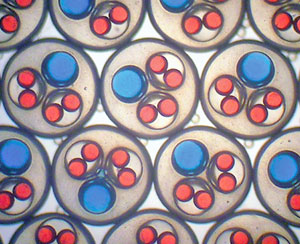
More than 100 embryos could be cultured in an area smaller than a credit card
The latest LOC article to be highlighted in Chemistry World is Michael Richardson‘s paper showing for the first time that an animal embryo can develop in a microfluidic environment. Their discovery could find application in high-throughput, low-cost assays for drug screening and life sciences research.
Zebrafish embryos are becoming important animal models, bridging the gap between cell culture assays and whole animal testing, and have found use in disease modelling and drug safety prediction assays. These assays are currently performed in microtitre plates and require periodic replacement of the buffer solution, which can cause stress or damage to the embryos. Now, Michael Richardson, from Leiden University, and coworkers have developed a microfluidic lab-on-a-chip device in which the buffer solution can flow continuously through the system, and have successfully raised zebrafish embryos under these conditions.
The team made the device from three layers of borosilicate glass with an array of temperature-controlled wells connected by channels; each well houses a single zebrafish embryo. The growth and development of embryos in the microchip was investigated following a five-day culture. Some minor phenotypic variations, such as reduced body length, were found in the microchip-raised embryos; however, there was no significant increase in other abnormalities compared with control experiments.
To find out more read Sarah Farley’s Chemisty World story here or download the paper:
Zebrafish embryo development in a microfluidic flow-through system
Eric M. Wielhouwer, Shaukat Ali, Abdulrahman Al-Afandi, Marko T. Blom, Marinus B. Olde Riekerink, Christian Poelma, Jerry Westerweel, Johannes Oonk, Elwin X. Vrouwe, Wilfred Buesink, Harald G. J. vanMil, Jonathan Chicken, Ronny van ‘t Oever and Michael K. Richardson
Lab Chip, 2011, Advance Article
DOI: 10.1039/C0LC00443J




 The prize is awarded annually to recognise an exemplary published paper for its impact on advancing science and technology in areas relevant to Corning’s strategic focus and expanding commercial opportunities for Corning.
 An article from LOC Editorial Board member
