Author Archive

One step at a time: new coating limits adsorption of non-protein drugs

Non-specific adsorption of molecules to surfaces is a significant issue associated with liquid-based microanalytical systems, such as polymeric microfluidic chips. In microanalytical systems physiological samples are separated into their components and then the amount of each component present can often be measured. What will happen though, if the biomarker that we are looking at is retained elsewhere in the system due to non-specific adsorption? The amount measured will be an underestimation: this means that we could, for example, misdiagnose a patient in a potentially life-threatening way.

No surprise then that a lot of attention is given to preventing non-specific adsorption. One universal approach is to coat the microchip surface with a chemical which limits the adsorption. The Holy Grail, of course, is a coating which has a broad utility and at the same time can be applied quickly to the surface – and stay on it!
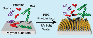
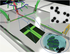
Niels Larsen and colleagues, from Technical University of Denmark, describe a potential breakthrough in Lab on a Chip a one-step procedure for surface modification with the polymer polyethylene glycol (PEG). The researchers use PEG coupled to benzophenone: a small molecule that, when exposed to UV light, reacts with the polymer that makes up the microchip, thus immobilizing the coating on the microchip channel wall surfaces.

Larsen and colleagues test how well this coating can inhibit the non-specific binding of a wide variety of molecules to the surface. They find that it limits the adsorption of proteins and DNA molecules. Most interestingly though, they discover that it is quite efficient in restricting the adsorption of many drugs of a varying degree of hydrophobicity, even in very low, physiologically relevant concentration regimes.

This HOT article is free to access for the next 4 weeks*, so read the detail by clicking the link below:

One-step polymer surface modification for minimizing drug, protein, and DNA adsorption in microanalytical systems
Esben Kjær Unmack Larsen and Niels B. Larsen
DOI: 10.1039/C2LC40750G

*Free access to individuals is provided through an RSC Publishing personal account. Registration is quick, free and simple

Published on behalf of Rafal Marszalek, Molecular BioSystems web science writer. Rafal is an Assistant Editor of Genome Biology at BioMed Central

Digg This
Reddit This
Stumble Now!
Share on Facebook
Bookmark this on Delicious
Share on LinkedIn
Bookmark this on Technorati
Post on Twitter
Google Buzz (aka. Google Reader)

HOT Communication: using a microfluidic platform to fine-tune the synthesis of chitosan nanoparticles for drug delivery

microfluidics, drug delivery, chitosan nanoparticlesA team in Switzerland and Iran led by Philippe Renaud at the Ecole Polytechnique Federale de Lausanne (EPFL), Switzerland, combine a microfluidic chip with hydrophobic modification of chitosan to produce monodisperse and well-defined chitosan nanoparticles capable of delivering hydrophobic anticancer drugs.

Chitosan is known for its biological properties, but is hydrophilic and must be modified to carry hydrophobic drugs. In this work, this is done using N-palmitoyl groups. A microfluidic method of making nanoparticles enables a number of parameters, such as flow ratio and mixing time, to be very finely tuned. This results in precise production of nanoparticles with the required properties.

This article demonstrates fine-tuning of hydrophobically-modified chitosan using a T-shaped PDMS microfluidic device.  For example, the self-assembly is triggered by a pH of 7.4 and this is controlled by tuning the mixing time. The researchers go on to demonstrate that their nanoparticles can encapsulate both hydrophobic and hydrophilic drugs with a sustainable release profile.

This fine-tuning approach will be applicable to production of other polymeric nanoparticles for nanomedicine. Read this communication, free to access for the next 4 weeks*, in full:

Microfluidic assisted self-assembly of chitosan based nanoparticles as drug delivery agents
Fatemeh Sadat Majedi, Mohammad Mahdi Hasani-Sadrabadi, Shahriar Hojjati Emami, Mohammad Ali Shokrgozar, Jules John VanDersarl, Erfan Dashtimoghadam, Arnaud Bertsch and Philippe Renaud
DOI: 10.1039/C2LC41045A

*Free access to individuals is provided through an RSC Publishing personal account. Registration is quick, free and simple

Digg This
Reddit This
Stumble Now!
Share on Facebook
Bookmark this on Delicious
Share on LinkedIn
Bookmark this on Technorati
Post on Twitter
Google Buzz (aka. Google Reader)

Lab on a Chip Issue 5 is online! Towards point-of-care biosensors, an RNAi screening method and a review of microfluidics for liposome production

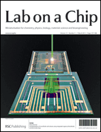
The front cover of Issue 5 illustrates work from Peter Ertl and colleagues at AIT, Austria, in which they combine organic photodiodes and electrode structures in a lab on a chip to develop the ability to quickly determine cell number to standardise microfluidic cell culture and simultaneously measure cell adhesion and interactions.

Standardization of microfluidic cell cultures using integrated organic photodiodes and electrode arrays
Verena Charwat, Michaela Purtscher, Sandro F. Tedde, Oliver Hayden and Peter Ertl
DOI: 10.1039/C2LC40965H


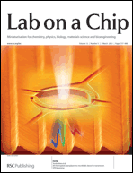
Research by scientists at the National Research Council and McGill University, Canada, features on the striking inside front cover. Teodor Veres and co-workers take a step towards the development of point-of-care nanoplasmonic biosensors with their all-polymeric transmission surface plasmon resonance sensor. It uses a simple and widely available technique and they demonstrate its application in sensing glycoprotein sCD44 at picomolar to nanomolar concentrations.

All-thermoplastic nanoplasmonic microfluidic device for transmission SPR biosensing
Lidija Malic, Keith Morton, Liviu Clime and Teodor Veres
DOI: 10.1039/C2LC41123G


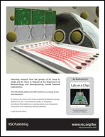
The back cover advert highlights research from the groups of Anup Singh and Oscar Negrete at Sandia National Laboratories, USA, on their advantageous microfluidic RNA interference screening method, which they validate using vesicular stomatitis virus and Rift Valley fever virus.

Microfluidic platforms for RNA interference screening of virus–host interactions
Benjamin R. Schudel, Brooke Harmon, Vinay V. Abhyankar, Benjamin W. Pruitt, Oscar A. Negrete and Anup K. Singh
DOI: 10.1039/C2LC41165B


This issue also features Research highlights from Ali Khademhosseini on biomimetic honeycombs, microfluidic vessel chips and BioMEMS, and a critical review written by Andrew deMello and Dirk van Swaay at ETH Zurich on the microfluidic technologies available for producing liposomes:

Research highlights
Gulden Camci-Unal, Šeila Selimović, Mehmet R. Dokmeci and Ali Khademhosseini
DOI: 10.1039/C3LC90002A

Microfluidic methods for forming liposomes
Dirk van Swaay and Andrew deMello
DOI: 10.1039/C2LC41121K


Find the issue in full here

Digg This
Reddit This
Stumble Now!
Share on Facebook
Bookmark this on Delicious
Share on LinkedIn
Bookmark this on Technorati
Post on Twitter
Google Buzz (aka. Google Reader)

ELISA cheap on paper: new nitrocellulose-based microchips for a versatile immunosorbent assay

The enzyme-linked immunosorbent assay (ELISA) is a complex biochemical test used as a diagnostic tool for the detection of, for example, HIV, hepatitis viruses, malaria and E. coli infections. Although it is extremely useful and versatile, many of its features limit its utility: the assay itself is complicated, the reagents expensive, and robotic instrumentation is usually needed to perform experiments with high-throughput.

Microfluidic chips provide solutions to some of these problems. They can be used as portable devices; they are much faster than traditional laboratory equipment and use less of the reagents. Until a few years ago, however, their construction was cumbersome: they were made from organic polymers and their manufacturing was tricky and still quite expensive.

That changed when George Whitesides’s group in their Lab on a Chip article (Whitesides et al., Lab Chip, 2008, 8, 2146-2150) proposed that microfluidic chips can be made from paper rapidly and at low cost. Since then many methods of cheap production of paper microchips have been developed.

ELISA, microfluidics, paperIn this recent article published in Lab on a Chip, Yuzuru Takamura and his colleagues from the Japan Advanced Institute of Science and Technology (JAIST) explain how simple microfluidic devices constructed by polymer-printing of the chip design on the sheet of nitrocellulose can be used to perform ELISA.

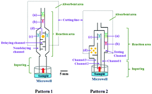
In normal ELISA, test multiple chemical reactions are separated by steps of rinsing. In his device Takamura separates reactions spatially: he deposits the antibodies required for subsequent steps along the main short channel and the substrate needed for the readout reaction in a longer side branch of the channel. When the sample solution travels through the chip, it first reacts with the enzyme-linked antibody; then this complex is caught by another antibody. Finally, the solution which travelled through the longer channel brings the substrate, which reacts with the enzyme and gives a color product – the observed signal. Thus, the rinsing steps are eliminated and the use of expensive antibodies limited to the minimum.

Takamura and colleagues demonstrate the application of this cheap, quick to make and easy to use ELISA-chip to the estimation of the levels of a known pregnancy hormone in urine. The possibilities of these devices, however, reach far beyond a simple pregnancy test: they may soon become a cheap detection tool for many diseases troubling developing countries.

This HOT article is free to access for 4 weeks*, just click on the link below:

Development of automated paper-based devices for sequential multistep sandwich enzyme-linked immunosorbent assays using inkjet printing
Amara Apilux, Yoshiaki Ukita, Miyuki Chikae, Orawon Chailapakul and Yuzuru Takamura
DOI: 10.1039/C2LC40690J

*Free access to individuals is provided through an RSC Publishing personal account. Registration is quick, free and simple

Published on behalf of Rafal Marszalek, Molecular BioSystems web science writer. Rafal is an Assistant Editor of Genome Biology at BioMed Central


Digg This
Reddit This
Stumble Now!
Share on Facebook
Bookmark this on Delicious
Share on LinkedIn
Bookmark this on Technorati
Post on Twitter
Google Buzz (aka. Google Reader)

HOT on the blog: A simple method for molding SU-8 microchannels on the benchtop

SU-8, microchannelsIn this HOT Lab on a Chip article, Daniel Kohane, Robert Langer and Michinao Hashimoto demonstrate a simple method of producing microchannels on the benchtop using polymers other than the popular PDMS.

Alternative polymers to PDMS have been previously shown to have better compatibility with organic solvents, making microfluidic channels more open to different solvents and applications.  Often PDMS surfaces are modified with more solvent-resistant materials such as glass, requiring modification of the channel dimensions. Using these more resistant materials without PDMS usually means a more involved and complex process.

This paper increases the range of materials that can be used in benchtop fabrication of microchannels, using PDMS as a base to support the polymer film.  They first carry out molding, making patterns of microchannels on the polymer film. The PDMS-supported polymer is then released and then an additional polymer surface can be added to create a closed channel.

Importantly, they show that SU-8 epoxy can be used with this method. It has been used before to make microchannels, but, as mentioned above, the methods required were more complex, needing a cleanroom for each layer of micropatterning. This approach means that SU-8 microchannels can be much more easily and quickly produced using benchtop replica molding.

All of our HOT articles are free to access for 4 weeks*, so click on the link below to read this one today:

Benchtop fabrication of microfluidic systems based on curable polymers with improved solvent compatibility
Michinao Hashimoto, Robert Langer and Daniel S. Kohane
DOI: 10.1039/C2LC40888K

*Free access to individuals is provided through an RSC Publishing personal account. Registration is quick, free and simple

Digg This
Reddit This
Stumble Now!
Share on Facebook
Bookmark this on Delicious
Share on LinkedIn
Bookmark this on Technorati
Post on Twitter
Google Buzz (aka. Google Reader)

Starting the week with a HOT article: sorting droplets by number of encapsulated particles

droplet sortingDroplet microfluidics has recently been used for encapsulating a number of different objects, including cells and microbeads. This enables analysis, detection and screening of the encapsulated material. The control of encapsulation to get droplets containing an equal number of particles is important, as often a researcher may need singly occupied droplets, but some of the resulting droplets may be empty or have one or more particles inside with current techniques.

Sorting by particle number would therefore be very helpful for isolating the needed droplets. For larger droplets, sorting by size doesn’t work as they remain the same size whether empty or encapsulating a particle. Fluorescence intensity is also often used as the basis for droplet sorting. However no technique currently sorts by particle number alone.

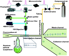
In this HOT article, collaborators in the USA and China led by Chang Lu develop a one-layer microfluidic device that sorts droplets based on number of particles.  It enables production of droplets with even number of encapsulated particles and the analysis of droplets with mixed particle numbers. It can sort up to 30 droplets a second very specifically. The droplets flow through a narrow channel and in this they are detected one-by-one by laser-induced fluorescence. A microcontroller uses this signal to make the sorting decision and a solenoid valve deflects the droplets into either waste or the collection channel. The microcontroller makes the decision based on the time interval between two travelling particles – if there are two particles in one droplet, the interval is smaller than the time taken for the droplet to pass through. It also counts the number of particles in one droplet.

This article free to access for the next 4 weeks* as with all of our HOT articles, so you can have a read and closer look at the technical diagrams by clicking on the link below:

Droplet sorting based on the number of encapsulated particles using a solenoid valve
Zhenning Cao, Fangyuan Chen, Ning Bao, Huacheng He, Peisheng Xu, Saikat Jana, Sunghwan Jung, Hongzhen Lian and Chang Lu
DOI: 10.1039/C2LC40950J

 *Free access to individuals is provided through an RSC Publishing personal account. Registration is quick, free and simple

Digg This
Reddit This
Stumble Now!
Share on Facebook
Bookmark this on Delicious
Share on LinkedIn
Bookmark this on Technorati
Post on Twitter
Google Buzz (aka. Google Reader)

LOC issue 4 – the Neuroengineering 2013 themed issue is online now!


Lab on a Chip’s 2013 Neuroengineering themed issue is introduced by guest editors David T. Eddington and Justin Williams, who also contributed the outside front cover design. This is followed by profiles of all of the contributors to the Neuroengineering themed issue.

Neuroengineering
David T. Eddington and Justin Williams
DOI: 10.1039/C3LC90003G


As well as plenty of cutting-edge primary research into lab on a chip technology for neuroengineering, Issue 4 includes two Critical Reviews.

A critical review of the development of engineered cell culture substrates and techniques for investigating axon development guidance by Santiago Costantino et al. at University of Montreal and McGill University, Canada, is highlighted on the inside front cover. They look at the opportunities that are now opening up due to these new technological developments, the biological insights that can now be gained and the breakthroughs waiting to happen in the near future.

Engineered cell culture substrates for axon guidance studies: moving beyond proof of concept
Joannie Roy, Timothy E. Kennedy and Santiago Costantino
DOI: 10.1039/C2LC41002H


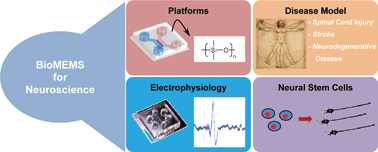
The second critical review from Noo Li Jeon et al. at Seoul National University, Korea, and The Salk Institute, USA, summarises the most recent technological developments of BioMEMs devices and their application to neuroscience research. They look at platforms for disease culture, modelling disease in vitro, neuron electrophysiology and stem cell biology.

Advances in microfluidics-based experimental methods for neuroscience research
Jae Woo Park, Hyung Joon Kim, Myeong Woo Kang and Noo Li Jeon
DOI: 10.1039/C2LC41081H


The HOT articles in this issue include:

Integration of pre-aligned liquid metal electrodes for neural stimulation within a user-friendly microfluidic platform
Nicholas Hallfors, Asif Khan, Michael D. Dickey and Anne Marion Taylor
DOI: 10.1039/C2LC40954B

Inherent amplitude demodulation of an AC-EWOD (electrowetting on dielectric) droplet
Myung Gon Yoon, Sang Hyun Byun and Sung Kwon Cho
DOI: 10.1039/C2LC41043E

One-step polymer surface modification for minimizing drug, protein, and DNA adsorption in microanalytical systems
Esben Kjær Unmack Larsen and Niels B. Larsen
DOI: 10.1039/C2LC40750G


To learn all about the exciting advances happening in applying microtechnology to neuroengineering, read the full issue here

Digg This
Reddit This
Stumble Now!
Share on Facebook
Bookmark this on Delicious
Share on LinkedIn
Bookmark this on Technorati
Post on Twitter
Google Buzz (aka. Google Reader)

Focus on circulating tumor cells in microfluidics

This important Focus article looks at the most up to date research on the isolation and characterisation of circulating tumor cells (CTCs) using microfluidic techniques.

CTCs are produced when tumor cells break off and enter the circulatory system. This makes them important targets for diagnosis and monitoring. However they are only present in extremely small quantities in the blood, meaning plenty of technical difficulties arise when trying to accurately characterise and assess them in a blood sample.

This Focus highlights: 

  1. Considerations in design of CTC isolation, separation and detection systems
  2. Disadvantages of macroscale systems
  3. Magnetic-based microfluidic systems
  4. Affinity chromatography-based microfluidic systems
  5. Size and deformability-based microdevices
  6. Dielectrophoretic-based microdevices

Tony Jun Huang et al. based at The Pennsylvania State University, Massachusetts Institute of Technology (MIT) and Harvard Medical School, USA, finish by discussing the possible combination of the above systems and the future of such technology in terms of clinical relevance for different cancers and technological development.

We’ve made this article free to access for the next 4 weeks*, read it now by clicking the link below:

Probing circulating tumor cells in microfluidics
Peng Li, Zackary S. Stratton, Ming Dao, Jerome Ritz and Tony Jun Huang
DOI: 10.1039/C2LC90148J

 *Free access to individuals is provided through an RSC Publishing personal account. Registration is quick, free and simple

Digg This
Reddit This
Stumble Now!
Share on Facebook
Bookmark this on Delicious
Share on LinkedIn
Bookmark this on Technorati
Post on Twitter
Google Buzz (aka. Google Reader)

First HOT article of the week – a simpler method for measuring ultralow interfacial tension

Surface tension is an important property of all liquids and influences many of their characteristics, such as droplet formation. Surface tension at the interface of two different liquids is interfacial tension. An example of this is the separation of oil and water in solution.

There are other solutions of dissimilar liquids that have extremely small interfacial tensions, 1000 times less than that of oil and water. Measuring this accurately by traditional techniques, such as force tensiometry methods, is a problem. Although many more methods, have been proposed to improve upon this, a simple and accurate technique is yet to be found.

Howard Stone has now led a team at Princeton University and Harvard University, USA, in producing the first microfluidic tensiometer with the capability of measuring ultralow interfacial tension. Their development uses only a small amount of reagents and magnetic particles followed by application of a mathematical model to calculate the interfacial tension.

The measurement is taken by observing the distance between the center of the magnetic particle and the flowing interface. The paramagnetic beads may pass through the interface or be trapped by it due to the balance of magnetic and interfacial forcesonly when this ratio passes a threshold value do the particles pass through.  The method uses these threshold values to then calculate the ultralow interfacial tension.

This simple, miniaturised tensiometer has application in many different systems, with advantages for the applications of ultralow interfacial tension including enhanced oil recovery and microemulsions.

Microfluidic ultralow interfacial tensiometry with magnetic particles
Scott S. H. Tsai, Jason S. Wexler, Jiandi Wan and Howard A. Stone
DOI: 10.1039/C2LC40797C

*Free access to individuals is provided through an RSC Publishing personal account. Registration is quick, free and simple

Digg This
Reddit This
Stumble Now!
Share on Facebook
Bookmark this on Delicious
Share on LinkedIn
Bookmark this on Technorati
Post on Twitter
Google Buzz (aka. Google Reader)

Point-of-care diagnostics using capillary forces – a HOT article!

Point-of-care diagnostics is the aim for much medical research. Often the problem is the ease of preparation of the biological sample, which usually requires multiple steps before the diagnostic test can even begin. Automation of sample preparation has been explored and another option is pressure-driven microfluidics. However, this requires complex external equipment to run the chip only suitable for use in the laboratory away from the patient.

To get true point-of-care diagnostics, sample preparation has to be fast, simple and cheap.

Stationary microfluidics is now being explored.  Liquid is present in fixed positions on the chip and it is magnetic particles that move between the points holding liquid. This often involves separating the liquids using an oil barrier, but this has disadvantages of over-complicating the device by having more than one liquid (the test solution), meaning an additional very careful pipetting step. Not all test solutions or all diagnostic tests would be compatible with this oil barrier, for example, purification of protein. Oil-free versions are now being designed, usually “open droplet” devices, but this again needs accurate pipetting and risks contamination of the sample. Using a large tube limits the miniaturisation capability.

Menno Prins has led a team based at Philips Research and Eindhoven University of Technology, The Netherlands, to solve this problem. They demonstrate technology called Magneto–Capillary Valve (MCV) technology. There is no need for an oil phase.  The confinement of the liquid occurs by capillary forces and they can be separated by a gas. The device operation is a balancing act between magnetic and capillary forces.

In this HOT article, they give numerous device architectures and clearly demonstrate the advantages of this technique in purifying and enriching nucleic acids and proteins. They look in detail at how the device works and its applicability for a wide range of problems in point-of-care-diagnostics.

This research is best read in detail, and as this HOT article is free to access for the next 4 weeks*, you can give it a read now by clicking on the link below:

Magneto-capillary valve for integrated purification and enrichment of nucleic acids and proteins
Remco C. den Dulk, Kristiane A. Schmidt, Gwénola Sabatté, Susana Liébana and Menno W. J. Prins
DOI: 10.1039/C2LC40929A

*Free access to individuals is provided through an RSC Publishing personal account. Registration is quick, free and simple

Digg This
Reddit This
Stumble Now!
Share on Facebook
Bookmark this on Delicious
Share on LinkedIn
Bookmark this on Technorati
Post on Twitter
Google Buzz (aka. Google Reader)