On the flow topology inside droplets moving in rectangular microchannels
A Microfluidic Linear Node Array for the Study of Protein-Ligand Interaction
Visualizing oil displacement with foam in a microfluidic device with permeability contrasts
Researchers at Virginia Tech create an elegant device to perform DNA amplification starting from whole cells by taking advantage of diffusivity differences in PCR components.
Diffusion can be friend or foe in the microscale regime, depending on the application. For active mixing, relying on diffusion can lengthen reaction time and thereby decrease reaction efficiency. But for separating reaction products, low ratios of convection to diffusion (Péclet number) enable control over elements based on their diffusivity[1]. Professors Luke Achenie and Chang Lu from the chemical engineering department at Virginia Tech took advantage of this diffusion-enabled control to combine cell lysis and PCR reactions in ‘one pot’ with temporal separation of how components add to the chamber due to diffusivity differences. Separation of cell lysis and DNA amplification steps in PCR is important as many traditional chemical reagents for cell lysis inhibit polymerases used in PCR and Phusion polymerases tolerant to surfactant lysis reagents are incompatible with downstream SYBR green dyes.
The device consists of a single reaction chamber connected on both sides to two separate loading chambers. A hydration line ensures minimal evaporation during the PCR cycle in the main chamber. The loading chambers are opened in sequence to let molecules into the reaction chamber via two-layer control valves. The substantial difference in reagent diffusivity in the lysis and amplification processes allow diffusion gradients to drive molecules from new solutions contacting the reaction chamber and replace reagents from previous steps without disturbing the DNA of interest. Taq polymerase and proteins are two orders of magnitude larger in diffusivity than typical (50 kb) DNA fragments, while primers, dNTPs, and lysis buffers are three orders smaller. Relying solely on diffusion to deliver reagents to the main chamber increases the time of the reaction, but this can be addressed by elevating the temperature or increasing concentration of starting reagents in the loading chambers.
The authors showed the functionality of their device with purified human genomic DNA as well as single cells. This work opens up new capabilities to perform multi-step preparation and amplification assays for DNA in a single chamber starting directly from few cells to a single cell.
Download the full article today – for free*
Diffusion-based microfluidic PCR for “one-pot” analysis of cells
Sai Ma, Despina Nelie Loufakis, Zhenning Cao, Yiwen Chang, Luke E Achenie and Chang Lu
DOI:10.1039/C4LC00498A
References: [1] T. M. Squires and S. R. Quake, Reviews of Modern Physics, 2005, 77, 977.
*Access is free through a registered RSC account until 19th September 2014 – click here to register
About the Webwriter
Sasha is a PhD student in bioengineering working with Professor Beth Pruitt’s Microsystems lab at Stanford University. Her research focuses on evaluating relationships between cell geometry, intracellular structure, and force generation (contractility) in heart muscle cells. Outside the lab, Sasha enjoys hiking, kickboxing, and interactive science outreach.
The µTAS 2014 Conference is featuring an art in science competition titled Under the Looking Glass: Art from the World of Small Science
Deadline 27th October 2014
Since the earliest publications of the scientific world, the aesthetic value of scientific illustrations and images has been critical to many researchers. The illustrations and diagrams of earlier scientists such as Galileo and Da Vinci have become iconic symbols of science and the scientific thought process. In current scientific literature, many scientists consider the selection of a publication as a “cover article” in a prestigious journal to be very complimentary.
Are you attending the µTAS 2014 Conference?
Would you like your image to be featured on the cover of Lab on a Chip?
Would you like to win a financial reward?
To draw attention to the aesthetic value in scientific illustration while still conveying scientific merit, NIST and Lab on a Chip are sponsoring this annual award. Applications are encouraged from authors in attendance of the µTAS Conference and the winner will be selected by a panel of senior scientists in the field of µTAS. Applications must show a photograph, micrograph or other accurate representation of a system that would be of interest to the µTAS community and be represented in the final manuscript or presentation given at the Conference. They must also contain a brief caption that describes the illustration’s content and its scientific merit. The winner will be selected on the basis of aesthetic eye appeal, artistic allure and scientific merit. In addition to having the image featured on the cover of Lab on a Chip, the winner will also receive a financial award at the Conference.
Art Award Submission Process – Easy 3 Step Process
Step 1. Sign-In to the Electronic Form Using Your Abstract/Manuscript Number
Step 2. Fill in Remaining Information on Electronic Submission Form
Step 3. Upload Your Image
For full guidelines, have a look on the competition website.
Good Luck!
Guest editor, George Whitesides, introduces this series of Insights in Lab on a Chip’s 200th editorial.
Collectively, these Insights demonstrate how the emphasis in LOC science and technology is shifting from foundational areas, such as methods of micofabrication and the physics of microscale flows, to serious explorations of uses and to demonstrations of applications. It is this research that provides the incentive for further and more extensive industrial engineering development and ultimately the incorporation into products. We hope you enjoy reading the collection as much as we did.
Frontiers
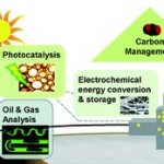
 Energy: the microfluidic frontier
David Sinton
Lab Chip, 2014, 14, 3127-3134
DOI: 10.1039/C4LC00267A
 Physics and technological aspects of nanofluidics
Lyderic Bocquet and Patrick Tabeling
Lab Chip, 2014, 14, 3143-3158
DOI: 10.1039/C4LC00325J
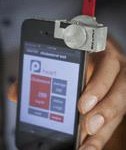
 Smartphone technology can be transformative to the deployment of lab-on-chip diagnostics
David Erickson, Dakota O’Dell, Li Jiang, Vlad Oncescu, Abdurrahman Gumus, Seoho Lee, Matthew Mancuso and Saurabh Mehta
Lab Chip, 2014, 14, 3159-3164
DOI: 10.1039/C4LC00142G
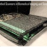
 Biomedical imaging and sensing using flatbed scanners
Zoltán Göröcs and Aydogan Ozcan
Lab Chip, 2014, 14, 3248-3257
DOI: 10.1039/C4LC00530A
Dr. Sangeeta N. Bhatia is winner of the 2014 Corning Inc./Lab on a Chip Pioneers of Miniaturisation Lectureship
The 9th ‘Pioneers of Ministurisation‘ Lectureship, is for extraordinary or outstanding contributions to the understanding or development of miniaturised systems and will be presented to Dr Bhatia at the µTAS 2014 Conference in San Antonio, Texas in October. Dr Bhatia will receive a certificate, $5000 and will give a short lecture at the µTAS Conference, later this year.
About the winner
Dr Bhatia conducts research at the intersection of engineering, medicine, and biology to develop novel platforms for understanding, diagnosing, and treating human disease. Her ‘tiny technologies’ interface living cells with synthetic systems, enabling new applications in tissue regeneration, stem cell differentiation, medical diagnostics and drug delivery. She and her colleagues were the first to demonstrate that microfabrication technologies used in semiconductor manufacturing could be used to organize cells of different types to produce a tissue with emergent properties. Dr. Bhatia’s findings have produced high-throughput-capable human microlivers, which model human drug metabolism, drug-induced liver disease, and interaction with human pathogens. Her group also develops nanoparticles and nanoporous materials that can be designed to assemble and communicate to diagnose and treat a variety of diseases, including cancer.
Dr. Bhatia co-authored the first undergraduate textbook on tissue engineering and has published more than 150 manuscripts, that have been cited over 13,500 times. She and her 150+ trainees have contributed to more than 40 issued or pending patents and launched 9 biotechnology companies with close to 100 products. She is a frequent advisor to governmental organizations and consults widely for academia and industry.
Dr. Bhatia holds a B.S. from Brown University; an M.S. in mechanical engineering from MIT; a Ph.D. in biomedical engineering from MIT; and an M.D. from Harvard Medical School and currently she directs the Laboratory for Multiscale Regenerative Technologies at MIT. She is a Howard Hughes Medical Institute Investigator and the John J. and Dorothy Wilson Professor of Health Sciences and Technology and Electrical Engineering and Computer Science at MIT. She is a member of the Institute for Medical Engineering and Science and the Koch Institute for Integrative Cancer Research at MIT, a senior member of the Broad Institute, and a biomedical engineer at Brigham & Women’s Hospital. Dr. Bhatia is an elected Fellow of the Massachusetts Academy of Sciences, Biomedical Engineering Society, American Institute for Medical and Biological Engineering, and the American Society for Clinical Investigation.
We would like to congratulate Dr Bhatia on this achievement!
The 2013 Pioneers of Miniaturisation Lectureship was awarded to Shuichi Takayama, University of Michigan.
The 2013 Pioneers of Miniaturisation Prize went to Shuichi Takayama!
Lab on a Chip joined forces with Corning Incorporated to award the eighth Pioneers of Miniaturisation Lectureship, including a certificate of recognition and a prize of $5000.
The lectureship was presented at the µTAS 2013 Conference in Freiburg, Germany. The Pioneers of Miniaturisation Lectureship recognises outstanding achievements and significant contributions to the understanding and advancement of micro- and nano-scale science. This year, the Lectureship was awarded to Professor Schuichi Takayama at the University of Michigan, USA.
Shu has made seminal contributions and provided true vision in advancing scientific developments and technologies that have increased our understanding of phenomena at the micro- and nano scale. Not only was he the first to report an organ on a chip, in his pioneering paper (PNAS 2007), but he has also developed bone-on-a-chip and stem cell-on-a-chip as well as establishing various organ-on-a-chip platforms. Amnosgt his many achievements, Shu has improved handling of sperm, eggs an embryos during the in vitro fertilization processes by designing integrated microfluidic systems.
Shu has published several papers in Lab on a Chip – click on the links to download his 2014 papers:
Elevating Sampling
Joseph M. Labuz and Shuichi Takayama
DOI: 10.1039/C4LC00125G, Frontier
From themed collection Lab on a Chip: Insights Issue
Defined topologically-complex protein matrices to manipulate cell shape via three-dimensional fiber-like patterns
Christopher Moraes, Byoung Choul Kim, Xiaoyue Zhu, Kristen L. Mills, Angela R. Dixon, M. D. Thouless and Shuichi Takayama
DOI: 10.1039/C4LC00122B, Paper
From themed collection Open access articles from Lab on a Chip
Control of soft machines using actuators operated by a Braille display
Bobak Mosadegh, Aaron D. Mazzeo, Robert F. Shepherd, Stephen A. Morin, Unmukt Gupta, Idin Zhalehdoust Sani, David Lai, Shuichi Takayama and George M. Whitesides
DOI: 10.1039/C3LC51083B, Paper
We are delighted to announce that our 2013 Impact Factor* has risen to 5.748!
Lab on a Chip provides a unique forum for the publication of significant and original work related to miniaturisation (on or off chips) at the micro- and nano- scale across a variety of disciplines. We would like to thank all of our Board members, authors, readers and reviewers for their continued support.
Contribute to our next Impact Factor – submit your latest piece of high impact work with us here.
Interested in other Royal Society of Chemistry journals? Click here to see how well they did.
*The Impact Factor provides an indication of the average number of citations per paper. Produced annually, Impact Factors are calculated by dividing the number of citations in a year by the number of citeable articles published in the preceding two years. Data based on 2013 Journal Citation Reports®, (Thomson Reuters, 2014).
In a recent paper in Lab on a Chip, a group of British researchers reported a ‘lab-in-a-briefcase’ for detection and quantification of the prostate cancer biomarker PSA in human serum and whole blood. Their lab-in-a-briefcase is a small container with a set of coated plastic capillaries, a pre-loaded microwell plate with reagents and a film scanner.
The researchers stress that their system is cheap and easy to handle, which would make it very useful for performing diagnostics in low resource areas. In addition, their lab-in-a-briefcase demonstrates the potential for point-of-care tests for prostate cancer, which would allow easy screening by non-experts in a non-clinical setting.
The concept of a lab-in-a-briefcase may have more far-reaching implications, though. Most lab-on-a-chip assays and microfluidic systems are usually developed in the context of interdisciplinary research collaborations. One research department may develop a new system, while another department has the – often unstable – samples that are used to demonstrate proof-of-concept. This complexity means that projects can quickly become logistic nightmares.
Multi-site collaborations make the portability and the standardized format that are found in the lab-in-a-briefcase and related technologies very important. It doesn’t matter if the application domain of a project is physics, biochemistry or biology. Developing a portable, standardized set-up with good documentation, automated analysis and easy read-out can contribute greatly to the success of a multi-disciplinary microfluidic engineering project, because it promotes collaboration and a wider application of the technology early on.
All of this means that the lab-in-a-briefcase is not just a niche product that is only useful for cheap point-of-care diagnostics in low resource areas. It is a design concept that anyone in the realm of microfluidic engineering needs to understand. Perhaps the concept is also applicable to the project you’re currently working on?
Go check it out for yourselves – you can download this paper fro free* for a limited time only!