Archive for September, 2011

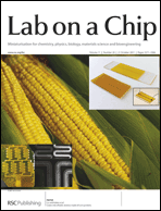
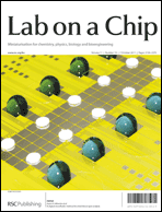
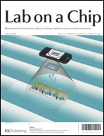
On the cover: mobile phones to read ELISA results, microfluidic devices made from corn and voltage-expandable liquid crystal surfaces

On the cover of Issue 20 we have hot articles from Ye and Demirci et al., Liu and Kokini et al. and Ren and Wu et al.

The outside front cover depicts Bin Ye and Utkan Demirci‘s paper where they have demonstrated that a mobile phone can be used to image and process the results of an ELISA (enzyme-linked immunosorbent assay) test on a microchip, reducing previously bulky equipment to a size where it could be used at the bedside.

Integration of cell phone imaging with microchip ELISA to detect ovarian cancer HE4 biomarker in urine at the point-of-care
ShuQi. Wang, Xiaohu Zhao, Imran Khimji, Ragip Akbas, Weiliang Qiu, Dale Edwards, Daniel W. Cramer, Bin Ye and Utkan Demirci
Lab Chip, 2011, 11, 3411-3418

The inside front cover highlights green microfluidic research from Gang Logan Liu and Jozef L. Kokini, where they have used a by-product from corn – zein – instead of the traditional plastics to produce a microfluidic device.  This article was also highlighted in Chemistry World.

Green microfluidic devices made of corn proteins
Jarupat Luecha, Austin Hsiao, Serena Brodsky, Gang Logan Liu and Jozef L. Kokini
Lab Chip, 2011, 11, 3419-3425

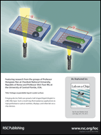
And on the back cover is work from Hongwen Ren and Shin-Tson Wu where they report a novel approach which can extensively spread a liquid crystal interface, which opens a route to new voltage controllable, polarization-insensitive, and broadband liquid photonic devices.

Voltage-expandable liquid crystal surface
Hongwen Ren, Su Xu and Shin-Tson Wu
Lab Chip, 2011, 11, 3426-3430

Digg This
Reddit This
Stumble Now!
Share on Facebook
Bookmark this on Delicious
Share on LinkedIn
Bookmark this on Technorati
Post on Twitter
Google Buzz (aka. Google Reader)

Top ten most accessed articles in August

This month sees the following articles in Lab on a Chip that are in the top ten most accessed:

Automated cellular sample preparation using a Centrifuge-on-a-Chip
Albert J. Mach, Jae Hyun Kim, Armin Arshi, Soojung Claire Hur and Dino Di Carlo
Lab Chip, 2011, 11, 2827-2834
DOI: 10.1039/C1LC20330D

Benchtop micromolding of polystyrene by soft lithography
Yuli Wang, Joseph Balowski, Colleen Phillips, Ryan Phillips, Christopher E. Sims and Nancy L. Allbritton
Lab Chip, 2011, 11, 3089-3097
DOI: 10.1039/C1LC20281B

Next-generation integrated microfluidic circuits
Bobak Mosadegh, Tommaso Bersano-Begey, Joong Yull Park, Mark A. Burns and Shuichi Takayama
Lab Chip, 2011, 11, 2813-2818
DOI: 10.1039/C1LC20387H

Beyond PDMS: off-stoichiometry thiol–ene (OSTE) based soft lithography for rapid prototyping of microfluidic devices
Carl Fredrik Carlborg, Tommy Haraldsson, Kim Öberg, Michael Malkoch and Wouter van der Wijngaart
Lab Chip, 2011, 11, 3136-3147
DOI: 10.1039/C1LC20388F

A high-performance microsystem for isolating circulating tumor cells
Xiangjun Zheng, Luthur Siu-Lun Cheung, Joyce A. Schroeder, Linan Jiang and Yitshak Zohar
Lab Chip, 2011, 11, 3269-3276
DOI: 10.1039/C1LC20331B

Paper on a disc: balancing the capillary-driven flow with a centrifugal force
Hyundoo Hwang, Seung-Hoon Kim, Tae-Hyeong Kim, Je-Kyun Park and Yoon-Kyoung Cho
Lab Chip, 2011, Advance Article
DOI: 10.1039/C1LC20445A

Double-emulsion drops with ultra-thin shells for capsule templates
Shin-Hyun Kim, Jin Woong Kim, Jun-Cheol Cho and David A. Weitz
Lab Chip, 2011, 11, 3162-3166
DOI: 10.1039/C1LC20434C

Rapid spatial and temporal controlled signal delivery over large cell culture areas
Jules J. VanDersarl, Alexander M. Xu and Nicholas A. Melosh
Lab Chip, 2011, 11, 3057-3063
DOI: 10.1039/C1LC20311H

Flexible microfluidic devices with three-dimensional interconnected microporous walls for gas and liquid applications
Po Ki Yuen and Michael E. DeRosa
Lab Chip, 2011, 11, 3249-3255
DOI: 10.1039/C1LC20157C

Microchip-based immunomagnetic detection of circulating tumor cells
Kazunori Hoshino, Yu-Yen Huang, Nancy Lane, Michael Huebschman, Jonathan W. Uhr, Eugene P. Frenkel and Xiaojing Zhang
Lab Chip, 2011, Advance Article
DOI: 10.1039/C1LC20270G

Why not take a look at the articles today and blog your thoughts and comments below.

Fancy submitting an article to Lab on a Chip? Then why not submit to us today or alternatively email us your suggestions.

Digg This
Reddit This
Stumble Now!
Share on Facebook
Bookmark this on Delicious
Share on LinkedIn
Bookmark this on Technorati
Post on Twitter
Google Buzz (aka. Google Reader)

HOT: label-free plasmonic microarray for high-throughput protein screening

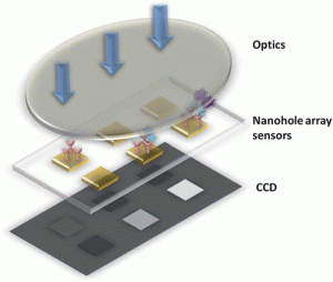
Microarrays offer the opportunity for the high-throughput study of protein-protein interactions that may uncover drug targets and provide early detection of various diseases. Most current technologies rely on fluorescent labelling of proteins, but this is costly, time-consuming and can be inaccurate due to steric interference, quenching and photo-bleaching.

Hatice Altug, Boston University, and colleagues from MIT have now found a way to accomplish label free detection by using a dual-colour filter method to image large scale plasmonic nanohole arrays.  Their high-density detection platform has over 1 million sensors and has reliably demonstrated massively multiplexed detection of antibody bindings with reduced image acquisition time.

Download the paper to read how they did it, it’s free to access for the next 4 weeks:

Large-scale plasmonic microarrays for label-free high-throughput screening
Tsung-Yao Chang, Min Huang, Ahmet Ali Yanik, Hsin-Yu Tsai, Peng Shi, Serap Aksu, Mehmet Fatih Yanik and Hatice Altug
Lab Chip, 2011, Advance Article
DOI: 10.1039/C1LC20475K

Digg This
Reddit This
Stumble Now!
Share on Facebook
Bookmark this on Delicious
Share on LinkedIn
Bookmark this on Technorati
Post on Twitter
Google Buzz (aka. Google Reader)

Corn microchips

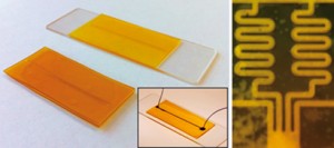
Left: macro images of zein-glass and zein-zein microfluidic devices. The inset shows a colourant-filled zein-glass microfluidic device with tubings. Right: serpentine channels for mixing two different chemical fluids

Gang Logan Liu and colleagues at the University of Illinois Urbana-Champaign, have demonstrated that by using lithography and solvent or vapour deposition bonding, thin films of zein (a protein extracted from corn by-products) can be used to construct microfluidic channels, grids and wells as a green alternative to the plastic materials currently in use.

The group tested their zein microfluidic devices using several types of fluid, including fluorescent dyes and solutions of microbeads. They found that the devices did not leak and the channels could easily be viewed through a microscope. They also extended their work to produce a concentration gradient generator, mixing dye and solvent within serpentine channels to create several different concentration mixtures.

Liu anticipates being able to use the simple bonding of the zein films to make multilayer microfluidic devices in the future.

Interested? Read Tamsin Phillips’ full Chemistry World article here or download the Lab on a Chip paper:

Green microfluidic devices made of corn proteins
Jarupat Luecha, Austin Hsiao, Serena Brodsky, Gang Logan Liu and Jozef L. Kokini
Lab Chip, 2011, Advance Article
DOI: 10.1039/C1LC20726A

Digg This
Reddit This
Stumble Now!
Share on Facebook
Bookmark this on Delicious
Share on LinkedIn
Bookmark this on Technorati
Post on Twitter
Google Buzz (aka. Google Reader)

Making nerve pathways in chips for brain studies

The study of brain development and degeneration is hindered by a lack of physiologically realistic models. French scientists have now developed a way to reconstruct neuronal networks in a microfluidic system to more closely mimic the directional neuronal pathways found in the brain.

Current experimental brain models include neuronal cell cultures and whole animal models; however, the former lack the complex architecture found in vivo, and the latter restrict studies at the cellular level. Aiming to bridge the gap between these models, Jean-Louis Viovy of the Curie Institute, Paris, and coworkers have developed a microfluidic device that allows for the growth of oriented and functional synaptic connections in vitro.

The device consists of two cell culture chambers connected by microchannels, through which axons – nerve fibres that conduct impulses away from the body of the nerve cell – can penetrate to form neuronal networks. In previous setups, replication of the unidirectional networks found in vivo could not be achieved since axons were sent from each chamber to the one opposite, travelling in both directions across the microchannel.

Inspired by the observation that axons can be mechanically constrained, the team modified the device to include asymmetric, funnel-shaped microchannels, termed ‘axon diodes’, to allow axons to grow from only one chamber to the other and not the opposite way. The concept was verified by experiments with mouse cortical neurons, in which the axon projection was 97 per cent selective for the ‘correct’ direction.

Neuronal networks have been grown in microfluidic chambers to replicate neural pathways in the brain

Next, by seeding cortical neurons (from the outer part of the brain) on the emitting side of the device and striatal neurons (from the inner part of the brain) on the receiving side, the team demonstrated the reconstruction of an active neuronal pathway involving two different neuronal subtypes. Furthermore, these networks were routinely maintained for three weeks in vitro, which would allow for both short and long-term experimentation.

‘I was struck by the simplicity of the system, it is beautiful,’ remarks Bonnie Firestein, an expert in cellular neurobiology from Rutgers University, New Jersey, US. ‘It is very easy to make and to use, and allows the recreation of what happens in vivo in an in vitro system.’

Such a device has many potential applications in neurobiological research. An initial motivation for this work was the requirement of a model to study the progression of neuronal damage in degenerative diseases such as Alzheimer’s. Additionally, Viovy believes the system is also an important platform for research into brain development and cognitive science. ‘How neurons communicate regarding information transmission is also an area in which we currently lack a model of the kind we have proposed here,’ he says. The team are currently working on further increasing the complexity of the networks, to more accurately model the neuronal organisation of the brain.

Interested? Read Sarah Farley’s full Chemistry World article here or download the Lab on a Chip paper:

Axon diodes for the reconstruction of oriented neuronal networks in microfluidic chambers
Jean-Michel Peyrin, Bérangère Deleglise, Laure Saias, Maéva Vignes, Paul Gougis, Sebastien Magnifico, Sandrine Betuing, Mathéa Pietri, Jocelyne Caboche, Peter Vanhoutte, Jean-Louis Viovy and Bernard Brugg
Lab Chip, 2011, Advance Article
DOI: 10.1039/c1lc20014c
Digg This
Reddit This
Stumble Now!
Share on Facebook
Bookmark this on Delicious
Share on LinkedIn
Bookmark this on Technorati
Post on Twitter
Google Buzz (aka. Google Reader)

New YouTube Videos

View the new videos on the Lab on a Chip YouTube site using the links below:

A switchable digital microfluidic droplet dye-laser

Electrolysis in nanochannels for in situ reagent generation in confined geometries

Magnetic domain wall conduits for single cell applications

Digg This
Reddit This
Stumble Now!
Share on Facebook
Bookmark this on Delicious
Share on LinkedIn
Bookmark this on Technorati
Post on Twitter
Google Buzz (aka. Google Reader)

On the cover: a digital microfluidic method for dried blood spot analysis

On the front cover of Issue 19 is an article from Aaron Wheeler et al. on their new method for the analysis of dried blood spot samples. The method has the potential to offer automation of dried blood samples, which are useful for a number of clinical and pharmaceutical applications due to the small sample sizes involved and ease of storage.  The team have developed a prototype microfluidic system to quantify amino acids in which analytes are extracted, mixed with internal standards, derivatized, and reconstituted for analysis by tandem mass spectrometry.

This hot article was also recently reported on in C&EN.

A digital microfluidic method for dried blood spot analysis
Mais J. Jebrail, Hao Yang, Jared M. Mudrik, Nelson M. Lafrenière, Christine McRoberts, Osama Y. Al-Dirbashi, Lawrence Fisher, Pranesh Chakraborty and Aaron R. Wheeler
Lab Chip, 2011, 11, 3218-3224
DOI: 10.1039/C1LC20524B

Digg This
Reddit This
Stumble Now!
Share on Facebook
Bookmark this on Delicious
Share on LinkedIn
Bookmark this on Technorati
Post on Twitter
Google Buzz (aka. Google Reader)

New YouTube Videos

View the new videos on the Lab on a Chip YouTube site using the links below:

Photoreversible fragmentation of a liquid interface for micro-droplet generation by light actuation

DNA-templated assembly of droplet-derived PEG microtissues

A microchip-based model wound with multiple types of cells

Automated cellular sample preparation using a Centrifuge-on-a-Chip

Double-emulsion drops with ultra-thin shells for capsule templates

Pyroelectric Adaptive Nanodispenser (PYRANA) microrobot for liquid delivery on a target

Classification of cell types using a microfluidic device for mechanical and electrical measurement on single cells

Microfluidic baker’s transformation device for three-dimensional rapid mixing

Integrated sieving microstructures on microchannels for biological cell trapping and droplet formation

Microfluidic platform for electrophysiological studies on Xenopus laevis oocytes under varying gravity levels

Digg This
Reddit This
Stumble Now!
Share on Facebook
Bookmark this on Delicious
Share on LinkedIn
Bookmark this on Technorati
Post on Twitter
Google Buzz (aka. Google Reader)

RSC e-membership launched

This week, the RSC launched a new product, RSC e-membership, allowing anyone to access an electronic version of Chemistry World through a MyRSC account and to enjoy the benefits of electronic networking via this professional online community for £20/year.

Subscribers to this do not benefit from the professional recognition or any of the other many services and discounts available to RSC Members, but it allows chemists from around the world, many already members of another chemical society in their own country, to benefit from the highly-esteemed content in Chemistry World and the networking opportunities offered from MyRSC, which now stands at over 11,000 members. The RSC e-membership also allows subscribers to join a virtual specialist interest group on MyRSC. If you are interested in joining, please visit www.rsc.org/emembership.

Digg This
Reddit This
Stumble Now!
Share on Facebook
Bookmark this on Delicious
Share on LinkedIn
Bookmark this on Technorati
Post on Twitter
Google Buzz (aka. Google Reader)