Author Archive

Nanoparticle sulfidation transformations

A study by Gregory Lowry and colleagues from Carnegie Mellon University suggests that Copper Oxide (CuO) nanoparticles (NPs) are sulfidized in the environment, affecting their resulting properties. Some of the affected properties, such as solubility are relevant to the toxicity of these nanoparticles in the environment.

Many nanoparticles are transformed in the environment; it is often the transformed materials which cause concern with regards to nanoparticle toxicity. There have been several papers highlighting how important it is to research the properties of nanoparticle transformations. A recent review by Nasia Von Moos provides an overview of what is currently known about environmental transformations of nanomaterials in freshwater systems, a recent paper by Julián A. Gallego-Urrea discusses the transformations which TiO2 nanoparticles undergo once they reach the aquatic environment and this research paper reports that sulfidation is an important transformation for some metal oxide nanoparticles, such as CuO NPs.

Why is sulfidation in the environment important?

Copper-based NPs are being used in semiconductors, heat transfer fluids, catalysts, batteries and many more products and technologies. Their wide spread uses will likely lead to subsequent release into the environment, raising concerns about their potential toxicity. It has already been demonstrated that CuO NPs are toxic to many organisms including crustaceans, algae and fish; although Cu2+ is more toxic to most of these organisms. It is therefore essential to determine what the products of sulfidized CuO NPs are and if they are more or less toxic to the environment when compared with pristine CuO.

Sulfidation of CuO

Cuo NPs were characterized and sulfidized in water by inorganic sulfide. Characterization of the resulting products showed that CuO is sulfidized to several copper sulphide species including crystalline CuS (covellite), amorphous (CuxSy) species and copper sulphate hydroxide species. In previous studies it has been demonstrated shown that sulfidation decreases the solubility and metal availability of Ag and ZnO NPs. This study however shows that the sulfidation of CuO NPs breaks the trend. Sulfidation actually increased the dissolved fraction of copper compared to pristine CuO NPs. This increased release of Cu2+ and CuS nanoclusters from sulfidized NPs compared to CuO suggests that toxicity studies with pristine CuO may be misleading in environments where sulfidation is likely to occur, demonstrating that it is prudent to use environmentally transformed nanoparticles in fate, transport and toxicitiy studies rather than focusing soley on the prisitne materials. Access the full article for free* by clicking the link below.

Sulfidation of copper oxide nanoparticles and properties of resulting copper sulfide
Rui Ma, John Stegemeier, Clement Levard, James Dale, Clinton W Noack, Tittany Yang, Gordon Brown and Gregory Lowry
DOI: 10.1039/C4EN00018H

*Access is free through a registered RSC account – click here to register

The author recommends further studies which are still needed to:

  1. Identify the nature of the CuxSy nanoclusters.
  2. Assess the toxicity of sulfidized CuO NPs and CuxSy nanoclusters.
  3. Assess the stability of very small metal sulphide clusters (Ag, Zn and Cu) against oxidation under environmental and biological conditions.
  4. Assess how sulfidation of CuO NPs occurs in situ at relevant CuO/S concentration ratios and how this affects their bioavailability under realistic exposure scenarios.
Digg This
Reddit This
Stumble Now!
Share on Facebook
Bookmark this on Delicious
Share on LinkedIn
Bookmark this on Technorati
Post on Twitter
Google Buzz (aka. Google Reader)

Vicki Grassian is the 2014 John Jeyes Award Winner

John Jeyes 2014 Award WinnerProfessor Vicki Grassian, Chair of Environmental Science: Nano has won the 2014 John Jeyes Award for her pioneering contributions to the chemistry of environmental interfaces, heterogeneous atmospheric chemistry and the environmental implications of nanomaterials.

It is truly an honor to receive this award Grassian said.

The John Jeyes Award, founded in 1975, is a biennial award for chemistry in relation to the environment. Vicki Grassian, Founding Director of the Nanoscience and Nanotechnology Institute at the University of Iowa, has mentored over one hundred students and postdocs in her laboratory, many of them now having their own research programs focused on energy and the environment.

The John Jeyes Award is wonderful recognition of the research that has been done by the students and postdoctoral associates who have worked in my laboratoryGrassian said.

We would like to congratulate Vicki on her achievement and take this opportunity to thank Vicki for her pioneering contributions to Environmental Science: Nano, bringing together a variety of communities to publish their work on nanoscience and the implications for the environment, health and sustainability.

Vicki’s latest Environmental Science: Nano paper Iron oxide nanoparticles induce Pseudomonas aeruginosa growth, induce biofilm formation and inhibit antimicrobial peptide function (C3EN00029J) is included in a dedicated themed collection of papers celebrating the 2014 RSC Prize and Award winners. All articles in this collection are free* to access until 6th June.

*Access is free through a registered RSC account – click here to register

Digg This
Reddit This
Stumble Now!
Share on Facebook
Bookmark this on Delicious
Share on LinkedIn
Bookmark this on Technorati
Post on Twitter
Google Buzz (aka. Google Reader)

Applications of porous nanomaterials

Sean Lehman and Sarah Larsen from the University of Iowa review zeolite and mesoporous silica nanomaterials with emphasis on connections to the environment.

In recent years, there has been a great deal of interest in zeolites and mesoporous silica nanomaterials (MSNs). Zeolites are widely used in industry for applications such as catalysis, separations and gas adsorption, however the authors believe that these porous nanomaterials have a largely unrealized commercial potential for environmental applications.

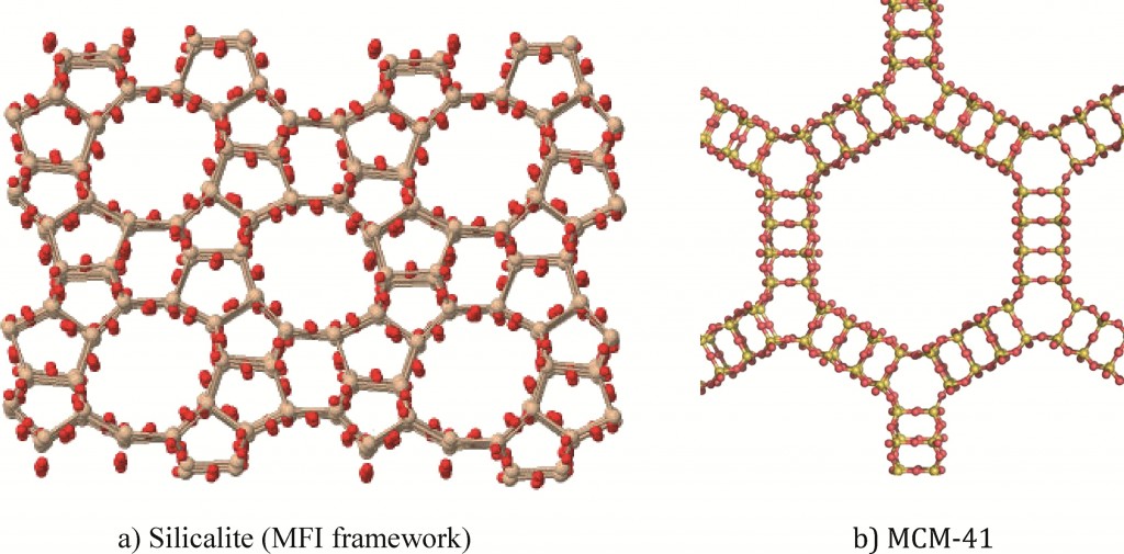
Structures of porous nanomaterials

This review article covers three major areas:

  1. Greener synthesis of zeolite and MSNs
  2. Potential of zeolite and MSNs for environmental applications
  3. The biological toxicity of zeolite and MSNs

Due to cost and reduced thermal stability MSNs are not as extensively applied as zeolite; however they are currently being investigated for potential environmental and biomedical applications. Their varied physiochemical properties open up a wide range of potential applications. The more applications that these porous nanomaterials have in industry, the greater the interested in developing greener synthesis for them and reducing their toxicity. With two measurements which are on the nanoscale, pore size as well as particle size, zeolites and MSN make very interesting nanomaterials.

This review describes both the environmental applications, including environmental catalysis and adsorption of environmental contaminants, and implications of zeolite and MSNs. Due to concerns that increased use of these materials translates to increased exposures, toxicity studies of both nanomaterials are also reviewed.

To read the full review for free* click the link below:

Zeolite and Mesoporous Silica Nanomaterials: Greener Syntheses, Environmental Applications and Biological Toxicity

Sean E Lehman and Sarah C Larsen
DOI: 10.1039/C4EN00031E, Critical Review

Zeolites and MSNs are silicate or aluminosilicate nanomaterials with well-defined pore networks; there are however some differences between the two porous nanomaterials.

Properties of zeolites:

  • Crystalline aluminosilicates (or silicates)
  • Regular arrangements of micropores
  • High surface areas
  • Exchangeable cations

Properties of MSN:

  • Amorphous silica materials
  • Regualr arrangement of mesopores
  • Very high surface area

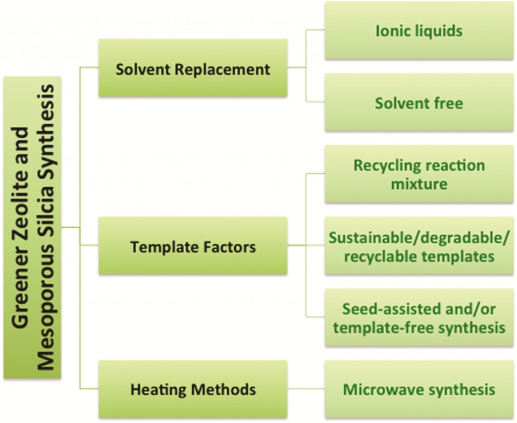
The first area discussed in this review is the synthesis of zeolite and MSNs using green synthetic routes. The green strategies can be organized into three main categories: solvent, template and heating. The diagram below demonstrates the strategies for the greener synthesis of zeolites and mesoporous silica.

Greener synthesis of zeolites and mesoporous silica

*Access is free through a registered RSC account – click here to register

Digg This
Reddit This
Stumble Now!
Share on Facebook
Bookmark this on Delicious
Share on LinkedIn
Bookmark this on Technorati
Post on Twitter
Google Buzz (aka. Google Reader)

Mass Spectrometry Proteomics Characterize Nanoparticle Protein Corona

The adventitious protein adsorption on to nanoparticles (NPs), commonly known as the NP-protein corona is in the limelight for many good reasons including its influence on the NP uptake, accumulation and ultimate cellular fate. The consequences of this phenomenon can work in different ways. NP-protein corona can lower the toxicity of NPs when compared to the “bare” NPs but at the same time facilitate crossing biological barriers that can trigger activation of specific regulatory pathways. The particle size, composition and surface properties of NPs are known to influence the composition of the NP-protein corona. Thus, Korin Wheeler, from Santa Clara University, and coworkers investigate the protein populations across the NP-protein corona using silver nanoparticles (AgNPs) in the presence of Yeast (Saccharomyces cerevisiae) proteins (YPE) with the aid of mass spectrometry (MS) proteomics.”

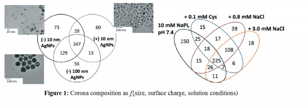
Experiments were designed to probe the effects of AgNP size, surface charge and solution conditions. Two different sizes of AgNPs (10 nm vs 100 nm) with anionic (citrate) and cationic (branched polyethyleneimmine – BPEI) coatings were selected. Protein corona formation was conducted under 0.8 mM NaCl, modeled after freshwater salinity; 3.0 mM NaCl, mimicking mitochondrial salinity; and 0.1 mM Cys, which was shown to mediate NP toxicity. After incubating AgNPs with the YPE, bound and unbound proteins were separated via centrifugation, digested with trypsin followed by LC-MS/MS analysis.

Over 500 different proteins were identified as bound and unbound but no trends were reported linking the molecular weight, pI, length, or the amino acid composition of proteins to their enrichment in the corona. However, AgNP surface charge played a stronger role with those having similar coatings sharing majority of corona population. This highlighted electrostatic modulation of protein affinity for AgNPs under low ionic strength conditions. Further studies with NaCl and cys to simulate more environmentally and biologically relevant conditions revealed more changes in the protein corona populations and modifications in their solution phase behavior in terms of aggregation and sedimentation. However, under all the variable factors there is a population of ubiquitous proteins that get enriched in the corona as shown in the Figure below.

The findings of this research is expected to contribute towards better understanding the bio physicochemical factors mediating NP-corona formation, their characterization and the development of predictive models within the environment.

To access the full article, download a copy for free* by clicking the link below.

Silver nanoparticle protein corona composition compared across engineered particle properties and environmentally relevant reaction conditions

Richard Eigenheer, Erick R. Castellanos, Meagan Y. Nakamoto, Kyle T. Gerner, Alyssa M. Lampe and Korin E. Wheeler
DOI: 10.1039/C4EN00002A

*Access is free through a registered RSC account – click here to register

Digg This
Reddit This
Stumble Now!
Share on Facebook
Bookmark this on Delicious
Share on LinkedIn
Bookmark this on Technorati
Post on Twitter
Google Buzz (aka. Google Reader)

Tracing Silver Nanoparticles

To understand the fate of silver nanoparticles (AgNPs) in the environment they need to be traced in complex natural samples. Often, studies that interrogate the environmental fate of AgNPs require AgNP concentrations that exceed realistic environmental levels. Potentially, this produces unreliable results, as high particle concentration could induce and identify distinct patterns of particle behaviour that is not relevant for ‘realistic’ environmental concentrations.  Adam Laycock, from Imperial College London, and colleagues investigate whether the same effects are also observed at low, environmentally relevant AgNP concentrations.

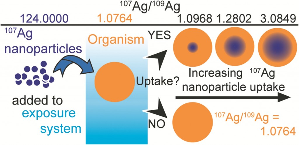
Stable isotope labelling is an attractive method of labelling which relies on the detection of changes when a contaminant is introduced to a system. Previous methods of tracing nanomaterials have had significant drawbacks. For example, fluorescent coatings can affect the surface chemistry of the nanomaterials causing dissociation. Radiolabelling, another technique used, involves the use of specialist equipment and licenses are required for the handling of radioactive material. Recent studies have shown that stable isotope tracing can be applied to nanomaterials – but first, the labelled NPs must be specifically prepared from a single, highly enriched stable isotope form of the elements.

In this study existing protocols were examined to develop techniques for the optimized preparation of isotopically labelled AgNPs. Three protocols were applied to produce particles with a variety of target sizes, with enriched 107Ag and natural Ag. The results show that the methods are suitable for small scale synthesis of stable labeled AgNPs at yields of approximately 80%. The labelling process does not generate unusual particle properties, demonstrating that isotopically modified AgNPs are equivalent to AgNPs with a natural isotope composition.

The authors finalise their study by presenting a series of calculations which reveal that stable isotope labelling can increase the detection sensitivity of AgNPS by at least a factor of 40, and possibly by up to 4000x in comparison to commonly employed bulk Ag concentration measurements. This approach of tracing nanomaterials is highly versatile, the label cannot be lost by dissociation or degradation, the element remains traceable and the use of enriched stable isotopes provides an extremely selective and sensitive means of elemental tracing, even in the presence  of high natural background levels.

To read the full experiment for free*, download the paper now:

Synthesis and characterization of isotopically labeled silver nanoparticles for tracing studies
Adam Laycock, Björn Stolpe, Isabella Römer, Agnieszka Dybowska, Eugenia (Éva) Valsami-Jones, Jamie R Lead and Mark Rehkamper
DOI: 10.1039/C3EN00100H

*Access is free through a registered RSC account – click here to register

Digg This
Reddit This
Stumble Now!
Share on Facebook
Bookmark this on Delicious
Share on LinkedIn
Bookmark this on Technorati
Post on Twitter
Google Buzz (aka. Google Reader)

Water flea’s assist in the mission to develop sustainable nanomaterials

Researchers are continuously on the lookout for materials which are more environmentally friendly and sustainable in order to improve emerging technologies. Nanoparticles (NPs) are the basis for a variety of emerging technologies used for industrial, biomedical and environmental applications, but their release into the environment is still a cause for concern. What if a nanomaterial, that has minimal negative environmental impact, was available? Jared Bozich from the University of Wisconsin and colleagues have published a research paper demonstrating that surface chemistry has the potential to increase or decrease negative biological impacts of NPs.

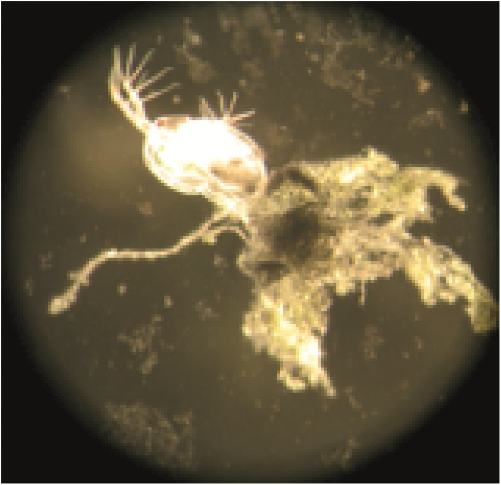
The surfaces of NPs are typically modified with surface functional groups that control properties such as stability. In this research, the acute and chronic toxicity of well characterized gold nanoparticles (AuNPs), functionalized with ligands of differing charges were investigated in Daphnia magna. D. magna (more commonly called water fleas) are widely accepted as a model organism for assessing the toxicity of environmental contaminates and experience reduced reproduction, growth and increased mortality with exposure to toxic substances. D.magna were exposed to concentrations of four types of functionalized AuNps.

The results showed that initial particle charge significantly impacted overall toxicity, with positively charged particles being more toxic than their negatively-charged counterparts. This could be explained by the increased cellular uptake of positively charged particles due to their high levels of attraction with cellular membrane. This creates a hole in the membrane due to the densely populated charge on the NP surface – allowing the particle to enter the intracellular matrix and continue to cause damage. Another interesting result of this study was that the smaller the particle, the higher the toxicity. This can be explained by a similar theory; after cellular uptake the smaller particles are able to cross the gut lumen of the daphnis, potentially further interacting with D. magna cells and causing damage.

The results of this study identify mechanisms for AuNP toxicity by examining NP toxicity with different charges, using an environmental relevant organism. NPs have the potential to be highly beneficial to society, but in order to minimize the environmental implications the mechanisms that govern the toxicity of NPs need to be betters elucidated. This study demonstrates how the charge and identity of a ligand can influence AuNp toxicity.

To read the full article, download your free* copy now:

Surface chemistry, charge and ligand type impact the toxicity of gold nanoparticles to Daphnia magna

Jared Bozich, Samuel E Lohse, Marco D Torelli, Catherine Murphy, Robert Hamers and Rebecca Klaper

DOI: 10.1039/C4EN00006D

*Access is free through a registered RSC account – click here to register

Digg This
Reddit This
Stumble Now!
Share on Facebook
Bookmark this on Delicious
Share on LinkedIn
Bookmark this on Technorati
Post on Twitter
Google Buzz (aka. Google Reader)

The bioavailability of nanomaterials in the aquatic environment

With the level of engineered nanomaterials (EMNs) in the environment continuously increasing, there are rising concerns with regards to their potential environmental impact. In recent years a more accurate understanding of particle behaviour in complex systems has been gained.  Numerous studies have investigated the environmental hazards of ENMs, but the link between these two aspects is less developed. There are still considerable knowledge gaps with respect to ENMs bioavailability in the environment.

A review by Nadia Von Moos, from the University of Geneva, and colleagues provides an overview of what is currently known about environmental transformations of nanomaterials as well as their interactions with, and their toxicity towards bacteria and microalgae.

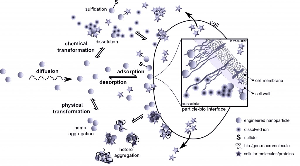
This diagram shows the processes at the medium – bio-interface underlying the bioavailability of ENMs to aquatic microorganisms (AMO). The bioavailability is dependent on many processes, such as chemical and physical transformations, adsorption and desorption, internalization, intracellular fate, agglomeration, dissolution and surface transformations. The importance of ENMs’ material characteristics has been reviewed before, but this review emphasises the environmental factors affecting the above processes. A quantitative understanding of ENM bioavailability requires insights into their behaviour during transport from the ambient medium to the AMO interface and of the processes underlying adsorption, internalization as well as intracellular fate – all of which are discussed in this review.

This review clearly concludes that there are still considerable knowledge gaps with respect to the effects of agglomeration on bioavailability, exact uptake routes, intracellular compartmentalization as well as dissolved organic matter-protein competition on the surface of internalized engineered nanoparticles. This review can be used to guide future research efforts in nanomaterial hazard and risk assessment. To read more, download your free* copy by clicking the link below:

Bioavailability of inorganic nanoparticles to planktonic bacteria and aquatic microalgae in freshwater

Nadia Von Moos, Paul Bowen and Vera I Slaveykova

DOI: 10.1039/C3EN00054K

*Access is free through a registered RSC account – click here to register

Digg This
Reddit This
Stumble Now!
Share on Facebook
Bookmark this on Delicious
Share on LinkedIn
Bookmark this on Technorati
Post on Twitter
Google Buzz (aka. Google Reader)

Bimetallic Fe nanoparticles to the Rescue

Who doesn’t like power? We all do. That’s why we love modifying nanomaterial surfaces. It gives us the power to control their properties. In the case of Fe nanoparticles (Fe NP), which are heavily used in environmental remediation, partial deposition of secondary metals such as Pd, Ni, Cu or Pt results in bimetallic clusters with enhanced reaction efficiencies towards treating contaminated groundwater and soils. Nevertheless one has to be cautious about how these modifications affect the nanoparticle toxicity. In fact, Fe NPs are known to possess antibacterial and antifungal properties; potential mechanisms of toxicity are membrane disruption and oxidative damage from the reactive oxygen species (ROS).

Therefore, E-J. Kim and colleagues from the Pohang University of Science and Technology have investigated the mechanism of toxicity for 4 different bimetallic Fe NPs: Fe/Cu, Fe/Ni, Fe/Pd and Fe/Pt, toward Escherichia coli along with bare Fe NPs. Synthesis of NPs was completed in-house and characterization data showed 50-70 nm primary particles with homogenous secondary metal coatings and zerovalent Fe cores.

The initial experiments consisted of testing cell viability upon exposure to NPs using CFU assay, ROS production using DCF-DA fluorescence dye (3hr post exposure), peroxidase activity using antioxidant enzyme glutathione peroxidase (GPx) and NP dissolution to determine the role of oxidative stress on the cell death. The results obtained are summarized below.

Therefore, the team concluded an alternative mechanism of toxicity via membrane disruption using a spectroscopic approach with FTIR spectroscopy, TEM imaging and anion release profiles. The TEM images showed higher levels of uptake for Fe/Cu in contrast to Fe/Pd. FTIR spectra of bacterial cells showed peaks corresponding to the C-O/C-O-C, PO2– and CH3– stretches that disappear upon exposure to NPs. Finally, the anion release profiles were found to be the most consistent with the cell viability indicating that NP-mediated membrane permeability and/or membrane damage is the major mechanism of toxicity. Higher amounts of PO32- and Cl were observed for cell cultures exposed to Fe/Cu while they were lowest for Fe/Pd and Fe/Pt. Interestingly Fe/Ni caused the highest release of SO42- indicating the disruption of sulfate transport system.

Overall, Fe/Pd was concluded to have the least toxicity and superior performance in terms of environmental decontamination while Fe/Cu NPs remained at the other end of the spectrum. The authors suggest this work may provide a platform for environmental engineers to design treatment strategies for environmental remediation with less harmful side effects over unmodified Fe NPs.

To read more about the full article, download a copy for free* by clicking the link below:

Comparative toxicity of bimetallic Fe nanoparticles toward Escherichia coli: mechanism and environmental implications

Eun-Ju Kim, Thao Le Thanh and   Yoon-Seok Chang

DOI: 10.1039/C3EN00057E

*Access is free through a registered RSC account – click here to register

Digg This
Reddit This
Stumble Now!
Share on Facebook
Bookmark this on Delicious
Share on LinkedIn
Bookmark this on Technorati
Post on Twitter
Google Buzz (aka. Google Reader)

Iron oxide nanoparticles: The smaller the particle, the bigger the bacterial growth

Iron-containing nanoparticles are beneficial in a wide range of applications, but little is known about the effects they have on bacterial pathogens.  Jennifer Borcherding and colleagues from the University of Iowa, including Vicki Grassian, the Environmental Science: Nano Editorial Board Chair, have completed research to show that iron oxide nanoparticles can induce bacterial growth and biofilm formation as well as inhibit antimicrobial peptide (AMP) function.

Iron-containing nanoparticles are generally thought to be non-toxic; however these studies suggest that the effect of co-exposures of nanoparticles with known pathogens and their impact on host innate immunity should be taken into consideration when screening nanoparticle toxicity. The use of iron-containing nanoparticles in industry is increasing, therefore the potential for iron-containing particle exposure, as particulate matter in air, is also increasing. This is a concern to human health, as particulate matter has been associated with increased respiratory exacerbations, pneumococcal infections, otitis media and eye infections.

Three main experiments were carried out to investigate the effect of iron-containing nanoparticles on bacterial pathogens.

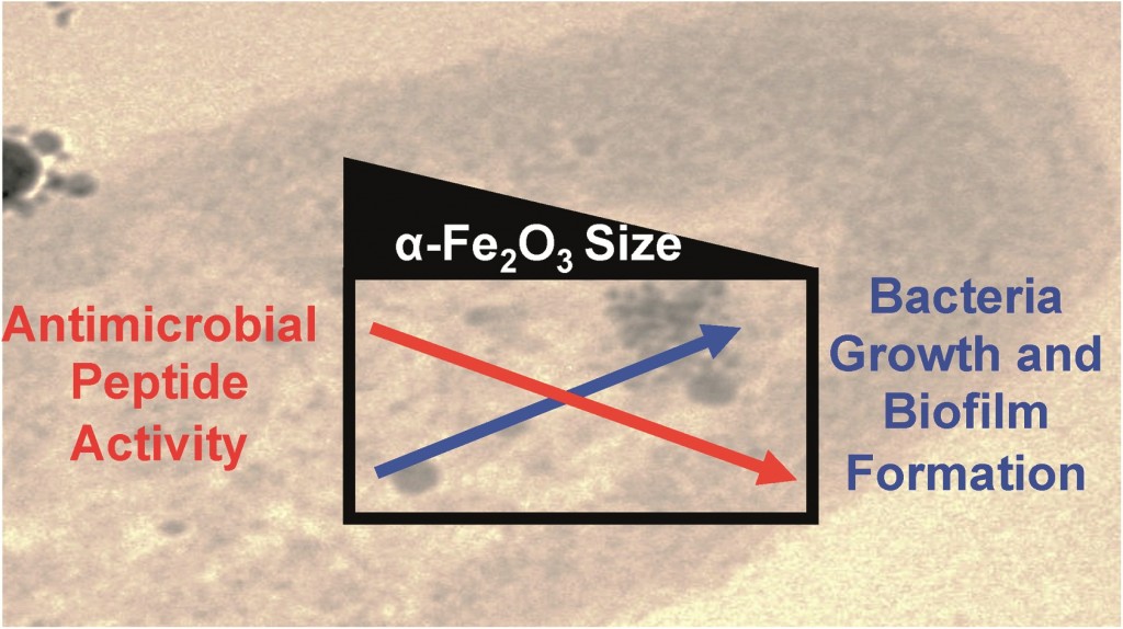
  1. Pseudomonas aeruginosa (PA01), a known pathogen to humans, animals and plants, was exposed to iron oxide nanoparticles of different size ranges. These results showed that the smallest particles induced the greatest amount of growth.
  2. Biofilms were grown in the presence of iron oxide particles and aluminium oxide particles of similar sizes. These results showed that biofilm formation was increased more in the presence of iron oxide particles than in the presence of aluminium oxide particles.
  3. The effects of iron-containing particles on AMP activity was determined by incubating physiologically relevant concentration of AMPs and iron oxide particles, of different sizes, and testing the effects. It was shown that the smaller iron-containing particles provided the greatest amount of bioavailable iron and inhibited AMP activity the most.

This study showed that the smaller the particle, the larger the effect for bacterial growth, biofilm formation and AMP function impairment. This is because the smaller particles have large surface areas and increased dissolution. The research has demonstrated that iron oxide nanoparticles provide a source of bioavailable iron and play an important role in bacterial growth.

Download the full article for free* by clicking the link below:

Iron Oxide Nanoparticles Induce Pseudomonas Aeruginosa Growth and Inhibit Antimicrobial Peptide Function

Jonas Baltrusaitis, Jennifer Borcherding, Haihan Chen, Larissa Stebounova, Chia-Ming Wu, Gayan Rubasinghege, Imali Mudunkotuwa, Juan Carballo, Joseph Zabner, Vicki Grassian and Alejandro Comellas

DOI: 10.1039/C3EN00029J

*Access is free through a registered RSC account – click here to register

Digg This
Reddit This
Stumble Now!
Share on Facebook
Bookmark this on Delicious
Share on LinkedIn
Bookmark this on Technorati
Post on Twitter
Google Buzz (aka. Google Reader)

BiOX photocatalysts: a bright future for environmental remediation

The rapid growth of urbanized and industrialized areas across the globe is an ecological concern. The subject of environmental remediation is an increasingly important focus of research, with much work undertaken to discover ways of effectively treating toxic pollutants in an environmentally sound and sustainable manner. A principal development within this field has been the advent of nanomaterial-based photocatalysts for degrading organic pollutants, which provide a potentially rapid, cheap and relatively green alternative to conventional physical (e.g. adsoption, ultrafiltration) and chemical (UV radiation, H2O2 oxidation) methods of treating and removing  pollutants.

BiOX (X= Cl, Br and I) materials are an emerging group of high performance semi-conducting nanomaterials for this purpose thanks to their desirable optical properties. Much research has focussed on the photocatalytic behaviour and performance of these materials under sunlight irradiation. This critical review by Liqun Ye and co-workers at Nanyang Normal University in China provides an overview of the recent developments in the research of heterogeneous chemistry and photochemistry of BiOX, relevant to their use in the photocatalytic removal of contaminants from air and water.

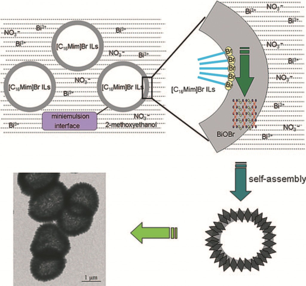
The review provides a concise description of key structural and optical properties that allow BiOX to act as efficient photocatalyts, outlines the different synthetic methods used for the production of these materials and describes how these different synthetic routes influence the morphology and photocatalytic activity of the different BiOX produced.  The synthetic formation mechanisms are also described and depicted.

Additionally, the review describes the photocatalytic mechanisms involved in the treatment of a wide range of pollutants (including VOCs, dyes, alcohols, heavy metals, bacteria NOx and PVC). This includes discussions of photocatalytic activity, selectivity and stability, as well as providing detailed pollutant degradation pathways and description of intermediate species.

While BiOX displays excellent catalytic behaviour, modifications are still required to optimise their practical application for remediation under natural solar irradiation. These modification methods (e.g. the use of cocatalysts, doping, coupling, dye sensitization, graphene, defects, surface plasmon resonance and solid solutions) are discussed in detail. The review also includes a discussion of different BiOX facet effects and describes methods of facet confirmation within these crystalline structures.

This paper is comprehensive review of the current understanding within an innovative field of research that has the potential to improve environmental safety and protection. Furthermore, the authors provide a guideline for the most efficient production of highly active BiOX photocatalysts and outline key uncertainties and questions remaining in this field, indicating potential directions for future research in this area, in both practical and theoretical perspectives.

To access the full review download your free* copy by following the link below:

Recent Advances on BiOX (X = Cl, Br and I) Photocatalysts: Synthesis, Modification, Facet Effect and Mechanisms
Liqun Ye, Yurong Su, Xiaoli Jin and Haiquan Xie
DOI: 10.1039/C3EN00098B

*Access is free through a registered RSC account – click here to register

Digg This
Reddit This
Stumble Now!
Share on Facebook
Bookmark this on Delicious
Share on LinkedIn
Bookmark this on Technorati
Post on Twitter
Google Buzz (aka. Google Reader)