Archive for April, 2014

Mass Spectrometry Proteomics Characterize Nanoparticle Protein Corona

The adventitious protein adsorption on to nanoparticles (NPs), commonly known as the NP-protein corona is in the limelight for many good reasons including its influence on the NP uptake, accumulation and ultimate cellular fate. The consequences of this phenomenon can work in different ways. NP-protein corona can lower the toxicity of NPs when compared to the “bare” NPs but at the same time facilitate crossing biological barriers that can trigger activation of specific regulatory pathways. The particle size, composition and surface properties of NPs are known to influence the composition of the NP-protein corona. Thus, Korin Wheeler, from Santa Clara University, and coworkers investigate the protein populations across the NP-protein corona using silver nanoparticles (AgNPs) in the presence of Yeast (Saccharomyces cerevisiae) proteins (YPE) with the aid of mass spectrometry (MS) proteomics.”

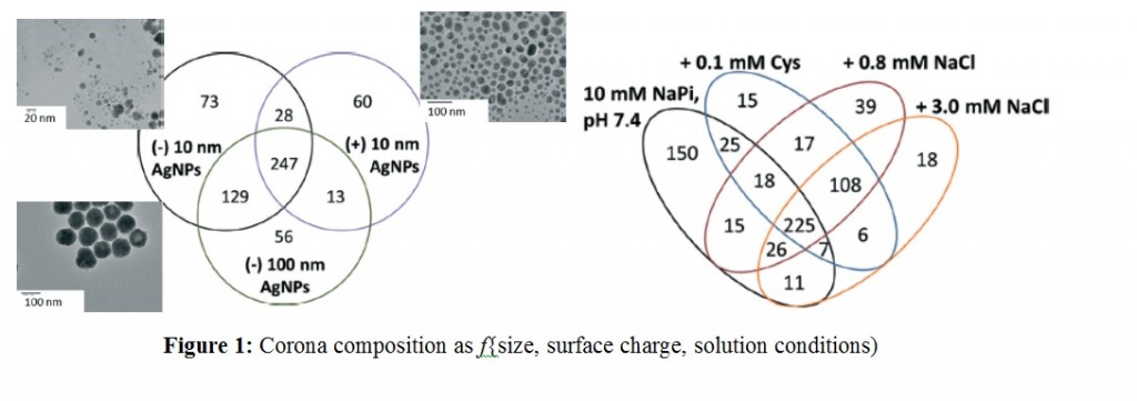
Experiments were designed to probe the effects of AgNP size, surface charge and solution conditions. Two different sizes of AgNPs (10 nm vs 100 nm) with anionic (citrate) and cationic (branched polyethyleneimmine – BPEI) coatings were selected. Protein corona formation was conducted under 0.8 mM NaCl, modeled after freshwater salinity; 3.0 mM NaCl, mimicking mitochondrial salinity; and 0.1 mM Cys, which was shown to mediate NP toxicity. After incubating AgNPs with the YPE, bound and unbound proteins were separated via centrifugation, digested with trypsin followed by LC-MS/MS analysis.

Over 500 different proteins were identified as bound and unbound but no trends were reported linking the molecular weight, pI, length, or the amino acid composition of proteins to their enrichment in the corona. However, AgNP surface charge played a stronger role with those having similar coatings sharing majority of corona population. This highlighted electrostatic modulation of protein affinity for AgNPs under low ionic strength conditions. Further studies with NaCl and cys to simulate more environmentally and biologically relevant conditions revealed more changes in the protein corona populations and modifications in their solution phase behavior in terms of aggregation and sedimentation. However, under all the variable factors there is a population of ubiquitous proteins that get enriched in the corona as shown in the Figure below.

The findings of this research is expected to contribute towards better understanding the bio physicochemical factors mediating NP-corona formation, their characterization and the development of predictive models within the environment.

To access the full article, download a copy for free* by clicking the link below.

Silver nanoparticle protein corona composition compared across engineered particle properties and environmentally relevant reaction conditions

Richard Eigenheer, Erick R. Castellanos, Meagan Y. Nakamoto, Kyle T. Gerner, Alyssa M. Lampe and Korin E. Wheeler
DOI: 10.1039/C4EN00002A

*Access is free through a registered RSC account – click here to register

Digg This
Reddit This
Stumble Now!
Share on Facebook
Bookmark this on Delicious
Share on LinkedIn
Bookmark this on Technorati
Post on Twitter
Google Buzz (aka. Google Reader)

Tracing Silver Nanoparticles

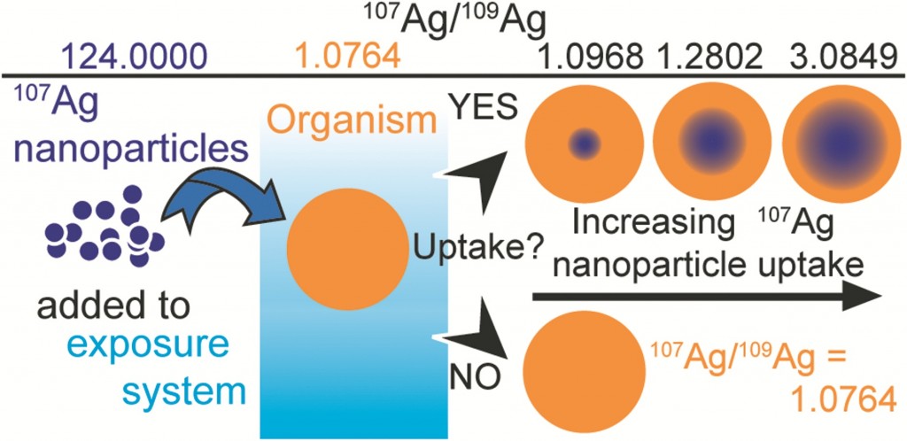
To understand the fate of silver nanoparticles (AgNPs) in the environment they need to be traced in complex natural samples. Often, studies that interrogate the environmental fate of AgNPs require AgNP concentrations that exceed realistic environmental levels. Potentially, this produces unreliable results, as high particle concentration could induce and identify distinct patterns of particle behaviour that is not relevant for ‘realistic’ environmental concentrations.  Adam Laycock, from Imperial College London, and colleagues investigate whether the same effects are also observed at low, environmentally relevant AgNP concentrations.

Stable isotope labelling is an attractive method of labelling which relies on the detection of changes when a contaminant is introduced to a system. Previous methods of tracing nanomaterials have had significant drawbacks. For example, fluorescent coatings can affect the surface chemistry of the nanomaterials causing dissociation. Radiolabelling, another technique used, involves the use of specialist equipment and licenses are required for the handling of radioactive material. Recent studies have shown that stable isotope tracing can be applied to nanomaterials – but first, the labelled NPs must be specifically prepared from a single, highly enriched stable isotope form of the elements.

In this study existing protocols were examined to develop techniques for the optimized preparation of isotopically labelled AgNPs. Three protocols were applied to produce particles with a variety of target sizes, with enriched 107Ag and natural Ag. The results show that the methods are suitable for small scale synthesis of stable labeled AgNPs at yields of approximately 80%. The labelling process does not generate unusual particle properties, demonstrating that isotopically modified AgNPs are equivalent to AgNPs with a natural isotope composition.

The authors finalise their study by presenting a series of calculations which reveal that stable isotope labelling can increase the detection sensitivity of AgNPS by at least a factor of 40, and possibly by up to 4000x in comparison to commonly employed bulk Ag concentration measurements. This approach of tracing nanomaterials is highly versatile, the label cannot be lost by dissociation or degradation, the element remains traceable and the use of enriched stable isotopes provides an extremely selective and sensitive means of elemental tracing, even in the presence  of high natural background levels.

To read the full experiment for free*, download the paper now:

Synthesis and characterization of isotopically labeled silver nanoparticles for tracing studies
Adam Laycock, Björn Stolpe, Isabella Römer, Agnieszka Dybowska, Eugenia (Éva) Valsami-Jones, Jamie R Lead and Mark Rehkamper
DOI: 10.1039/C3EN00100H

*Access is free through a registered RSC account – click here to register

Digg This
Reddit This
Stumble Now!
Share on Facebook
Bookmark this on Delicious
Share on LinkedIn
Bookmark this on Technorati
Post on Twitter
Google Buzz (aka. Google Reader)

Water flea’s assist in the mission to develop sustainable nanomaterials

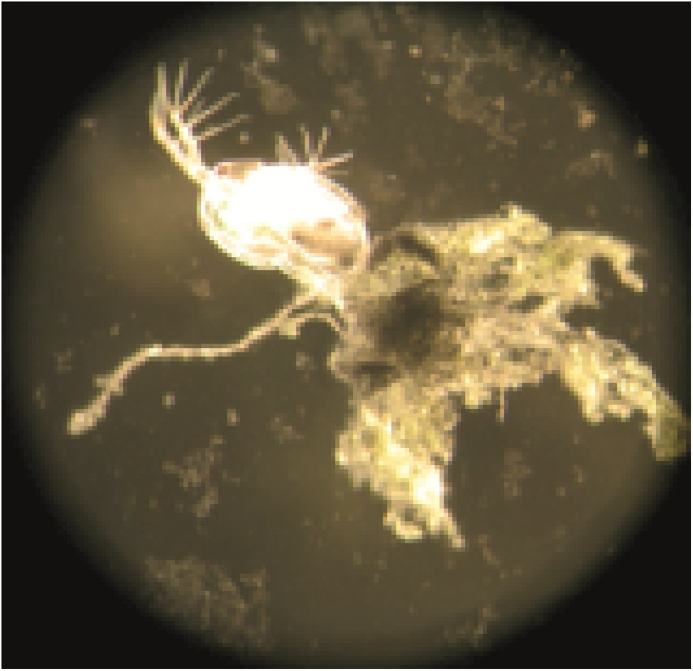
Researchers are continuously on the lookout for materials which are more environmentally friendly and sustainable in order to improve emerging technologies. Nanoparticles (NPs) are the basis for a variety of emerging technologies used for industrial, biomedical and environmental applications, but their release into the environment is still a cause for concern. What if a nanomaterial, that has minimal negative environmental impact, was available? Jared Bozich from the University of Wisconsin and colleagues have published a research paper demonstrating that surface chemistry has the potential to increase or decrease negative biological impacts of NPs.

The surfaces of NPs are typically modified with surface functional groups that control properties such as stability. In this research, the acute and chronic toxicity of well characterized gold nanoparticles (AuNPs), functionalized with ligands of differing charges were investigated in Daphnia magna. D. magna (more commonly called water fleas) are widely accepted as a model organism for assessing the toxicity of environmental contaminates and experience reduced reproduction, growth and increased mortality with exposure to toxic substances. D.magna were exposed to concentrations of four types of functionalized AuNps.

The results showed that initial particle charge significantly impacted overall toxicity, with positively charged particles being more toxic than their negatively-charged counterparts. This could be explained by the increased cellular uptake of positively charged particles due to their high levels of attraction with cellular membrane. This creates a hole in the membrane due to the densely populated charge on the NP surface – allowing the particle to enter the intracellular matrix and continue to cause damage. Another interesting result of this study was that the smaller the particle, the higher the toxicity. This can be explained by a similar theory; after cellular uptake the smaller particles are able to cross the gut lumen of the daphnis, potentially further interacting with D. magna cells and causing damage.

The results of this study identify mechanisms for AuNP toxicity by examining NP toxicity with different charges, using an environmental relevant organism. NPs have the potential to be highly beneficial to society, but in order to minimize the environmental implications the mechanisms that govern the toxicity of NPs need to be betters elucidated. This study demonstrates how the charge and identity of a ligand can influence AuNp toxicity.

To read the full article, download your free* copy now:

Surface chemistry, charge and ligand type impact the toxicity of gold nanoparticles to Daphnia magna

Jared Bozich, Samuel E Lohse, Marco D Torelli, Catherine Murphy, Robert Hamers and Rebecca Klaper

DOI: 10.1039/C4EN00006D

*Access is free through a registered RSC account – click here to register
Digg This
Reddit This
Stumble Now!
Share on Facebook
Bookmark this on Delicious
Share on LinkedIn
Bookmark this on Technorati
Post on Twitter
Google Buzz (aka. Google Reader)