Professor Mike Ashfold picks out his Editor’s Choice on photochemistry and spectroscopy

Mike AshfoldPCCP Advisory Board members are occasionally invited to select a few personal ‘highlights’ from recent issues of the journal. I received such a call in autumn 2012, and readily agreed to try and pick out ten particularly noteworthy papers in the general areas of photochemistry and spectroscopy.hydrogen bonding in electronically excited states

I had long penciled in the Christmas break as the first realistic opportunity for this task. I have been involved with PCCP in one way or another since its launch in 1999, so I should know better than most just how successful a journal it has now become. But it very soon became apparent that I would need to constrain my selection more tightly than I had originally imagined. PCCP publishes a lot of papers (1843 in 2012, many of which impinge on areas of photochemistry and spectroscopy, and many of which certainly merit very careful study. So, my first self-imposed (and completely arbitrary) constraint was that only papers carrying a (print) publication date of 2012 would be eligible for inclusion in my ‘Editor’s Choice’ list. This still left the challenge of somehow picking out less than one in every hundred papers!

The next obvious challenge was that Vol. 14 contains several themed issues, two of which – on Ultrafast chemical dynamics and on Hydrogen bonding in electronically excited states – could easily provide more than ten personal highlights.  In both cases, the guest editors did an exemplary job securing many new, high quality articles. I thoroughly recommend both of these themed issues to all who share my scientific interests, and claim writer’s prerogative by counting each as just one of my ten! I applied one more filter, and excluded from consideration the several excellent papers published in PCCP by Bristol colleagues during 2012.

The 2012 collection includes many notable articles offering new insights into photo-induced processes (e.g. photofragmentations, studies of intramolecular energy flow, photoionization, etc) and into the dynamics of bimolecular encounters – some with unprecedented definition of the initial (reactant) and final (product) quantum states. From the many excellent gas phase studies, I eventually selected:

M.P. Grubb, M.L. Warter and S.W. North, ‘Stereodynamics of multistate roaming’, which reported clear correlations between the velocity vector v and the angular momentum vector j of the NO fragments from NO3 photolysis, implying that the final intra-molecular abstraction in this roaming ‘reaction’ is constrained to occur at planar geometries;

C. Logé and U. Boesl, ‘Laser mass spectrometry with circularly polarized light: two-photon circular dichroism’ demonstrating two-photon circular dichroism of isolated gas phase molecules (cyclopentadienone) by multiphoton ionization measurements;

and the perspective article C.D. Lin and Junliang Xu, ‘Imaging ultrafast dynamics of molecules with laser-induced electron diffraction’, as a new route to imaging the ultrafast dynamics of small molecules with sub-Å spatial and a few-fsec temporal resolution.

My personal interests are now evolving to include photo-induced processes in solution, with particular emphasis on studies that reveal dynamical information about such processes. Obviously, there already exists a very rich and extensive literature pertaining to this field – which I am working hard to absorb – but the 2012 issues of PCCP provide many further articles that I have now come to regard almost as essential reading.  The following five complete my ‘Editor’s Choice’ list:

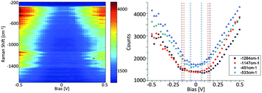
B. Abel, U. Buck, A. L. Sobolewski and W. Domcke, ‘On the nature and signatures of the solvated electron in water’ – a perspective article that reviews recent measurements of the binding energy of hydrated electrons by liquid-jet photoelectron spectroscopy, and considers their implications for our understanding of electron solvation in aqueous environments;

R.R. Frontiera, C. Fang, J. Dasgupta and R.A. Mathies, ‘Probing structural evolution along multidimensional reaction coordinates with femtosecond stimulated Raman spectroscopy’. Another excellent perspective article, in which the authors demonstrate how the exquisite time and frequency resolution afforded by fsec stimulated Raman spectroscopy has allowed them to follow structural evolutions in an isomerization, and in selected electron and proton transfer reactions;

O. Braem, T.J. Penfold, A. Cannizzo and M. Chergui, ‘A femtosecond fluorescence study of vibrational relaxation and cooling dynamics of UV dyes’– that reports a fsec broad-band fluorescence up-conversion study that explores details of the (contrasting) vibrational relaxation dynamics of two UV chromophores;

K.M. Lange, A. Kothe and E.F. Aziz, ‘Chemistry in solution: recent techniques and applications using soft X-ray spectroscopy’.  Another perspective article, this one highlighting some of the new opportunities for studying the structure and dynamics of chemical and biochemical systems in solution offered by time-resolved soft X-ray absorption and emission spectroscopies;

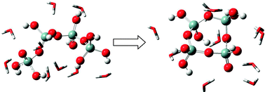
F. Zamponi, J. Stingl, M. Woerner and T. Elsaesser, ‘Ultrafast soft-mode driven charge relocation in an ionic crystal’  – a short paper that illustrates some of the exciting structural and dynamical insights offered by the latest generation fsec X-ray laser pulses in powder diffraction measurements of an ionic solid (potassium dihydrogen phosphate in this case).

Read Professor Ashfold’s Editor’s choice selection for free for a limited period:

Stereodynamics of multistate roaming
Michael P. Grubb, Michelle L. Warter and Simon W. North
DOI: 10.1039/C2CP40235A

Laser mass spectrometry with circularly polarized light: two-photon circular dichroism
Christoph Logé and Ulrich Boesl
DOI: 10.1039/C2CP41405H

Imaging ultrafast dynamics of molecules with laser-induced electron diffraction
C. D. Lin and Junliang Xu
DOI: 10.1039/C2CP41606A

On the nature and signatures of the solvated electron in water
B. Abel, U. Buck, A. L. Sobolewski and W. Domcke
DOI: 10.1039/C1CP21803D

Probing structural evolution along multidimensional reaction coordinates with femtosecond stimulated Raman spectroscopy
Renee R. Frontiera, Chong Fang, Jyotishman Dasgupta and Richard A. Mathies
DOI: 10.1039/C1CP22767J

Chemistry in solution: recent techniques and applications using soft X-ray spectroscopy
Kathrin M. Lange, Alexander Kothe and Emad F. Aziz
DOI: 10.1039/C2CP24028A

A femtosecond fluorescence study of vibrational relaxation and cooling dynamics of UV dyes
Olivier Braem, Thomas J. Penfold, Andrea Cannizzo and Majed Chergui
DOI: 10.1039/C2CP23167K

Ultrafast soft-mode driven charge relocation in an ionic crystal
F. Zamponi, J. Stingl, M. Woerner and T. Elsaesser
DOI: 10.1039/C2CP24072F

And don’t forget to check out his themed issue highlights:

Ultrafast chemical dynamics 
Guest Editors: Klaas Wynne (University of Glasgow, UK) and Neil T. Hunt (University of Strathclyde, UK).

Hydrogen bonding in electronically excited states 
Guest Editors: Guang-Jiu Zhao and Ke-Li Han (Dalian Institute of Chemical Physics, China)

Digg This
Reddit This
Stumble Now!
Share on Facebook
Bookmark this on Delicious
Share on LinkedIn
Bookmark this on Technorati
Post on Twitter
Google Buzz (aka. Google Reader)

Surface-Enhanced Raman Spectroscopy – how much do we understand?

Despite 35 years of research and over 10 000 publications on surface-enhanced Raman spectroscopy (SERS), a universal agreement regarding the physical mechanism responsible for the effect still appears to be elusive. Is SERS in actual fact better understood than the impression given by a number of tenacious misconceptions? Professor Martin Moskovits believes so.

surface enhanced Raman spectroscopyIn his recent PCCP Perspective, Moskovits discusses several important topics in SERS, which are often subject to dispute or contradictions in the current literature, including its physical origin. He explains plasmonic theory, the mechanism that underlies the current understanding, and reasons that alternatively proposed mechanisms, such as chemical enhancement, have not only impeded the establishment of a universally accepted understanding, but have hindered the use of SERS in applications such ultrasensitive chemical and biological sensors.

SERS is a surface-sensitive technique that enhances Raman signals from Raman-active analyte molecules absorbed onto specially prepared metal surfaces.  SERS is sufficiently sensitive to allow the detection of single molecules.

SERS theory, as it is commonly applied, is a classical theory largely based on electrostatics. To obtain a more complete understanding of SERS, a theory based on quantum mechanics, and approached as a dynamical theory, needs developing, Moskovits explains in his review. He suggests that in the meantime, researchers should deal with the surface chemistry that arises in a given experiment on a molecule-by-molecule basis.

Read the details in this HOT PCCP Perspective today:

Persistent misconceptions regarding SERS
Martin Moskovits
DOI: 10.1039/C2CP44030J

If you enjoyed this perspective, you may also be interested in our web collection on SERS.

Digg This
Reddit This
Stumble Now!
Share on Facebook
Bookmark this on Delicious
Share on LinkedIn
Bookmark this on Technorati
Post on Twitter
Google Buzz (aka. Google Reader)

Coherent transient spectroscopy with continuous wave quantum cascade lasers

Rapid passage effects have been demonstrated in a Lamb dip experiment for the first time, by researchers from Oxford, UK.

Grant Ritchie’s team conducted Lamb dip spectroscopy on a low pressure sample of NO using a narrow-linewidth, high-power, single-mode quantum cascade laser. They were able to determine the laser linewidth, and that the power was sufficient to induce significant population transfer of up to 35%, from the widths of the Lamb dips. The population transfer efficiencies could be controlled by the laser chirp rate.

The lamb dip signals became increasingly asymmetric as the chirp rate was increased. At sufficiently high chirp rates rapid passage oscillations on the Lamb dips were observed due to the narrow laser linewidth, and were seen to be affected by the hyperfine splitting in the transition.

Furthermore, the team showed that it was possible to separate the asymmetry effect from the rapid passage oscillations by pumping a single velocity group using a second laser at a fixed frequency.

Read this HOT article today:

Coherent transient spectroscopy with continuous wave quantum cascade lasers
James M. R. Kirkbride, Sarah K. Causier, Elin A. McCormack, Damien Weidmann and Grant A. D. Ritchie
DOI: 10.1039/C2CP44116K

Digg This
Reddit This
Stumble Now!
Share on Facebook
Bookmark this on Delicious
Share on LinkedIn
Bookmark this on Technorati
Post on Twitter
Google Buzz (aka. Google Reader)

Simply a diamino–acridinium, yet ever so complicated

Thomas Just Sørensen is a guest web-writer for PCCP. He is currently a post-doctoral researcher at the University of Copenhagen, Denmark.

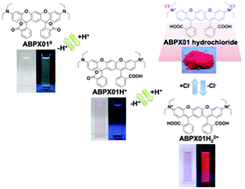
The commercial ATTO 465 dye, based on 9-alkylated proflavine, appears ever so simple: a 2,7-diamino-9-alkyl-acridinium fluorophore with a relatively remote carboxylate group. Even so Arden-Jacob and co-workers find that the photophysical behaviour is far from straightforward. This despite the fact that the cousin: ATTO 495 with the 2,7-bis(dialkylamino)-9-alkyl-acridinium dye, is perfectly simple.

ATTO 465 is interesting. As dye it is not spectacular: it absorbs light at roughly a fifth of the best possible and emits around half of the number of photons it absorbs. Nothing unusual here, but the shape of the spectra, the way the dye changes depending on which solvent that surround it, and mechanisms it uses to dissipate energy; that is unusual. And apparently the explanation lies with the hydrogen atoms on the peripheral amino groups?

A dye can be reduced to a group of electrons moving in a box of nanometer dimensions: a small antenna. The macroscopic counter-piece is a radio antenna, which contains a number of electrons that move in the volume of the antenna. If the dye is changes, the interaction between the nano-antenna and light changes. It can be a variation in colour or a difference in the amount of light emitted. ATTO 465 is a box where both the shape and the number of electrons can change. By bonding hydrogen to the amino groups electrons are removed, the bonding also twists the amino groups resulting in a differently shaped box. Unfortunately the ‘twisting’ is not discreet and many different shapes will be present at all times, as long as there are protons (hydrogen atoms) in the solution that surrounds ATTO 465. The result is the complex photophysical behaviour observed and discussed by Arden-Jacob and co-workers.

Much more detail can be found in the full PCCP article:

Ultrafast photoinduced dynamics of the 3,6-diaminoacridinium derivative ATTO 465 in solution

Jutta Arden-Jacob, Karl-Heinz Drexhage, Sergey I. Druzhinin, Maria Ekimova, Oliver Flender, Thomas Lenzer, Kawon Oum and Mirko Scholz
DOI: 10.1039/C2CP43493H

by Dr Thomas Just Sørensen

Digg This
Reddit This
Stumble Now!
Share on Facebook
Bookmark this on Delicious
Share on LinkedIn
Bookmark this on Technorati
Post on Twitter
Google Buzz (aka. Google Reader)

Novel SERS substrate allows fast cancer cell imaging

Fluorescence imaging is typically used to study biomolecules with high sensitivity and resolution, but many biological molecules are fluorescent themselves, limiting the potential of this technique. On the other hand, surface enhanced Raman spectroscopy (SERS) is capable of high sensitivity but is not inhibited by the problems of autofluorescence.

Folate receptors (FRs) are known to be overexpressed in many cancer cells and scientists in China have taken advantage of this, making graphene oxide–silver nanoparticle composites functionalised with folic acid (which can bind to the FRs). These were introduced into cancer cells and the localisation was then visualised using SERS mapping (the graphene oxide signal is very strong and can be used as an identifier of the composite). Only the FR-positive cells show a signal on the SERS map.

The method is very fast with an integration time of 0.06s per pixel.

Read the full details of this HOT PCCP article today:

Graphene oxide based surface-enhanced Raman scattering probes for cancer cell imaging
Zhiming Liu, Zhouyi Guo, Huiqing Zhong, Xiaochu Qin, Mingming Wan and Biwen Yang
DOI: 10.1039/C2CP43715E

Digg This
Reddit This
Stumble Now!
Share on Facebook
Bookmark this on Delicious
Share on LinkedIn
Bookmark this on Technorati
Post on Twitter
Google Buzz (aka. Google Reader)

Grabbing attention: Graphene oxide and its ability to remove radioactive waste

In a recent PCCP paper, scientists have shown that graphene oxide has a surprising ability to quickly remove radioactive material from contaminated water.

This exciting article, by James Tour, Stepan Kalmykov and co-workers, has been featured in Futurity, UPI.com and Business Standard.

They found that atom-thick flakes of graphene oxide can bind quickly to natural and human-made radionuclides and condense them into solids for easy removal from contaminated water. The flakes are soluble in liquids and easily produced in bulk. 

Although detection of radionuceotide contamination is relatively simple, their removal from aqueous media is significantly challenging and costly. It is hoped that the findings from this collaborative effort from researchers at Rice University and Lomonosov Moscow State could be put into use in the cleanup of contaminated sites like the Fukushima nuclear plants damaged by the 2011 earthquake and tsunami.

Read this HOT PCCP article today: 

Graphene oxide for effective radionuclide removal
Anna Yu. Romanchuk, Alexander S. Slesarev, Stepan N. Kalmykov, Dmitry V. Kosynkin and James M. Tour
DOI: 10.1039/C2CP44593J

Digg This
Reddit This
Stumble Now!
Share on Facebook
Bookmark this on Delicious
Share on LinkedIn
Bookmark this on Technorati
Post on Twitter
Google Buzz (aka. Google Reader)

Handedness in the solid state

Thomas Just Sørensen is a guest web-writer for PCCP. He is currently a post-doctoral researcher at the University of Copenhagen, Denmark.

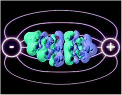
Chiral structures interact with circularly polarised light. Gennaro Pescitelli and co-workers have investigated the effects of the close contact between handed molecules in micro-crystals in their interaction with light. The well-rounded study explains—as promised–the specific case, but does it take us closer to a general structure-property relationship regarding the preferential absorption of one handedness of light? And is such as a correlation even possible?

Table of contents imageIn ”Intermolecular exciton coupling and vibronic effects in solid-state circular dichroism: a case study” by Gennaro Pescitelli, Daniele Padula and Fabrizio Santoro, the finer details of the interaction between circularly polarised light and chiral matter is discussed. In this specific case, homochiral microcrystals are investigated experimentally and theoretically. The results presented are impressive, as the theoretical model fits and explains the experimental observation, but why is this study so important? What is this chirality? And which answers are we looking for?

Chirality and the word chiral derive from the Greek word cheir or hand: chiral can be read as handed and chirality as handedness. The definition of a chiral object is that it cannot be superimposed on its mirror image, just as our hands. Chirality is hugely important as every biological building block that makes up our hands, and indeed the rest of us, is handed. It comes in a left-hand and a right-hand form.

As it is the entire biological machinery on earth is almost exclusively left-handed. How this came about, we do not know, but it is suggested that we, in our part of space, have an abundance of circularly polarised light emitters of a certain handedness, and that this has caused the handedness of our biology. Chirality and circularly polarised light are linked. Each can be described of having a specific handedness and one handedness of light interacts with one handedness of chiral matter. An abundance of right-handed light in our part of the universe may have induced the single handedness of our biology.

In order to figure out how the homo-chirality of the biosphere on earth arose, we need to understand how circularly polarised light and chiral material interacts; exactly what Gennaro Pescitelli and co-workers are investigating. Studies like this, where experiment and highly advanced theory is used to correlate observations and the chiral structure of the material, are needed if we are to understand how chirality in a molecular framework absorbs one specific handedness of light.

We understand how regular material (not handed) and normal light (linearly polarised) interact, and how to design materials that interact with light in a desired fashion. We cannot yet design materials that absorb a specific handedness of light. The paper  ”Intermolecular exciton coupling and vibronic effects in solid-state circular dichroism: a case study” by Gennaro Pescitelli, Daniele Padula and Fabrizio Santoro takes us one step closer, it was published in of Physical Chemistry Chemical Physics 2013, 15, 795-802.

by Dr Thomas Just Sørensen

Read this fascinating study today:

Intermolecular exciton coupling and vibronic effects in solid-state circular dichroism: a case study
Gennaro Pescitelli, Daniele Padula and Fabrizio Santoro
DOI: 10.1039/C2CP43660D

Digg This
Reddit This
Stumble Now!
Share on Facebook
Bookmark this on Delicious
Share on LinkedIn
Bookmark this on Technorati
Post on Twitter
Google Buzz (aka. Google Reader)

Predicting properties of ionic liquids

It is now possible to accurately predict the density, refractive index and molar polarisability of any imidazolium-based ionic liquid thanks to the recent work of Schröder et al.

Given the availability of at least one million simple ionic liquids, predicting which ionic liquid is best suited to a given application is a significant task. Molar polarisability is a key factor in describing solvation effects and, in principle, can be determined by various quantum-mechanical methods. However, these methods are Polarisabilities of ionic liquidstime-consuming and can only be applied to a small subset of ionic liquids.

Schroder et al. used a Designed Regression Analysis to deconstruct the molar polarisability and molar volume into atomic contributions in this recent PCCP article. They used their approach to explore how the refractive indices of various imidazolium-based ionic liquids were influenced by the length of the alkyl chains.

Read the full PCCP article today:

Polarisabilities of alkylimidazolium ionic liquids
Christian Schröder, Katharina Bica, Maggel Deetlefs and Kenneth R. Seddon
DOI: 10.1039/C3CP43867H

Digg This
Reddit This
Stumble Now!
Share on Facebook
Bookmark this on Delicious
Share on LinkedIn
Bookmark this on Technorati
Post on Twitter
Google Buzz (aka. Google Reader)

Micro-structure of solvents

Thomas Just Sørensen is a guest web-writer for PCCP. He is currently a post-doctoral researcher at the University of Copenhagen, Denmark.

We know much about the energetics in solutions and of species in solution. We can directly measure most parameters, when we measure energy. When it comes to the structure of solution or in solutions, we know next to nothing. Indirect measurements are our main source of information, as we cannot see the fluctuating solution structures. Miyuki Tanaka and co-workers have performed one of the most informative studies using indirect methods to probe solvent structures that I have come across.

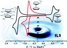
In a recent paper in PCCP by Miyuki Tanaka, Tomoaki Yago and Masanobu Wakasa the microscopic structure of ionic liquid solvents is investigated. The diffusion of single particles is compared to the macroscopic measure of viscosity. They find that the ionic liquids are ‘more sticky’, when looking at the diffusion of single molecules, than more commonly used solvents; although the viscosities of the two solvents are identical.

In “Local structure of ionic liquids probed by self-quenching of thiobenzophenone” the movement of single microscopic particles in solution (molecular diffusion), are compared to measurement determining how difficult a macroscopic object moves in the same solution (the viscosity). A single particle is excited using light and gains energy. The energy can be released the by collision with another particle in the solution.  By following the evolution of the population of excited particles the molecular movements can be followed.

The result presented in this paper is another piece of the puzzle we have to assemble in order to understand the microstructure of solvents, and the conundrum of solvation. Solvents are everywhere, most ubiquitous is water. We have a limited understanding of the structure of pure solvents, and know even less about the structure of complex solutions. The collection of small molecules that constitute most liquids can have an astoundingly complex structure. A structure that we have to know and understand, if we are to comprehend the complex condensed phases such as the cells that make up most living things.

At the most advanced facility for structural investigation of matter, the SLAC national accelerator laboratory, an entire team of elite scientists has been assembled ‘just’ to elucidate the structure of water. The method applied by Tanaka and co-workers is a much simpler route to understanding molecular movements in solution. When a large library of different solvents has been investigated, we will be able to deduce effects of specific solvation. Following this, we may be able to explain exactly why the ionic liquids are ‘more sticky’ than traditional solvents.

Local structure of ionic liquids probed by self-quenching of thiobenzophenone” by Miyuki Tanaka, Tomoaki Yago and Masanobu Wakasa was published in Physical Chemistry Chemical Physics (PCCP) at the beginning of 2013: M. Tanaka, T. Yago, M. Wasaka Phys. Chem. Chem. Phys.2013, 15, 787-794

By Dr Thomas Just Sørensen

Digg This
Reddit This
Stumble Now!
Share on Facebook
Bookmark this on Delicious
Share on LinkedIn
Bookmark this on Technorati
Post on Twitter
Google Buzz (aka. Google Reader)

New PCCP Associate Editor: Dr Ajayaghosh

New PCCP Associate Editor Dr Ayyappanpillai AjayaghoshWe are extremely pleased to annouce that Dr Ayyappanpillai Ajayaghosh is a new Associate Editor for Physical Chemistry Chemical Physics (PCCP). His research interests include supramolecular chemistry, chemosensors, low band-gap polymers, fluorescent gels, organic nanostructures and photoresponsive systems.

Dr Ajayaghosh is a CSIR Outstanding Scientist at the National Institute for Interdisciplinary Science and Technology (NIIST), India. He completed his PhD at the University of Calicut after graduating from Kerala University with a BSc. He has received numerous awards including  the Infosys Prize (2012) for his outstanding contribution in the field of physical sciences, and in particular for his work that has advanced supramolecular chemistry.

Dr Ajayagosh will begin handling papers soon and so we encourage you to submit to his editorial office.

Read Dr Ajayaghosh’s paper in the PCCP themed issue on materials innovation through interfacial physics and chemistry:

Excitation energy migration in oligo(p-phenylenevinylene) based organogels: structure-property relationship and FRET efficiency
Chakkooth Vijayakumar, Vakayil K. Praveen, Kalathil K. Kartha and Ayyappanpillai Ajayaghosh
DOI: 10.1039/C0CP02110E

Digg This
Reddit This
Stumble Now!
Share on Facebook
Bookmark this on Delicious
Share on LinkedIn
Bookmark this on Technorati
Post on Twitter
Google Buzz (aka. Google Reader)