Patterned 3D cornea replacements

Biomaterials Science web writer, Debanti Sengupta, highlights a HOT article from the journal

Hasirci and colleagues have developed a new type of engineered corneal construct using a combination of cells derived from the cornea as well as organized elastin-based biomaterials.  

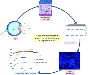
The stroma is a layer in our cornea that consists of highly organized layers of collagen fibers and keratocytes, a special type of corneal cell.  The cells and the collagen layers are aligned with one another and follow a specific pattern of organization.   This organized structure allows the cornea to be transparent and to have the mechanical strength necessary to function correctly.

This study used specific biomaterials that draw inspiration from nature.  Specifically, the researchers used elastin, a commonly found biomaterial, as a template, and created a biomaterial with a similar protein composition. They modified a traditional elastin-based sequence to include the amino acid chain YIGSR, which is known to help cells attach to biomaterial structures.  They blended their elastin-based biomaterial with collagen, and used a silicon template to mold these biomaterials into sheets with surfaces that contain micron-scale grooves or micropatterns that cells can sense and align to. 

The biomaterial sheets were dehydrated completely under vacuum, causing the films to form internal bonds or crosslinks.  The crosslinking confers mechanical stability to the sheets, ensuring that the sheets do not degrade too quickly in response to enzymes secreted by keratocytes.  To test this idea, the biomaterial sheets were then subjected to a collagen-degrading enzyme, collagenase II.  The crosslinked sheets were better able to resist degradation for 24 hours as compared to their non-crosslinked counterparts.  This suggested that the keratocytes could form organized layers on the crosslinked biomaterials before degrading away the original structures. To further test these materials, the biomaterial sheets were then immersed in a saline solution for 4 weeks to study non-enzyme based degradation, and the crosslinked sheets again resisted degradation better than the non-crosslinked sheets. The crosslinked sheets were also able to transmit light, which is crucial for their application as corneal replacements.

Finally, the researchers tested these biomaterial sheets with human corneal keratocytes. The cells were able to survive on the biomaterial and grew well on the micropatterned biomaterial sheets. The cells also followed the alignment of the micropatterns on the surface of the biomaterials. The researchers then placed multiple layers of the micropatterned scaffolds together such that each successive patterned layer was perpendicular to the previous layer, and ‘glued’ the scaffolds together using droplets of collagen.  Cells were then seeded onto each layer using a syringe. They found that the keratocytes remained aligned for a period of 21 days in culture. The material was transparent even with the presence of cells for up to 4 weeks. In fact, the presence of keratocytes contributed to the construct’s overall transparency.

This work is an elegant demonstration of how biomaterials and cells can be combined to produce a viable tissue-engineered construct that may one day be used as corneal replacement therapy.

A collagen-based corneal stroma substitute with micro-designed architecture
Cemile Kilic, Alessandra Girotti, J. Carlos Rodriguez-Cabello and Vasif Hasirci  
Biomater. Sci., 2014, Advance Article DOI: 10.1039/C3BM60194C

Debanti Sengupta recently completed her PhD in Chemistry from Stanford University.  She is currently a Siebel postdoctoral scholar at the University of California, Berkeley.  

Follow the latest journal news on Twitter @BioMaterSci or go to our Facebook page.

To keep up-to-date with all the latest research, sign-up to our RSS feed or Table of contents alert.

Digg This
Reddit This
Stumble Now!
Share on Facebook
Bookmark this on Delicious
Share on LinkedIn
Bookmark this on Technorati
Post on Twitter
Google Buzz (aka. Google Reader)

Honeycomb surfaces for stem cell differentiation

Biomaterials Science web writer, Brian Aguado, highlights a HOT article from the journal

The ability for a stem cell to differentiate into a specific cell type depends on the cell’s surrounding microenvironment. Variables such as substrate stiffness, presence of certain growth factors, fluid flow, and extracellular matrix composition in the cell culture environment all play a role in the cell’s ability to differentiate. Additionally, there is great interest in studying the effects of surface topology on stem cell differentiation in vitro to more accurately mimic the in vivo microenvironment. Using various micro-fabrication technologies, 2D and 3D substrates with varying topographic features including grooves, pits, pores, and posts can be fabricated and tested with cells to evaluate cell response.

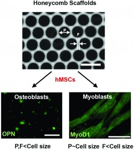
Researchers at Tohoku University in Japan have fabricated “honeycomb” topological substrates to regulate the differentiation of mesenchymal stem cells. The substrates are manufactured using a chloroform solution of polystyrene and an amphiphilic co-polymer, poly(N-dodecyl acrylamide-co-6-acrylamidehexanoic acid), or dioleoylphosphatidylethanolamine (DOPE). The solutions were cast on a glass slide and exposed to a flow of humid air to generate polystyrene films that resemble honeycomb from a beehive. The honeycomb substrates had varying pore size and strut width depending on the chloroform evaporation time.

After obtaining four different honeycomb films with varying pore size and strut width, human mesenchymal stem cells (hMSCs) were seeded on the surfaces to evaluate cell differentiation into osteo-specific (bone) and myo-specific (muscle) cells. To determine the cell differentiation state, cells were stained with osteoblast and myoblast markers, osteopontin and MyoD1, respectively.

Interestingly, when both the pore size and strut width were smaller than hMSC size, cells showed globular morphologies and increased expression of osteopotin (consistent with osteo-specific differentiation). On the other hand, when the pore size was the same size as hMSCs, the cells expressed MyoD1 and had elongated morphologies (consistent with myo-specific differentiation). Given the promising morphological and staining results, further studies are required to fully characterize the molecular mechanisms of the differentiation process.

This study demonstrates that geometries of honeycomb topographical substrates may have a direct influence on stem cell differentiation. Surface topology of biomaterial surfaces must be taken into account for future designs of cell culture environments for stem cell differentiation studies.

Honeycomb-shaped surface topography induces differentiation of human mesenchymal stem cells (hMSCs): uniform porous polymer scaffolds prepared by the breath figure technique
Takahito Kawano, Madoka Sato, Hiroshi Yabu and Masatsugu Shimomura 
Biomater. Sci., 2014, Advance Article DOI: 10.1039/C3BM60195A

Brian Aguado is currently a Ph.D. Candidate and NSF Fellow in the Biomedical Engineering department at Northwestern University. He holds a B.S. degree in Biomechanical Engineering from Stanford University and a M.S. degree in Biomedical Engineering from Northwestern University. When he’s not in the lab, Brian enjoys traveling, cooking, swimming, and spending time with family and friends. Read more about Brian’s research publications here.

To keep up-to-date with all the latest research, sign-up to our RSS feed or Table of contents alert.

Digg This
Reddit This
Stumble Now!
Share on Facebook
Bookmark this on Delicious
Share on LinkedIn
Bookmark this on Technorati
Post on Twitter
Google Buzz (aka. Google Reader)

Bioactive hydrogel beads for iPS cell expansion

Biomaterials Science web writer, Brian Aguado, highlights a HOT article from the journal


Induced pluripotent stem (iPS) cells continue to hold great promise for cell transplantation therapies for numerous debilitating diseases. For instance, a patient’s skin cells can be “re-programmed” into iPS cells and converted into heart, liver, or bone cells (to name a few). These cells can then be used to regenerate specific tissues, and would theoretically bypass the patient’s immune response since the original cells come directly from the patient.

However, the scalability and growth of iPS cell in vitro continues to be a significant challenge. To address this problem, researchers at the University of Tokyo and École Polytechnique Fédérale de Lausanne have engineered bioactive hydrogel beads to significantly increase the expansion of iPS cells in vitro while maintaining their pluripotency.

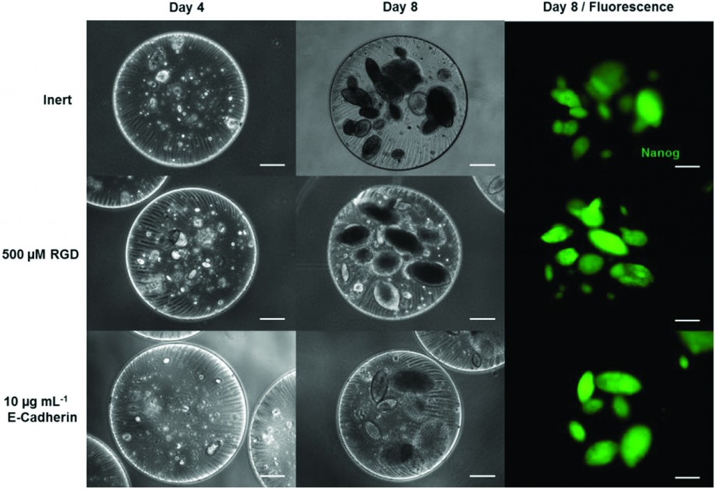
Mouse iPS cells were readily encapsulated and expanded in alginate/polyethylene glycol (PEG) gel beads using an effective three-step protocol.  First, iPS cells were added to a precursor solution of functionalized PEG and alginate. The precursor solution was loaded into a linear extrusion vessel and allowed to drip into a calcium chloride bath to crosslink the alginate and form gel beads. Second, the maleimide-functionalized 4-armed PEG in the beads was further crosslinked with di-thiolated linear PEG using a Michael addition reaction. Finally, the fully-crosslinked beads were coated with poly(L-lysine) to prevent volumetric swelling of the beads, since swelling imparts harmful mechanical stress to the encapsulated cells during the culture process.

Interestingly, the encapsulated iPS cells were pluripotent after 8 days in culture (confirmed with Nanog expression and colony morphology). To make the beads “bioactive,” the PEG networks were functionalized with cell adhesion components (RGD peptide or E-cadherin) to enhance iPS cell expansion. Overall, increasing the concentration of RGD binding sites in the bead offers up to an 80% increase in iPS cell proliferation compared to non-bioactive bead controls.

Additional experiments demonstrate that the expression of pluripotency markers and primitive endoderm markers is a function of bead design. Notably, the pluripotency marker Rex1 was significantly more expressed in beads containing RGD or E-Cadherin compared to non-bioactive beads. Furthermore, endoderm markers such as Gata4 and HNF4 were down regulated in beads containing specific concentrations of RGD. These results support the conclusion that the design of the bioactive bead has direct effects on the pluripotency of encapsulated iPS cells.

Ultimately, bioactive hydrogel beads are a promising solution for increasing iPS cell expansion in vitro, and are well suited for use in future regenerative medicine applications.

Development of bioactive hydrogel capsules for the 3D expansion of pluripotent stem cells in bioreactors
Yoji Tabata, Ikki Horiguchi, Matthias P. Lutolf and Yasuyuki Sakai  
Biomater. Sci., 2014, Advance Article DOI: 10.1039/C3BM60183H

Brian Aguado is currently a Ph.D. Candidate and NSF Fellow in the Biomedical Engineering department at Northwestern University. He holds a B.S. degree in Biomechanical Engineering from Stanford University and a M.S. degree in Biomedical Engineering from Northwestern University. When he’s not in the lab, Brian enjoys traveling, cooking, swimming, and spending time with family and friends. Read more about Brian’s research publications here.

To keep up-to-date with all the latest research, sign-up to our RSS feed or Table of contents alert.

Digg This
Reddit This
Stumble Now!
Share on Facebook
Bookmark this on Delicious
Share on LinkedIn
Bookmark this on Technorati
Post on Twitter
Google Buzz (aka. Google Reader)

Top 10 Most-accessed Biomaterials Science articles – Q3 2013

This month sees the following articles in Biomaterials Science that are in the top ten most accessed from July – September:

Mesoporous silica nanoparticles for the design of smart delivery nanodevices
Montserrat Colilla, Blanca González and María Vallet-Regí
Biomater. Sci., 2013,1, 114-134
DOI: 10.1039/C2BM00085G

Bio-inspired catechol conjugation converts water-insoluble chitosan into a highly water-soluble, adhesive chitosan derivative for hydrogels and LbL assembly
Kyuri Kim, Ji Hyun Ryu, Dong Yun Lee and Haeshin Lee
Biomater. Sci., 2013,1, 783-790
DOI: 10.1039/C3BM00004D

Influence of structure and properties of colloidal biomaterials on cellular uptake and cell functions
Zhengwei Mao, Xiangyan Zhou and Changyou Gao
Biomater. Sci., 2013,1, 896-911
DOI: 10.1039/C3BM00137G

Enzyme responsive materials: design strategies and future developments
Mischa Zelzer, Simon J. Todd, Andrew R. Hirst, Tom O. McDonald and Rein V. Ulijn
Biomater. Sci., 2013,1, 11-39
DOI: 10.1039/C2BM00041E

Preparation and biological characterization of hollow magnetic Fe3O4@C nanoparticles as drug carriers with high drug loading capability, pH-control drug release and MRI properties
Kai Cheng, Zhiyuan Sun, Yumei Zhou, Hao Zhong, Xiangkai Kong, Peng Xia, Zhen Guo and Qianwang Chen
Biomater. Sci., 2013,1, 965-974
DOI: 10.1039/C3BM60087D

Nucleic acid-directed self-assembly of multifunctional gold nanoparticle imaging agents
Ziyan Zhang, Yongjian Liu, Chad Jarreau, Michael J. Welch and John-Stephen A. Taylor
Biomater. Sci., 2013,1, 1055-1064
DOI: 10.1039/C3BM60070J

Enhanced drug delivery to hepatocellular carcinoma with a galactosylated core–shell polyphosphoester nanogel
Juan Wu, Tian-Meng Sun, Xian-Zhu Yang, Jing Zhu, Xiao-Jiao Du, Yan-Dan Yao, Meng-Hua Xiong, Hong-Xia Wang, Yu-Cai Wang and Jun Wang
Biomater. Sci., 2013,1, 1143-1150
DOI: 10.1039/C3BM60099H

Hyaluronan and self-assembling peptides as building blocks to reconstruct the extracellular environment in skin tissue
Daniela S. Ferreira, Alexandra P. Marques, Rui L. Reis and Helena S. Azevedo
Biomater. Sci., 2013,1, 952-964
DOI: 10.1039/C3BM60019J

Fibrinogen nanofibers for guiding endothelial cell behavior
Dencho Gugutkov, Johan Gustavsson, Maria Pau Ginebra and George Altankov
Biomater. Sci., 2013,1, 1065-1073
DOI: 10.1039/C3BM60124B

Traceless protein delivery with an efficient recyclable nanocarrier
Xuanjun Wu, Yunlong Song, Jiahuai Han, Liu Yang and Shoufa Han
Biomater. Sci., 2013,1, 918-923
DOI: 10.1039/C3BM60128E

Why not take a look at the articles today and blog your thoughts and comments below.

Fancy submitting an article to Biomaterials Science? Then why not submit to us today!

Digg This
Reddit This
Stumble Now!
Share on Facebook
Bookmark this on Delicious
Share on LinkedIn
Bookmark this on Technorati
Post on Twitter
Google Buzz (aka. Google Reader)

Creating functional muscle in a dish

Biomaterials Science web writer, Debanti Sengupta, highlights a HOT article from the journal

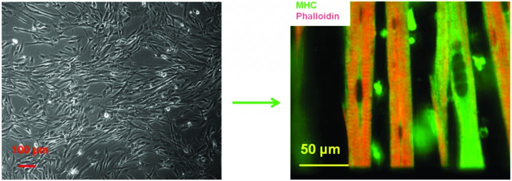
Hickman et al. have developed methods to successfully differentiate human muscle progenitor cells into functional, multinucleated myofibers. 

The first step towards replacing muscle lost due to diseases such as muscular dystrophy is to build functional model systems of healthy muscle tissue.  In order to do this with human cells, human muscle progenitor cells must be removed from the body, cultured in a dish, and treated with appropriate signaling molecules in order to appropriately differentiate into functional muscle fibers, known as myotubes or myofibers.  Hickman et al. have done precisely this – the authors demonstrate that it is possible to differentiate muscle progenitor cells into human myofibers outside of the body.  These myofibers are also shown to contract both in response to external electrical stimulation as well as spontaneously.

The researchers developed a special media cocktail that allowed these myofibers to grow in culture over a period of more than two weeks.  Importantly, the media cocktail did not contain serum, an undefined extract from animal blood that is often used to keep grow cells in culture outside of the body.  This has important implications for eventual clinical translation.  The researchers found that their differentiation medium produced myofibers that contained organized sarcomeres (the internal cell machinery that allows fibers to contract and do work). Muscle contraction generally occurs through the uptake of calcium by the cells from their surroundings.  Calcium uptake requires the presence of two different types of calcium channels, DHPR and RyR.  The researchers generated myofibers that demonstrated the presence of both of these channels.  Finally, these myofibers also contracted on their own, as well as in response to an applied electrical potential. 

This research marks an important step forward in muscle development research, and can be found here.


Spontaneous contraction of human myotubes in culture

In vitro differentiation of functional human skeletal myotubes in a defined system
Xiufang Guo, Keshel Greene, Nesar Akanda, Alec S. T. Smith, Maria Stancescu, Stephen Lambert, Herman Vandenburgh and James J. Hickman  
Biomater. Sci., 2013, Advance Article  DOI: 10.1039/C3BM60166H

Debanti Sengupta recently completed her PhD in Chemistry from Stanford University.  She is currently a Siebel postdoctoral scholar at the University of California, Berkeley.  

Follow the latest journal news on Twitter @BioMaterSci or go to our Facebook page.

To keep up-to-date with all the latest research, sign-up to our RSS feed or Table of contents alert.

Digg This
Reddit This
Stumble Now!
Share on Facebook
Bookmark this on Delicious
Share on LinkedIn
Bookmark this on Technorati
Post on Twitter
Google Buzz (aka. Google Reader)

Better bioceramics for bone generation: the importance of crystal structure

Biomaterials Science web writer, Ellen Tworkoski, highlights a HOT article from the journal

Bioactive ceramics are commonly used for the repair and replacement of damaged bone tissues.  However, there is still a limited understanding of how the inherent material properties of these ceramics can be used to predict their osteoinductive properties.  The development of models which relate these quantities could lead to the more efficient and intelligent design of materials that promote bone formation.

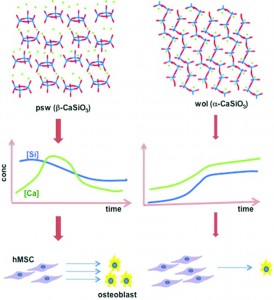
In a study that pushes this goal one step closer to reality, researchers from the University of Wisconsin, Madison, the University of Akron and the University of Michigan investigate the role which crystal structure plays in controlling the attachment and osteogenic differentiation of human mesenchymal stem cells (hMSCs).  The group studied hMSC viability and differentiation on two CaSiO3 polymorphs: pseudowollastonite (psw) and wollastonite (wol).  As polymorphs, these materials have identical chemical compositions and stoichiometries, and can be fabricated to have nearly equivalent surface roughness.  The only significant difference between these two materials is their crystal structure.  While the wol polymorph has a stable, open silicate chain structure, the psw polymorph has a highly strained silicate ring structure. 

Interestingly, significant differences in hMSC growth and differentiation were observed, with the psw polymorph exhibiting superior osteoinductive properties.  Although the hMSCs cultured on psw initially exhibited poor viability, they were able to recover and, after 20 days, exhibited a statistically higher cell count than the hMSCs cultured on wol.  Fine-grained calcium phosphate precipitate developed on the surfaces of both polymorphs but only the psw polymorph exhibited polycrystalline calcite aggregates as well as nodular amorphous precipitates which were identified as CaP, or bone nodules.

The group attributed these differences in hMSC behavior to the different soluble factor release rates exhibited by the two materials.  Psw has a much higher initial dissolution rate than wol, presumably because its highly strained structure makes it more susceptible to hydrolysis.  This resulted in a large initial release of silicon atoms, which caused the initial low cell viability, and calcium atoms, which caused increased calcite and calcium phosphate formation.

Overall, the study proved that psw is more osteoinductive than wol, thereby showing that crystal structure can play an enormous role in determining the osteoconductive and osteoinductive properties of a bioceramic.  The collected data mark a promising step forward in the creation of a model that can successfully predict osteoinductivity based on a bioceramic’s inherent material properties.

Crystal structures of CaSiO3 polymorphs control growth and osteogenic differentiation of human mesenchymal stem cells on bioceramic surfaces
Nianli Zhang, James A. Molenda, Steven Mankoci, Xianfeng Zhou, William L. Murphy and Nita Sahai  
Biomater. Sci., 2013, 1, 1101-1110 DOI: 10.1039/C3BM60034C

Ellen Tworkoski is a guest web-writer for Biomaterials Science.  She is currently a second year Ph.D. student in the biomedical engineering department at Northwestern University (Evanston, IL, USA).

Follow the latest journal news on Twitter @BioMaterSci or go to our Facebook page.

To keep up-to-date with all the latest research, sign-up to our RSS feed or Table of contents alert.

Digg This
Reddit This
Stumble Now!
Share on Facebook
Bookmark this on Delicious
Share on LinkedIn
Bookmark this on Technorati
Post on Twitter
Google Buzz (aka. Google Reader)

Multi-layer collagen sheets for soft tissue engineering

Biomaterials Science web writer, Brian Aguado, highlights a HOT article from the journal

Collagen has been widely used in scaffold fabrication protocols in the tissue-engineering field for decades, since the material is biocompatible and biodegradable. However, the structure of collagen molecules is often disrupted during various processing techniques (e.g. decellularization or electrospinning), which decreases the mechanical stiffness of the material. There is an urgent need to generate mechanically robust materials that integrate with the host tissue for soft tissue engineering applications.

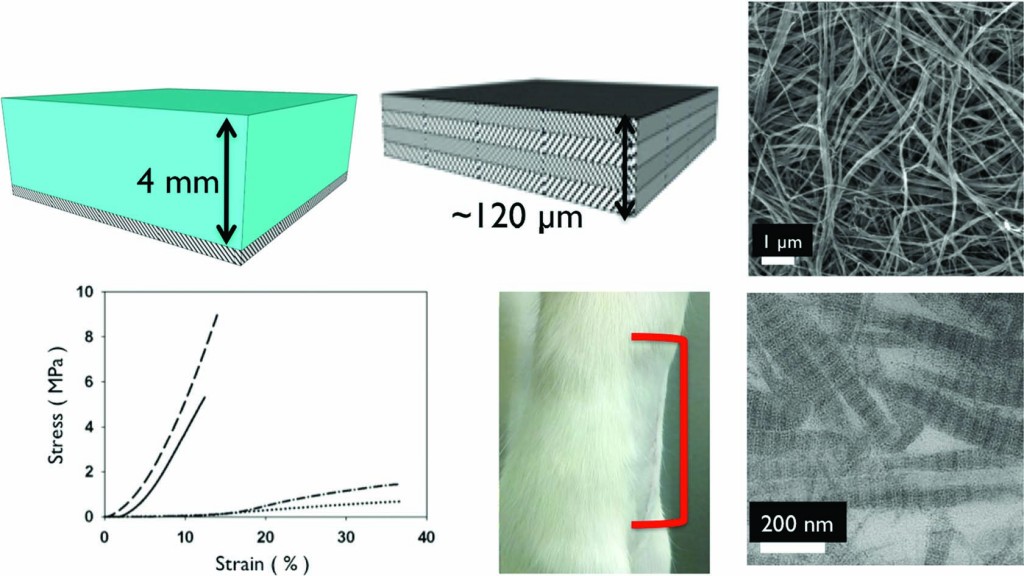
To address this issue, Elliot Chaikof’s group at Harvard Medical School has taken a “bottom-up approach” to fabricate thick collagen mats without damaging the structure of collagen molecules. Collagen gels were cast in a rectangular mold and dried to generate a sheet of collagen approximately 15-40 microns thick. Using a layer-by-layer fabrication strategy, subsequent collagen layers were cast on top of the previous layer and dried to form 120 μm thick sheets. The casting method successfully preserves the collagen molecule microstructure, and allows for tunable mechanical properties over several orders of magnitude as a function of concentration, thickness, and number of layers.

After elegantly characterizing the material properties, the collagen mats were used in an in vivo model of ventral hernia repair. The mats were compared to commercially available PermacolTM collagen sheets currently used for hernia repair. After surgically forming a hernia in the abdomen of rats, the collagen mats were sutured on the abdominal wall. After 3 months, the collagen mats showed significantly enhanced integration at the implant site over the PermacolTM controls.

Vivek Kumar, lead author on the study, explains that the collagen mats were 65% thinner than the 1 mm thick PermacolTM matrices – potentially accounting for the enhanced integration of the mat at the implant site. Even though the collagen mats were thinner than the commercially available collagen sheets, they were able to prevent re-herniation in all the rats tested due to the comparable mechanical properties of the collagen mat to the PermacolTM matrices.

Additionally, the layer-by-layer stacking method could be used to incorporate specific drugs in between the layers (much like making a sandwich) to reduce the risk of re-herniation and stimulate new tissue formation. Kumar also claims that these collagen mats can be used for a wide array of applications, such as vascular tissue replacements, artificial skin, and dura mater replacements.

 Interestingly, Kumar also suggests the method he developed is scalable. “We have cast much larger gels and much smaller gels successfully,” Kumar says. “The fabrication scheme is amenable to a variety of other geometries such as tubes and other intricate mold designs. The simplicity of our approach suggests good potential for use in industry – where often times a simpler fabrication method is better.”

This innovative research shows a simple method to create mechanically robust collagen mats that integrate exceptionally well with the surrounding tissue, and competes with current commercially available materials.

Collagen-based substrates with tunable strength for soft tissue engineering
Vivek A. Kumar, Jeffrey M. Caves, Carolyn A. Haller, Erbin Dai, Liying Liu, Stephanie Grainger and Elliot L. Chaikof 
Biomater. Sci., 2013, 1, 1193-1202 DOI: 10.1039/C3BM60129C

Brian Aguado is currently a Ph.D. Candidate and NSF Fellow in the Biomedical Engineering department at Northwestern University. He holds a B.S. degree in Biomechanical Engineering from Stanford University and a M.S. degree in Biomedical Engineering from Northwestern University. When he’s not in the lab, Brian enjoys traveling, cooking, swimming, and spending time with family and friends. Read more about Brian’s research publications here.

To keep up-to-date with all the latest research, sign-up to our RSS feed or Table of contents alert.
Digg This
Reddit This
Stumble Now!
Share on Facebook
Bookmark this on Delicious
Share on LinkedIn
Bookmark this on Technorati
Post on Twitter
Google Buzz (aka. Google Reader)

Using structural changes in carbon nanostructures to modify cellular responses

Biomaterials Science web writer, Ellen Tworkoski, highlights a HOT article from the journal

Carbon nanostructures have rapidly become subjects of increasing interest for use in biomedical applications ranging from drug delivery to bioimaging.  This is primarily due to their exceptionally high surface areas as well as their unique optical and electronic properties.  In this study, researchers from the Piramal Group in collaboration with the University of Florida investigated how the surface composition and structure of these materials affects their biological impact.

Specifically, the group investigated the protein binding capabilities, degree of cellular internalization, and cytotoxicity of carbon nanotubes (CNT) and graphene (G) sheets as well as CNT and G structures which had been conjugated with PAMAM dendrimers (G4) and polyethylene glycol (PEG).  These two polymers were chosen as conjugates as they are both known to improve the dispersibility and stability of various materials in solution.

Initial protein binding studies showed that the CNT and G conjugates exhibited weaker interactions with a model protein, bovine serum albumin (BSA), when compared to their non-conjugated counterparts.  These weaker interactions, which were thought to be primarily electrostatic in nature, could be extremely beneficial in limiting any possible functional interference which could arise from protein agglomeration on the nanostructure’s surface.  The group also studied the degree to which the different nanostructures were internalized within cells.  CNT conjugates were consistently found to be internalized in higher quantities than the G conjugates, presumably because their tubular shape promoted increased cell membrane penetration.  Finally, cytotoxicity studies illustrated that the conjugated nanostructures typically exhibited reduced toxicity levels compared to unmodified CNT or G, particularly at higher concentrations of the nanomaterial.

Overall, the study demonstrated that carbon nanostructures can have different biological activities depending on their shape and surface composition.  It also suggests that conjugation of carbon materials with polymers such as PEG and G4 might be an effective method for limiting protein binding and reducing cytotoxicity.


Structure effect of carbon nanovectors in regulation of cellular responses
Shashwat S. Banerjee, Archana Jalota-Badhwar, Prateek Wate, Sneha Asai, Khushbu R. Zope, Russel Mascarenhas, Dimple Bhatia and Jayant Khandare 
Biomater. Sci., 2013, Advance Article DOI: 10.1039/C3BM60082C

Ellen Tworkoski is a guest web-writer for Biomaterials Science.  She is currently a second year Ph.D. student in the biomedical engineering department at Northwestern University (Evanston, IL, USA).

Follow the latest journal news on Twitter @BioMaterSci or go to our Facebook page.

To keep up-to-date with all the latest research, sign-up to our RSS feed or Table of contents alert.

Digg This
Reddit This
Stumble Now!
Share on Facebook
Bookmark this on Delicious
Share on LinkedIn
Bookmark this on Technorati
Post on Twitter
Google Buzz (aka. Google Reader)

Treating cancer with nanoparticles

Biomaterials Science web writer, Debanti Sengupta, highlights a HOT article from the journal

Cancer therapeutics is a burgeoning field of research, particularly because chemotherapy has serious side effects that include the devastation of healthy tissue and the possibility that some cancerous tissue survives the radiation. Research is therefore underway to specifically target cancerous tissue by using targeted drug delivery systems.   We know that nanoparticles can accumulate in cancer tissue, and Wei Wang’s group takes advantage of this fact to create a cancer-specific nanoparticle-based drug delivery system. 

The researchers produce nanoparticles that contain UV-sensitive proteins.  When exposed to UV light, the nanoparticle proteins assemble with a cancer drug and a cancer cell-targeting protein fragment.  The researchers aim to create a nanoparticle-drug complex that specifically attaches to cancerous cells.   When fluorescent nanoparticles were injected into cancerous mice, the researchers found that the particles specifically accumulated in the cancerous tissue.  Further, these nanoparticles were able to effectively kill cancer cells in a dish.  Finally, the researchers showed that these nanoparticles could decrease the size of a tumor in cancerous mice.  This research demonstrates an important breakthrough in cancer treatment. 

Photo synthesis of protein-based drug-delivery nanoparticles for active tumor targeting
Jinbing Xie, Ying Li, Yi Cao, Chun Xu, Mao Xia, Meng Qin, Jiwu Wei and Wei Wang
Biomater. Sci., 2013, Advance Article DOI: 10.1039/C3BM60174A

Debanti Sengupta recently completed her PhD in Chemistry from Stanford University.  She is currently a Siebel postdoctoral scholar at the University of California, Berkeley.  

Follow the latest journal news on Twitter @BioMaterSci or go to our Facebook page.

To keep up-to-date with all the latest research, sign-up to our RSS feed or Table of contents alert.

Digg This
Reddit This
Stumble Now!
Share on Facebook
Bookmark this on Delicious
Share on LinkedIn
Bookmark this on Technorati
Post on Twitter
Google Buzz (aka. Google Reader)

Cargo delivery to adhering myoblast cells from liposome-containing poly(dopamine) composite coatings

Biomaterials Science web writer, Debanti Sengupta, highlights a HOT article from the journal

Most of us are accustomed to thinking of medicinal drugs as something we ingest or inject into our bodies.  However, if the drug can be introduced at the particular part of the body where it is needed the most, it will be more effective. Further, if we can slow down the release of the drug, we may be able to use less drugs for the same purpose.  With these long-term goals in mind, Brigitte Städler’s group has recently published a paper where they investigate the release of drugs from a biological coating.  This work can one day be used to modify the surface of implants in our body (commonly used examples include bone implants or pacemakers).  Modifying the surface of these implants would allow us to use them as drug depots in our body.  These implants can then release drugs more slowly into the bloodstream as opposed to all at once, thus making the drugs more effective.

The researchers used a structure known as a liposome – particles made out of fat that can potentially trap a drug in their centers – and surrounded them with a material that can be used to coat implant surfaces. They found that muscle cells could grow on these surfaces without dying, thus proving that the surfaces are not toxic. The liposomes were made out of a fat that fluoresces (emits light under certain conditions).  The muscle cells could absorb these fluorescent fats, which made the cells themselves fluorescent and allowed the cells to be tracked. Next, the researchers trapped a toxic drug in the liposomes, and found that the presence of the drug meant that they could control whether the cells survived or died.  While any kind of drug could in theory be trapped in the coating, a toxic drug could specifically be useful if the coating was near harmful or cancerous cells.

This work shows a great deal of promise in improving the way we deliver drugs to patients.

Cargo delivery to adhering myoblast cells from liposome-containing poly(dopamine) composite coatings
Martin E. Lynge, Boon M. Teo, Marie Baekgaard Laursen, Yan Zhang and Brigitte Städler 
Biomater. Sci., 2013, Advance Article DOI: 10.1039/C3BM60107B

Debanti Sengupta recently completed her PhD in Chemistry from Stanford University.  She is currently a Siebel postdoctoral scholar at the University of California, Berkeley.  

Follow the latest journal news on Twitter @BioMaterSci or go to our Facebook page.

To keep up-to-date with all the latest research, sign-up to our RSS feed or Table of contents alert.

Digg This
Reddit This
Stumble Now!
Share on Facebook
Bookmark this on Delicious
Share on LinkedIn
Bookmark this on Technorati
Post on Twitter
Google Buzz (aka. Google Reader)