2015 World Stem Cell Summit & RegMed Capital Conference

2015 World Stem Cell Summit & RegMed Capital Conference
December 10-12, 2015
Hyatt Regency Atlanta
Atlanta, Georgia, USA

The 11th annual World Stem Cell Summit and RegMed Capital Conference is organized by Genetics Policy Institute (GPI) & the Regenerative Medicine Foundation with the Georgia Center for Regenerative Engineering & Medicine (Georgia Tech, University of Georgia & Emory University), Mayo Clinic, Kyoto University Institute for Integrated Cell-Material Sciences (iCeMS), BioBridge Global, New York Stem Cell Foundation and Wake Forest Institute for Regenerative Medicine. The Summit forges collaborations to advance cell therapies, while creating a supportive environment of regulation, legislation, financing, reimbursement and patient advocacy.

Featuring over 250+ speakers, including Biomaterials Science co-Editor-in-Chief Norio Nakatsuji, Anthony Atala, and Todd McDevitt, and 65+ hours of Programming.  Tracks include:

Discovery, Translation & Clinical Trials * Innovation Showcase for Cell Manufacturing, Regenerative Engineering & BioBankingRegenerative Applications & ServicesRegMed Capital ConferenceEthics Law & SocietyHot Topics & Future Trends

The RegMed Capital Conference (RMCC) showcases 30+ regenerative medicine company presentations to the investment community. Also included are expert discussions related to funding sources, case studies & trials, IP advice, forecasts and investor insights.

The Summit includes compelling keynotes, plenary sessions, focus sessions. The diverse three-day program includes Expert Lunch Discussion roundtables; a centrally located Exhibit Hall, an Interdisciplinary Poster Forum, The Stem Cell Action Awards Dinner; as well as many networking and partnering opportunities.

View 2014 World Stem Cell Report Table of Content: Acces comprehensive information on the global stem cell research landscape, including articles on key issues affecting translational research.

Register and become a sponsor now.

Poster Abstract Deadline: November 20.  You can submit your abstracts here.

Digg This
Reddit This
Stumble Now!
Share on Facebook
Bookmark this on Delicious
Share on LinkedIn
Bookmark this on Technorati
Post on Twitter
Google Buzz (aka. Google Reader)

Announcing our newest Advisory Board member: Arabinda Chaudhuri

We are delighted to announce that Professor Arabinda Chaudhuri has joined the Advisory Board of Biomaterials Science.

Dr. Chaudhuri is currently Chief Scientist in the Biomaterials Group, LST DIV, CSIR-Indian Institute of Chemical Technology, India, having pursued his post-doctoral research at Harvard Medical School, USA (1991-1994). His group designs efficient receptor specific liposomal drug and gene delivery systems for use in anti-angiogenic cancer therapy and dendritic cell based cancer immunotherapy. He has published over 50 papers in a variety of leading journals such as Biomaterials, Journal of Controlled Release, Chemical Society Reviews and Journal of the American Chemical Society. He was elected as Fellow of the Indian Academy of Sciences in January, 2008.

Dr Chaudhuri  brings a wealth of expertise and we are excited to have him join the International Advisory Board of Biomaterials Science.

Digg This
Reddit This
Stumble Now!
Share on Facebook
Bookmark this on Delicious
Share on LinkedIn
Bookmark this on Technorati
Post on Twitter
Google Buzz (aka. Google Reader)

Top 10 Most-accessed Biomaterials Science articles – Q2 2015

This month sees the following articles in Biomaterials Science that are in the top ten most accessed from April – June 2015:

The synergistic effect of a hybrid graphene oxide–chitosan system and biomimetic mineralization on osteoblast functions
D. Depan, T. C. Pesacreta and R. D. K. Misra
Biomater. Sci., 2014,2, 264-274
DOI: 10.1039/C3BM60192G

Smart hydrogels as functional biomimetic systems
Han L. Lim, Yongsung Hwang, Mrityunjoy Kar and Shyni Varghese
Biomater. Sci., 2014,2, 603-618
DOI: 10.1039/C3BM60288E

The development, characterization, and cellular response of a novel electroactive nanostructured composite for electrical stimulation of neural cells
D. Depan and R. D. K. Misra
Biomater. Sci., 2014,2, 1727-1739
DOI: 10.1039/C4BM00168K

Electrospinning and additive manufacturing: converging technologies
Paul D. Dalton, Cédryck Vaquette, Brooke L. Farrugia, Tim R. Dargaville, Toby D. Brown and Dietmar W. Hutmacher
Biomater. Sci., 2013,1, 171-185
DOI: 10.1039/C2BM00039C

Stimuli-responsive functionalized mesoporous silica nanoparticles for drug release in response to various biological stimuli
Xin Chen, Xiaoyu Cheng, Alexander H. Soeriyadi, Sharon M. Sagnella, Xun Lu, Jason A. Scott, Stuart B. Lowe, Maria Kavallaris and J. Justin Gooding
Biomater. Sci., 2014,2, 121-130
DOI: 10.1039/C3BM60148J

Mesoporous silica nanoparticles for the design of smart delivery nanodevices
Montserrat Colilla, Blanca González and María Vallet-Regí
Biomater. Sci., 2013,1, 114-134
DOI: 10.1039/C2BM00085G

Design of thiol–ene photoclick hydrogels using facile techniques for cell culture applications
Lisa A. Sawicki and April M. Kloxin
Biomater. Sci., 2014,2, 1612-1626
DOI: 10.1039/C4BM00187G

Cell-derived matrices for tissue engineering and regenerative medicine applications
Lindsay E. Fitzpatrick and Todd C. McDevitt
Biomater. Sci., 2015,3, 12-24
DOI: 10.1039/C4BM00246F

DNA origami technology for biomaterials applications
Masayuki Endo, Yangyang Yang and Hiroshi Sugiyama
Biomater. Sci., 2013,1, 347-360
DOI: 10.1039/C2BM00154C

Hydrogel scaffolds as in vitro models to study fibroblast activation in wound healing and disease
Megan E. Smithmyer, Lisa A. Sawicki and April M. Kloxin
Biomater. Sci., 2014,2, 634-650
DOI: 10.1039/C3BM60319A

Why not take a look at the articles today and blog your thoughts and comments below.

Fancy submitting an article to Biomaterials Science? Then why not submit to us today!

Digg This
Reddit This
Stumble Now!
Share on Facebook
Bookmark this on Delicious
Share on LinkedIn
Bookmark this on Technorati
Post on Twitter
Google Buzz (aka. Google Reader)

Shining light on lung alveoli using photodegradable PEG hydrogels

Brian Aguado highlights a recent hot article in Biomaterials Science

When I was in elementary school, I remember having lots of fun making papier-mâché piñatas.  To form a spherical, hollow structure, I would inflate a balloon and layer papier-mâché on top. Once the papier-mâché dried, I popped the balloon with great satisfaction to leave behind a hollow sphere, which I then painted and filled with candy to complete my piñata. The best part of the whole process was enjoying the candy.

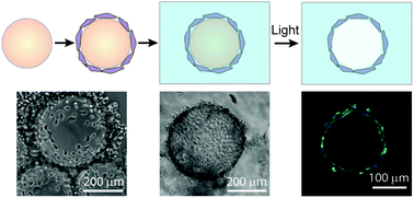
The Anseth Lab at the University of Colorado in Boulder has developed a clever biomaterials technique that reminds me of my favorite arts & crafts activity. In their Biomaterials Science paper Katherine Lewis et al. describe how they used photodegradable PEG microspheres (analogous to balloons) coated with lung epithelial cells (analogous to papier-mâché) to generate cyst structures that mimic lung alveoli in vitro. In alveoli, lung epithelial cells form tight junctions to create a barrier between the airway and blood vessels. To appropriately model this barrier in vitro, the photodegradable microspheres were functionalized with the adhesive peptide CRGDS to allow epithelial cell attachment to the surface of the microsphere. Subsequently, cell-coated microspheres were encapsulated in a PEG hydrogel with stiffness similar to lung tissue, which was functionalized with CRGDS and enzymatically-cleavable peptide cross-linkers. Finally, the photodegradable microspheres were degraded away with cell-compatible light to form cysts, similar to popping the balloon when making a piñata. The cells forming the spherical cysts retained their tight junctions because they also adhered to the surrounding encapsulating hydrogel. The morphology and cell–cell junctions of the cysts were elegantly characterized with confocal microscopy and immuno-staining to demonstrate barrier formation. These 3D models of alveolar cysts demonstrate yet another unique application of photodegradable PEG hydrogels. These cysts may be used to develop models of diseases including pulmonary fibrosis for in vitro screening of potential therapeutics. Discovering treatments to lung-associated diseases using this technology in the future would certainly be a sweeter success than enjoying candy from a piñata.

Check out the June cover article here: In vitro model alveoli from photodegradable microsphere templates by Katherine J.R. Lewis, Mark W. Tibbitt, Yi Zhao, Kelsey Branchfield, Xin Sun, Vivek Balasubramaniam, and Kristi S. Anseth


Brian AguadoDr. Brian Aguado (@BrianAguado) completed his Ph.D. in Biomedical Engineering from Northwestern University as an NSF fellow in 2015. He holds a B.S. degree in Biomechanical Engineering from Stanford University and a M.S. degree in Biomedical Engineering from Northwestern University. Read more about Brian’s research publications here.


Follow the latest journal news on Twitter @BioMaterSci or go to our Facebook page.

Digg This
Reddit This
Stumble Now!
Share on Facebook
Bookmark this on Delicious
Share on LinkedIn
Bookmark this on Technorati
Post on Twitter
Google Buzz (aka. Google Reader)

Polymers in Medicine and Biology

We are pleased to announce the Polymers in Medicine and Biology meeting to be held in Sonoma Wine Country, California, USA between September 14 – 17, 2015.

The meeting, co-organised by Buddy Ratner (University of Washington, USA), Kathryn Uhrich (Rutgers University, USA) and Judy Riffle (Virginia Tech, USA), will cover a broad range of topics over the 4 days, including advances in drug delivery, novel polymeric biomaterials, surfaces and biointerfaces, and implants and implantable materials.

The programme features a range of top speakers including Biomaterials Science Editor-in-Chief Phil Messersmith, Ali Khademhosseini, Joseph DeSimone, Martina Stenzel, David Grainger and Yukio Nagasaki.

The registration deadline is Monday August 3 2015.

Full location: Hilton Sonoma Wine Country, Santa Rosa, California, USA

Digg This
Reddit This
Stumble Now!
Share on Facebook
Bookmark this on Delicious
Share on LinkedIn
Bookmark this on Technorati
Post on Twitter
Google Buzz (aka. Google Reader)

Facile Synthesis of Antibacterial Graphene Films Doped with Silver Nanoparticles

The development of antibacterial materials that can guard against microbial infections during medical procedures has been a topic of increasing interest. In this paper, a team of researchers from Beijing University of Chemical Technology and the University of Bremen describe a facile synthesis method for a hybrid nanomaterial that has previously demonstrated excellent antibacterial activity: reduced graphene oxide films decorated with silver nanoparticles. The efficient, dimensionally scalable, and environmentally friendly nature of this method make it a potentially valuable tool for other groups interested in fabricating these hybrid films for applications ranging from medical biomaterials, to cell culture scaffolds, to drug delivery platforms, to environmental remediation.

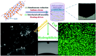
Simultaneous reduction and thermal evaporation-driven self assembly fabrication method used to create RGO/AgNP hybrid film
Schematic model for fabricating RGO/AgNP hybrid film

Zhang et al. created these hybrid films by using sodium citrate to simultaneously reduce aqueous graphene oxide (GO) and aqueous silver nitrate to reduced graphene oxide (RGO) and silver nanoparticles (AgNPs), respectively. This was followed by a prolonged incubation at 80°C to induce thermal evaporation-driven self-assembly of the hybrid RGO/AgNP films. The mass ratio of silver nitrate to GO in the initial solution could be adjusted to produce films with varying densities of AgNPs. In addition, this mass ratio as well the thermal evaporation duration provided some control over the thickness of the resulting hybrid film. The film’s size could be readily changed via adjustments to the dimensions of the reaction container and the film could be later transferred to other substrates without any additional post processing steps.

Subsequent characterisation of the film confirmed the successful reduction of GO as well as the formation of primarily spherical AgNPs between 12 and 30 nm in size on the surface of the film. The resulting material was found to be very hydrophilic and supported the adhesion and proliferation of mouse osteoblasts. An antibacterial assay measuring E. coli adhesion on the surface of silicon wafers coated with these RGO/AgNP films found that the few bacteria that did manage to adhere to the film’s surface exhibited damaged cell walls. A subsequent E. coli colony viability assay proved that no living bacterial colonies were present on the surface of the hybrid film, a result that could not be obtained when using either an uncoated silicon wafer or a pure RGO film.

The biocompatibility, hydrophilicity, durability, and antibacterial activity of these RGO/AgNP hybrid films make them intriguing candidates for a number of medical and environmental applications. The simple, dimension-scalable, and environmental friendly synthesis method presented by Zhang et al. in this paper opens up new avenues for the creation and widespread use of these versatile hybrid films.

Check out the full article:
Graphene film doped with silver nanoparticles: self-assembly formation, structural characterizations, antibacterial ability, and biocompatibility by Panpan Zhang, Haixia Wang, Xiaoyuan Zhang, Wei Xu, Yang Li, Qing Li, Gang Wei, and Zhiqiang Su

Ellen Tworkoski is a Web Writer for Biomaterials Science and is currently a graduate student in the biomedical engineering department at Northwestern University (Evanston, IL, US).

Follow the latest journal news on Twitter @BioMaterSci or go to our Facebook page.

Digg This
Reddit This
Stumble Now!
Share on Facebook
Bookmark this on Delicious
Share on LinkedIn
Bookmark this on Technorati
Post on Twitter
Google Buzz (aka. Google Reader)

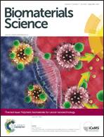
Polymeric biomaterials for cancer nanotechnology themed issue now online

We are delighted to announce that the Polymeric biomaterials for cancer nanotechnology themed issue is now available online.

Polymeric biomaterials for cancer nanotechnology

Guest Edited by Jianjun Cheng (University of Illinois at Urbana-Champaign, USA) and Suzie H. Pun (University of Washington, USA), this themed issue highlights the latest discoveries and innovations in polymeric biomaterials for cancer nanotechnology.

Polymeric biomaterials have been extensively used in nanomedicine formulations for cancer therapy. Preclinical and clinical studies have in general revealed that polymeric nanocarriers, when used for chemotherapeutic drug delivery, reduce systemic toxicity and thus mitigate adverse side effects of the drug. This themed issue contains reviews and research articles in the areas of: (i) expanding the available suite of polymeric biomaterials that can be reproducibly and controllably manufactured at a suitable scale, (ii) designing carriers with improved biodistribution to tumour sites, (iii) increasing tumour distribution and penetration of polymeric nanocarriers, and (iv) controlling efficient drug release at a desired location and with optimal kinetics.

Read all the themed issue papers here

A few articles from the themed issue are highlighted below:

Drug-free macromolecular therapeutics – a new paradigm in polymeric nanomedicines
Te-Wei Chu and Jindřich Kopeček
Biomater. Sci., 2015,3, 908-922

Lipid-coated polymeric nanoparticles for cancer drug delivery
Sangeetha Krishnamurthy, Rajendran Vaiyapuri, Liangfang Zhang and Juliana M. Chan
Biomater. Sci., 2015, 3, 923-936

Enhanced transcellular penetration and drug delivery by crosslinked polymeric micelles into pancreatic multicellular tumor spheroids
Hongxu Lu, Robert H. Utama, Uraiphan Kitiyotsawat, Krzysztof Babiuch, Yanyan Jiang and Martina H. Stenzel
Biomater. Sci., 2015, 3, 1085-1095

Polymeric assembly of hyperbranched building blocks to establish tunable nanoplatforms for lysosome acidity-responsive gene/drug co-delivery
Hui-Zhen Jia, Wei Zhang, Xu-Li Wang, Bin Yang, Wei-Hai Chen, Si Chen, Gang Chen, Yi-Fang Zhao, Ren-Xi Zhuo, Jun Feng and Xian-Zheng Zhang
Biomater. Sci., 2015,3, 1066-1077

Digg This
Reddit This
Stumble Now!
Share on Facebook
Bookmark this on Delicious
Share on LinkedIn
Bookmark this on Technorati
Post on Twitter
Google Buzz (aka. Google Reader)

Biomaterials Science celebrates its first (partial) Impact Factor

Biomaterials Science is pleased to announce its first (partial) Impact Factor of 3.831


Biomaterials Science is committed to publishing exceptional articles that explore the underlying science behind the function, interactions and design of biomaterials. Its impressive first (partial) Impact Factor of 3.831 is a strong assurance that research published in our new journal is highly visible and relevant to the biomaterials community. Take a look at these popular Biomaterials Science articles below:

Sustained delivery of bioactive neurotrophin-3 to the injured spinal cord
Irja Elliott Donaghue, Charles H. Tator and Molly S. Shoichet
Biomater. Sci., 2015, 3, 65-72

Hyperbranched PEG-based supramolecular nanoparticles for acid-responsive targeted drug delivery
Xiaofei Chen, Xuemei Yao, Chunran Wang, Li Chen and Xuesi Chen
Biomater. Sci., 2015, 3, 870-878

Angiopoietin-1 peptide QHREDGS promotes osteoblast differentiation, bone matrix deposition and mineralization on biomedical materials
Nicole T. Feric, Calvin C. H. Cheng, M. Cynthia Goh, Vyacheslav Dudnyk, Val Di Tizio and Milica Radisic
Biomater. Sci., 2014, 2, 1384-1398

A novel hanging spherical drop system for the generation of cellular spheroids and high throughput combinatorial drug screening
A. I. Neto, C. R. Correia, M. B. Oliveira, M. I. Rial-Hermida, C. Alvarez-Lorenzo, Ruis L. Reis and Joao F. Mano
Biomater. Sci., 2015, 3, 581-585

In vitro model alveoli from photodegradable microsphere templates
Katherine J. R. Lewis, Mark W. Tibbitt, Yi Zhao, Kelsey Branchfield, Xin Sun, Vivek Balasubramaniam and Kristi S. Anseth
Biomater. Sci., 2015, 3, 821-832

Noninvasive theranostic imaging of HSV-TK/GCV suicide gene therapy in liver cancer by folate-targeted quantum dot-based liposomes
Dan Shao, Jing Li, Yue Pan, Xin Zhang, Xiao Zheng, Zheng Wang, Ming Zhang, Hong Zhang and Li Chen
Biomater. Sci., 2015, 3, 833-841

Publishing your research in Biomaterials Science means that your article will be read and cited by your colleagues.

Our unique combination of high quality articles, outstanding Editorial and Advisory Board, free colour and flexible manuscript format make it clear to see why Biomaterials Science is a leading journal within the biomaterials field.

Our fast times to publication ensure that your research is reviewed and announced to the community rapidly.

From receipt, your research papers will be published in 68 days. Communications articles will be published in 53 days. (Data taken from average manuscript handling times between July 2014 – January 2015)

So join the many leading scientists that have already chosen to publish in Biomaterials Science and submit your research today to be seen with the best!

Submit your research
Digg This
Reddit This
Stumble Now!
Share on Facebook
Bookmark this on Delicious
Share on LinkedIn
Bookmark this on Technorati
Post on Twitter
Google Buzz (aka. Google Reader)

Organ-on-a-Chip World Congress & 3D-Printing in the Life Sciences

Biomaterials Science is pleased to announce that the Organ-on-a-Chip World Congress & 3D-Printing will be held at Wyndham Boston Beacon Hill in Boston, USA on the 8th – 9th July 2015.

Deadlines and dates

Registration is open, so why not sign up for this fascinating meeting now!

If you are interesting in presenting a poster you must submit your abstract by 30th June 2015. Abstracts for oral presentations are also being accepted.

Themes and topics

The 3D-printing field is expanding exponentially and it is starting to impact the life sciences arena.  The current interest in this space is the use of various bioinks to “print” parts of tissues in the goal and hope to bioprint organs as well as body parts in the future for regenerative medicine and other medical applications.

This conference explores 3D-printing in the life sciences through presentations from academic researchers as well as industry participants. Several companies involved in bioprinting and bioinks will be exhibiting at this conference. The companion conference track explores Organ-on-a-Chip/Body-on-a-chip, which employs the use of microfluidics and lab-on-a-chip (LOAC) technologies to build “cell clusters in 3D-format” in functionally-relevant patterns.  These patterns enable cellular function to be recapitulated ex vivo and has wide-ranging potential for drug discovery and development applications in the pharmaceutical and biotechnology industries.

Please contact event organisers Karen Saunders or Enal Razvi if you have any queries.

Head to the conference website to find out more about this 2-track event.

Join the conversation on Twitter: #OOAC2015 and let us know if you’re going @BioMaterSci.


Digg This
Reddit This
Stumble Now!
Share on Facebook
Bookmark this on Delicious
Share on LinkedIn
Bookmark this on Technorati
Post on Twitter
Google Buzz (aka. Google Reader)

Nanocarriers for cancer drug delivery

Chen et al. discuss the emerging antitumor applications of extracellularly reengineered polymeric nanocarriers.


Chen et al. write an informative and interesting review on the methods by which nanoparticle drug delivery vehicles are engineered using diverse triggers that result in drug release.

In the field of drug delivery, particularly to sites of tumour, there are many different considerations – the drug must be delivered to the site of the tumour, it must be intact when delivered, and it must act to destroy cancerous tissue while remaining as nontoxic as possible to healthy tissue. As a result, much research has been devoted to the development of core-shell drug delivery structures that consist of the drug in the nanoparticle core surrounded by a protective shell. This protective shell may be removed using both internal and external triggers. Many nanocarriers use PEG (polyethylene glycol)-based shells for ease of solubility and in order to prevent proteins from being absorbed onto the surface of the shell. However, additional materials are also increasingly used for the development of these materials.

The authors review the materials as well as common strategies used to remove the shell. Specifically, they summarise literature that exploits changes in pH, since the acidity of the tumour microenvironment differs from its healthy surroundings. Charge-reversal nanocarriers with a positively charged core and a negatively charged shell are also used. In addition, enzymes can degrade the external shell. An enzyme family known as matrix metalloproteinases, or MMPs, is commonly used for this purpose, but other enzymes are also beginning to be explored. Finally, these nanocarriers can also be assembled or de-assembled using interactions between the nanocarrier and the host body.

Emerging antitumor applications of extracellularly reengineered polymeric nanocarriers by Jinjin Chen, Jianxun Ding, Chunsheng Xiao, Xiuli Zhuang and Xuesi Chen

Debanti Sengupta


Debanti Sengupta completed her PhD in Chemistry in 2012 from Stanford University.  She was previously a Siebel postdoctoral scholar at the University of California, Berkeley, and is currently a postdoctoral scholar in Radiation Oncology at Stanford University. Follow her on Twitter @debantisengupta.

Follow the latest journal news on Twitter @BioMaterSci or go to our Facebook page.

Digg This
Reddit This
Stumble Now!
Share on Facebook
Bookmark this on Delicious
Share on LinkedIn
Bookmark this on Technorati
Post on Twitter
Google Buzz (aka. Google Reader)