Paper of the month: Magnetic delivery of Fe3O4@polydopamine nanoparticle-loaded natural killer cells suggest a promising anticancer treatment

Written by web writer Muhammad Ovais

Natural killer (NK) cells have the intrinsic ability to recognise and eliminate cancer cells along with the potential to inhibit metastasis. NK cells utilize a variety of ways to kill tumor cells, such as stimulating cytokine release, direct cytotoxicity and activating targeted cells apoptosis. Non-small cell lung cancer (NSCLC) has shown significant response to NK cell based immunotherapy in clinical settings. Recently, researchers have begun investigating ways to augment the recruitment and infiltration of NK cells into tumors for improved theranostics. Hence, it is vital to develop non-invasive methods for in vivo control and for the monitoring of the administered NK cells with tissue targeting ability. FDA approved superparamagnetic iron oxide nanoparticles (SPIONs) based magnetic resonance imaging (MRI) contrast agents have been proven biocompatible delivery vehicles and imaging probes for NK cells.

c8bm00588e

In the present work Jiang et al have synthesized nanoparticles (NPs) composed of a Fe3O4 core and polydopamine (PDA) shell, for tumor theranostics. The aim of this study was to develop magnetic NPs for an immune-cell delivery system to target NSCLC cells. The system stimulated the accumulation of NK cells at the tumor site via the placement of a tiny external magnetic device inside animals. The NK cells actively took up the Fe3O4@PDA NPs due to its physiological stability, while the biology of NK cells was not affected, owing to its biocompatible nature. In vivo studies demonstrated the reduced expression of Ki-67 and the elevated apoptosis of A549 cancer cells upon treatment with Fe3O4@PDA NP-labeled NK cells. Though there are some limitations associated with the invasive approach, the magnetic delivery of NP-NKs can be of promising value in clinical applications.

Tips/comments from the authors:

  • Coating of magnetic NPs with PDA played a vital role in its cellular uptake, as surface modification is an essential factor in the determination of the biocompatibility and the cellular absorption of magnetic NPs.
  • Due to the biocompatible nature of magnetic NPs, even high concentrations (100 μg/mL) did not induce apoptosis of NK cells.
  • With improved retention over time the delivery of Fe3O4@PDA NP-labeled NK cells can be expedited to the tumor via application of local magnetic field.
  • The position of the tumor and the implanted magnetic field should be close enough, while sufficient time should be given to the magnetic field for the achievement of potent therapeutic effect.
  • The non-invasive nature of three-dimensional (3-D) rotating magnetic fields or high gradient magnetic fields can enhance the magnetic strength in a central point for the effective accumulation of NPs.


Read the full article here: Magnetic delivery of Fe3O4@polydopamine nanoparticle-loaded natural killer cells suggest a promising anticancer treatment Biomater. Sci., 2018,6, 2714-2725


About the web writer

Muhammad OvaisMuhammad Ovais is a Web Writer for Biomaterials Science. Currently, he is a PhD candidate in Prof. Chunying Chen Lab at CAS Center for Excellence in Nanoscience, CAS Key Laboratory for Biomedical Effects of Nanomaterials and Nanosafety, National Center for Nanoscience and Technology (NCNST), Beijing. His research interest lies in the development of novel nano-delivery systems for cancer immunotherapy. He has published a total of ~30 research/review articles. You can find or contact him on ResearchGate, LinkedIn and Chunying Chen’s lab

Contact Email: movais@bs.qau.edu.pk

Twitter: https://twitter.com/OVAISBiotec

Digg This
Reddit This
Stumble Now!
Share on Facebook
Bookmark this on Delicious
Share on LinkedIn
Bookmark this on Technorati
Post on Twitter
Google Buzz (aka. Google Reader)

Sericin hydrogels promote skin wound healing

Severe skin injuries affect millions of patients each year worldwide. These lead to serious issues, such as the formation of nonfunctional large scar tissue and the loss of skin appendages (such as hair follicles and sebaceous glands; etc). This can cause the patients insufferable bodily discomfort and a poor quality of life. Current available treatments include autologous skin grafting, allotransplantation and artificial skin substitutes. However, it remains very challenging to functionally regenerate skin tissue after severe loss of the epidermis and dermis. Other limitations include the lack of donor skin, costly medical expenses, the chance of immune-rejection and unsatisfactory skin regeneration. Hence, the development of an efficient alternative skin substitute is highly desired.

10.1039/C8BM00934A

Sericin is a natural biomaterial derived from silk cocoons has been used previously for a variety of types of injury repair. Previously, Wang and coworkers utilized sericin made hydrogels or scaffolds for transected sciatic nerve regeneration, repair of ischemic stroke, and cartilage regeneration. In this present work, a photo-crosslinkable sericin hydrogel (SMH) for the repair of scarless skin and sebaceous glands regeneration is reported. The sericin hydrogel promotes such regeneration by the following mechanisms: (a) effective inhibition of inflammation; (b) promotion of angiogenesis by stimulating the growth factors like VEGF and EGF; (c) reduction of scar formation through regulating the expressions of TGF-β1 and TGF-β3; and (d) effective conscription of stem cells to injury sites, where they differentiate and regenerate into skin appendages. Overall, these results showcase the potential of this innovative bimodal tool for the development of new artificial skin substitutes for the clinical treatment of severe skin injuries.

Read the full article for free until 19th November

Sericin hydrogels promote skin wound healing with effective regeneration of hair follicles and sebaceous glands after complete loss of epidermis and dermis  Biomater. Sci., 2018, Advance Article DOI: 10.1039/C8BM00934A.


About the webwriter
Dr Sudip MukherjeeDr. Sudip Mukherjee is a Web Writer for Biomaterials Science. He is currently a Postdoctoral Research Associate at the Rice University. His research is involved in the development of advanced nanomaterials for drug/gene delivery in cancer theranostics, immunomodulatory applications & angiogenesis. He published a total of ~35 research articles/patents. He serves as International Advisory Board Member for ‘Materials Research Express‘, IOP Sciences. He is an associate member (AMRSC) of RSC, UK. He serves as reviewer for several international journals like ChemComm, J Mater Chem A, J Mater Chem B, Journal of Biomedical Nanotechnology, RSC Advances, IOP Nanotechnology, Biofabrication etc.

Contact Email: sudip.mukherjee@rice.edu

Twitter: https://twitter.com/sudip_88

Digg This
Reddit This
Stumble Now!
Share on Facebook
Bookmark this on Delicious
Share on LinkedIn
Bookmark this on Technorati
Post on Twitter
Google Buzz (aka. Google Reader)

Nominations open for the 2019 Biomaterials Science Lectureship

Do you know an early-career researcher who deserves recognition for their contribution to the biomaterials field?

Now is your chance to put them forward for the accolade they deserve!

Biomaterials Science is pleased to announce that nominations are now being accepted for its 2019 Lectureship award. This annual award was established in 2014 to honour an early-stage career scientist who has made a significant contribution to the biomaterials field.

The recipient of the award will be asked to present a lecture at the 2019 European Society for Biomaterials Annual Meeting, where they will also be presented with the award. The Biomaterials Science Editorial Office will provide financial support to the recipient for travel and accommodation costs.

The recipient will also be asked to contribute a lead article to the journal and will have their work showcased free of charge on the front cover of the issue in which their article is published.

Prof Zhen Gu (University of North Carolina at Chapel Hill and North Carolina State University)

Zhen Gu, winner of the 2018 Biomaterials Science Lectureship, receives his certificate from Executive Editor Neil Hammond


Previous winners

2018 – Zhen Gu, University of North Carolina at Chapel Hill & North Carolina State University, USA

2017 – Zhuang Liu, Soochow University, China

2016 – Fan Yang, Stanford University, USA

2015 – Joel Collier, Duke University, USA

2014 – Suzie Pun, University of Washington, USA

Eligibility

To be eligible for the lectureship, candidates should meet the following criteria:

  • Be an independent researcher, having completed PhD and postdoctoral studies
  • Be actively pursuing research within the biomaterials field, and have made a significant contribution to the field
  • Be at an early stage of their independent career (this should be within 12 years of attaining their doctorate or equivalent degree, but appropriate consideration will be given to those who have taken a career break, for example for childcare leave, or followed an alternative study path)

Although the Biomaterials Science Lectureship doesn’t explicitly reward support of or contributions to the journal, candidates with no history of either publishing in or refereeing for the journal would typically not be considered.

Selection

  • Eligible nominated candidates will be notified of their nomination, and will be asked to provide 3 recent articles that they feel represent their current research.
  • All eligible nominated candidates will be assessed by a shortlisting panel, made up of members of the Biomaterials Science Advisory Board and a previous lectureship winner.
  • The shortlisting panel will consider the articles provided by the candidates as well as their CVs and letters of nomination.
  • Shortlisted candidates will be further assessed by the Biomaterials Science Editorial Board, and a winner will be selected based on an anonymous poll.
  • Selection is not based simply on quantitative measures. Consideration will be given to all information provided in the letter of recommendation and candidate CV, including research achievements and originality, contributions to the biomaterials community, innovation, collaborations and teamwork, publication history, and engagement with Biomaterials Science.

Nominations

  • Nominations must be made via email to biomaterialsscience-rsc@rsc.org, and should include a short CV and a brief letter of nomination
  • Self-nomination is not permitted
  • Nominators do not need to be senior researchers, and we encourage nominations from people at all career levels
  • As part of the Royal Society of Chemistry, we believe we have a responsibility to promote inclusivity and accessibility in order to improve diversity. Where possible, we encourage each nominator to consider nominating candidates of all genders, races, and backgrounds.
  • Candidates outside of the stated eligibility criteria may still be considered
  • Nomination letters should be up to 1 page in length. They should particularly highlight contributions that the nominee has made to the field as an independent researcher, and any career breaks or alternative career paths that should be taken into consideration by the judging panel. Nomination of one candidate by multiple people in the same letter is accepted


Nominations should be submitted no later than 19th December 2018.


Digg This
Reddit This
Stumble Now!
Share on Facebook
Bookmark this on Delicious
Share on LinkedIn
Bookmark this on Technorati
Post on Twitter
Google Buzz (aka. Google Reader)

New hybrid nanoparticles for enhanced drug accumulation in glioblastoma cells

Glioblastoma is the most frequent and aggressive primary malignant tumor of the central nervous system. The efficacy of antineoplastic drugs that are able to cross the blood-brain barrier is limited mainly by various resistance mechanisms. Hence, local strategies have been developed to improve the therapeutic efficacy. Platinum derivatives and one among them, cisplatin, demonstrated promising results when locally administered into the brain of glioblastoma-bearing rats. A more specific vectorization of the active substance may further promote its accumulation within cancer cells, therefore improving its bioavailability. In this context, biocompatible and biodegradable copolymers that are approved for medical applications can be synthesized without any toxic organic solvents or excipients. Their amphiphilicity, namely the combination of both a hydrophilic and a hydrophobic sequence, is responsible for their spontaneous self-assembly in water. Such a formulation process is simple and flexible. The versatile structure of these drug delivery systems allows imaging moieties to be grafted onto their surface while encapsulating a drug within their core. The diagnosis of glioblastoma relies on magnetic resonance imaging (MRI) after the injection of gadolinium-based contrast agents. Thus, the injection of hybrid nanoplatforms that combine an MRI contrast agent and a drug would enable to non-invasively monitor the biodistribution of the treatment by analyzing the tumor response in real time. Personalized medicine and theranostic applications require similar strategies for the purposes of adjusting the treatment regimen to the patient response.


Such smart drug delivery systems were designed by Lajous and coworkers based on amphiphilic block copolymers. Gadolinium complexes were grafted at the end of the hydrophilic chain while a chemical modification of the other block allowed for cisplatin cross-linking with the copolymer backbone. The self-assembly of these functionalized copolymers in water resulted in stable cisplatin cross-linked nanoparticles with a mean size of 100.63 ± 12.04 nm consistent with biological investigations. High field MRI confirmed the intrinsic potential of these hybrid nanoparticles as alternative MRI contrast agents compared to conventional low molar mass Gd-DTPA complexes. Their infusion within the striatum of glioblastoma-bearing mice resulted in a signal that persisted over time. The accumulation of platinum compounds in human glioblastoma cells when treated with these drug delivery systems and the subsequent formation of Pt-DNA adducts was significantly increased in comparison with free cisplatin by up to 50-fold and 32-fold respectively. These results support the potential of this innovative bimodal tool for further applications.

Hybrid Gd3+/cisplatin cross-linked polymer nanoparticles enhance platinum accumulation and formation of DNA adducts in glioblastoma cell lines  Biomater. Sci., 2018, 6, 2386-2409


Read the full article now for free until 11 October


About the web writer

Dr. Sudip MukherjeeDr. Sudip Mukherjee is a Web Writer for Biomaterials Science. He is currently a Postdoctoral Research Associate at the Rice University. His research is involved in the development of advanced nanomaterials for drug/gene delivery in cancer theranostics, immunomodulatory applications & angiogenesis. He published a total of ~35 research articles/patents. He serves as International Advisory Board Member for ‘Materials Research Express‘, IOP Sciences. He is an Associate Member (AMRSC) of RSC, UK. He serves as reviewer for several international journals like ChemComm, J Mater Chem A, J Mater Chem B, Journal of Biomedical Nanotechnology, RSC Advances, IOP Nanotechnology, Biofabrication etc.


Digg This
Reddit This
Stumble Now!
Share on Facebook
Bookmark this on Delicious
Share on LinkedIn
Bookmark this on Technorati
Post on Twitter
Google Buzz (aka. Google Reader)

Chemically modified nanogels for superior bioavailability

The pharmacokinetics and pharmacodynamics of any drug or nanoparticles plays an important role towards the therapeutic activity, half-life of the therapeutic agent and frequency of dosing of the biomaterial in various diseases. Recently, the development of biopolymer nanogels has received tremendous attention due to their effective delivery of therapeutics, size uniformity, high drug encapsulation capacity, easy preparation, high biocompatibility and size tunability. However, the tendency of these nanogels to disassemble in the bloodstream is cause for concern, due to the interactions with serum proteins and excessive dilution volume that decreases the tumor targeting efficiency through EPR effects. In this regard, substantial research is needed to develop novel platform technologies for bioactive nanogels with enhanced bioavailability.

c8bm00396c

In this work, Auzély-Velty and co-workers, developed a novel and easy method to synthesize stable self-assembled hyaluronic acid (HA) nanogels, modified with a thermoresponsive ketone-functional copolymer by hydrazone formation. The cross-linking density played a crucial role in the nanogel stability and pharmacokinetics, and it was easily tuned by varying dihydrazide crosslinker to ketone ratio. Several physicochemical characterizations including cryo-transmission, dynamic light scattering and scanning electron microscopy were performed to analyze the size, morphology and stability of the nanogels. The authors showed the in vitro cellular uptake of the nanogels by CD44 receptor mediated pathway further confirmed the effectiveness of the cross-linking strategy. The modified nanogels demonstrated superior bioavailability in tumors, with enhanced blood circulation for over 24 hours, demonstrated in vivo in biodistribution studies with mouse tumor models. Overall, these nanogels are inexpensive, stable, easily tunable and biocompatible, thus holding promise for the discovery of a new class of molecules for cancer therapy application in near future.


This article is free to read and download until 13th August.

A versatile method for the selective core-crosslinking of hyaluronic acid nanogels via ketone-hydrazide chemistry: from chemical characterization to in vivo biodistribution Biomater. Sci., 2018, 6, 1754-1763


About the web writer

Dr. Sudip MukherjeeDr. Sudip Mukherjee is a Web Writer for Biomaterials Science. He is currently a Postdoctoral Research Associate at the Rice University. His research is involved in the development of advanced nanomaterials for drug/gene delivery in cancer theranostics, immunomodulatory applications & angiogenesis. He published a total of ~35 research articles/patents. He serves as International Advisory Board Member for ‘Materials Research Express‘, IOP Sciences. He is an associate member (AMRSC) of RSC, UK. He serves as reviewer for several international journals like ChemComm, J Mater Chem A, J Mater Chem B, Journal of Biomedical Nanotechnology, RSC Advances, IOP Nanotechnology, Biofabrication etc.

Contact Email: sudip.mukherjee@rice.edu
Twitter: https://twitter.com/sudip_88

Digg This
Reddit This
Stumble Now!
Share on Facebook
Bookmark this on Delicious
Share on LinkedIn
Bookmark this on Technorati
Post on Twitter
Google Buzz (aka. Google Reader)

2018 Biomaterials Science Lectureship

It is with great pleasure that we announce Prof. Zhen Gu (University of North Carolina at Chapel Hill and North Carolina State University) as the recipient of the 2018 Biomaterials Science Lectureship!

Professor Zhen GuThe Biomaterials Science Lectureship is an annual award that honours an early-career researcher for their significant contribution to the biomaterials field. The recipient is selected by the Biomaterials Science Editorial Board from a list of candidates nominated by the community.

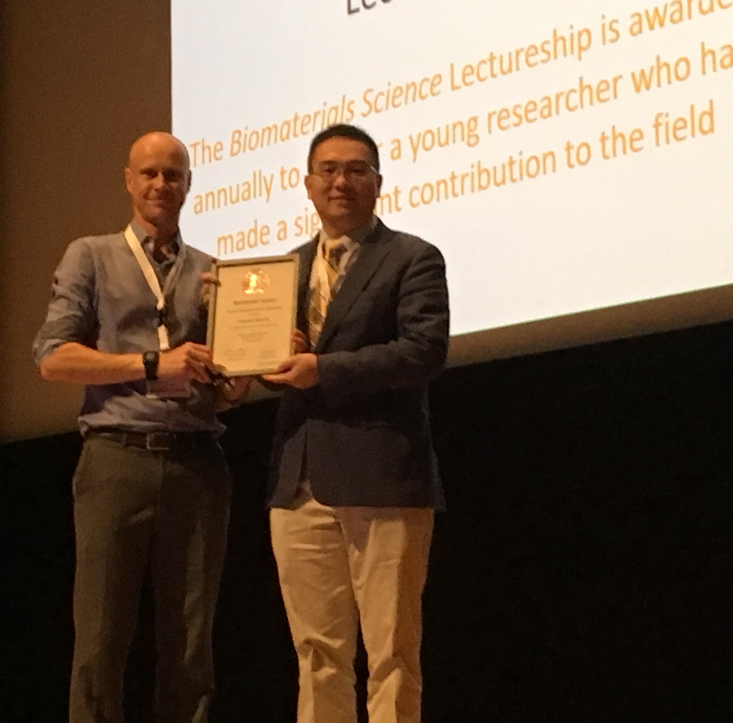
This year we are delighted to award the Lectureship to Professor Zhen Gu (University of North Carolina at Chapel Hill and North Carolina State University). He will present the Biomaterials Science lecture and receive his award at the European Society for Biomaterials Annual Meeting in Maastricht in September 2018.

Prof. Zhen Gu received his B.S. degree in Chemistry and M.S. degree in Polymer Chemistry and Physics from Nanjing University. In 2010, he obtained Ph.D. at the University of California, Los Angeles, under the guidance of Prof. Yi Tang in the Department of Chemical and Biomolecular Engineering. He was a Postdoctoral Associate working with Profs. Robert Langer and Daniel Anderson at MIT and Harvard Medical School during 2010 to 2012.

Prof. Zhen Gu is the recipient of the Young Investigator Award of the Controlled Release Society (CRS, 2017), Sloan Research Fellowship (2016), Pathway Award of the American Diabetes Association (ADA, 2015) and Young Innovator Award in Cellular and Molecular Engineering of the Biomedical Engineering Society (BMES, 2015). MIT Technology Review listed him in 2015 as one of the global top innovators under the age of 35 (TR35).

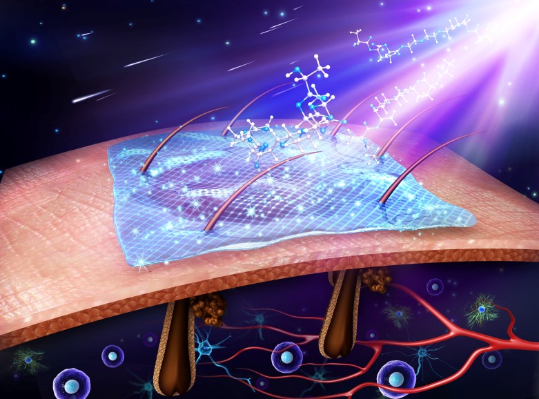
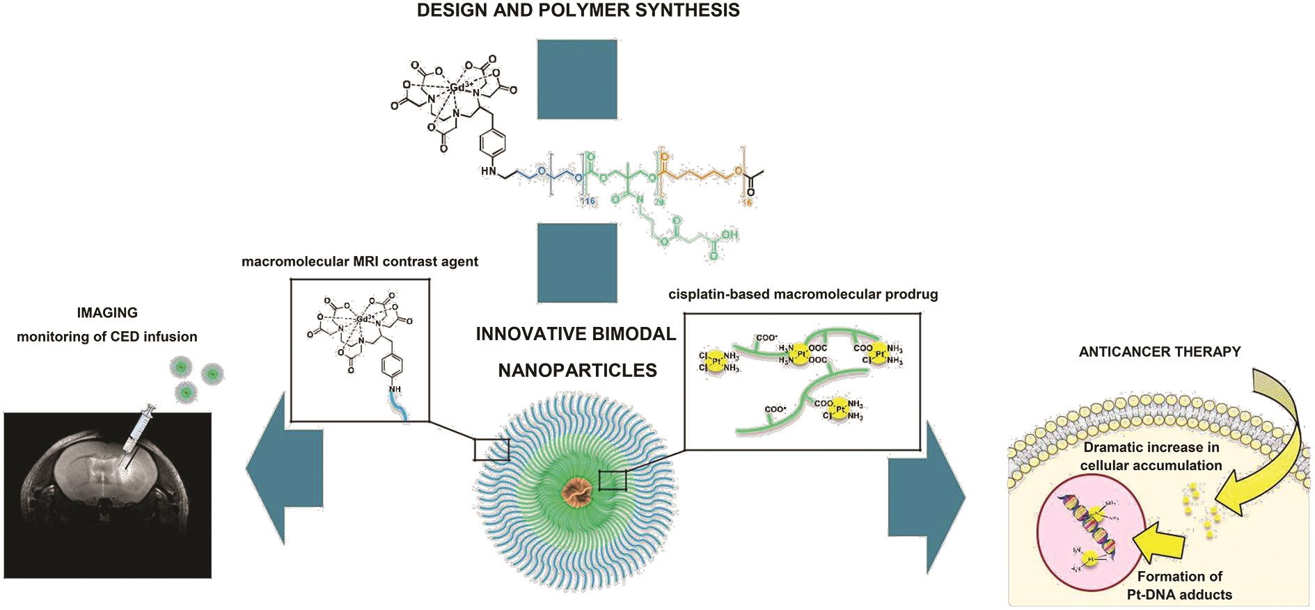
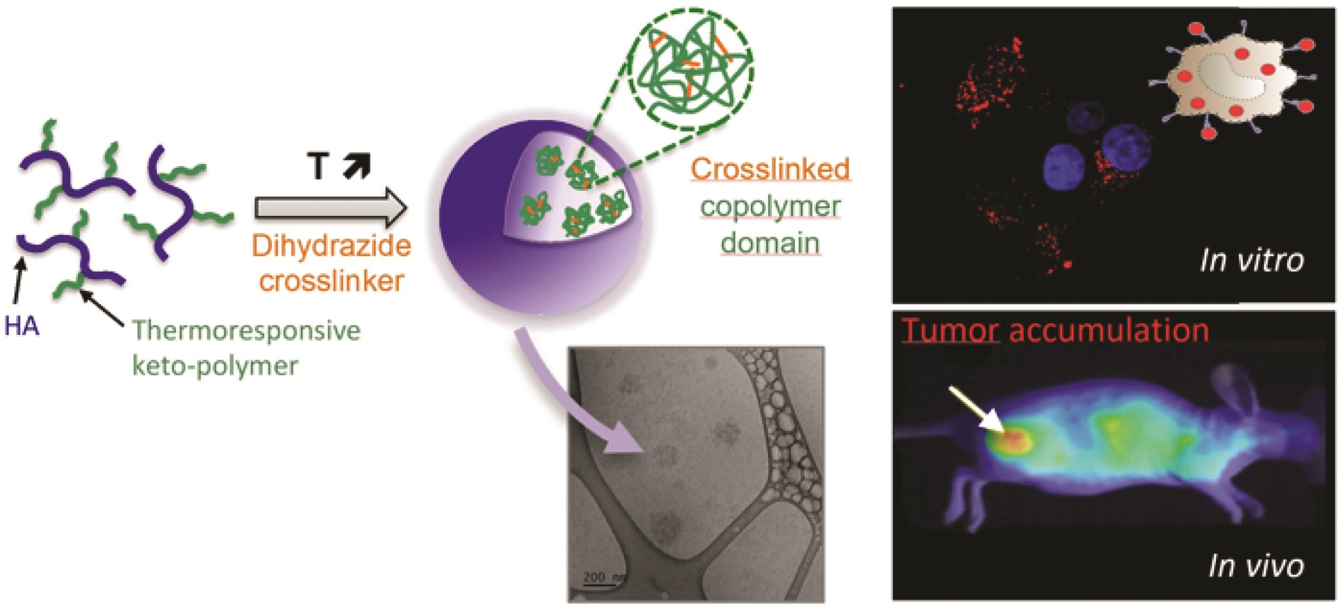
His group studies controlled drug delivery, bio-inspired materials and nanobiotechnology, especially for cancer and diabetes treatment.

To learn more about Zhen’s research, have a look at his recent publications in Biomaterials Science and our sister journals:

Engineering DNA scaffolds for delivery of anticancer therapeutics
Wujin Sun  and  Zhen Gu
Biomater. Sci., 2015,3, 1018-1024, Minireview

Advances in liquid metals for biomedical applications
Junjie Yan,  Yue Lu,  Guojun Chen,  Min Yang  and  Zhen Gu
Chem. Soc. Rev., 2018,47, 2518-2533, Tutorial Review

Investigation and intervention of autophagy to guide cancer treatment with nanogels
Xudong Zhang,  Xin Liang,  Jianjun Gu,  Danfeng Chang,b  Jinxie Zhang,  Zhaowei Chen,  Yanqi Ye,  Chao Wang,  Wei Tao,  Xiaowei Zeng,  Gan Liu,  Yongjun Zhang,  Lin Mei  and  Zhen Gu
Nanoscale, 2017,9, 150-163, Paper

Internalized compartments encapsulated nanogels for targeted drug delivery
Jicheng Yu,  Yuqi Zhang,  Wujin Sun,  Chao Wang,  Davis Ranson,  Yanqi Ye,  Yuyan Weng  and  Zhen Gu
Nanoscale, 2016,8, 9178-9184, Paper

Self-folded redox/acid dual-responsive nanocarriers for anticancer drug delivery
Yue Lu,  Ran Mo,  Wanyi Tai,  Wujin Sun,  Dennis B. Pacardo,  Chenggen Qian,  Qundong Shen,  Frances S. Ligler  and  Zhen Gu
Chem. Commun., 2014,50, 15105-15108, Communication

Please join us in congratulating Zhen on his award!


Digg This
Reddit This
Stumble Now!
Share on Facebook
Bookmark this on Delicious
Share on LinkedIn
Bookmark this on Technorati
Post on Twitter
Google Buzz (aka. Google Reader)

Cancer theranostics applications of Graphene Oxides

Graphene oxide (GO) has actively been used in various biomedical applications, including biomedical sensors, electronic sensors, functional composites and so on. In addition to having a large surface area, large-scale manufacturability and being dispersible in water, GO shows strong fluorescence and conjugation properties, owing to the abundance of oxygen functional groups on its surface.

Numerous methods have been developed to enable photon emission from GO sheets, such as reduction, labeling with fluorescent protein, and cleaving GO sheets into smaller fragments to produce graphene quantum dots (QDs), although this method does remove oxygen from the carbon lattice. This is a problem because reducing the oxygen content prevents the further functionalization of GO structures with biomolecules. Furthermore, such methods have been reported to cause cytotoxicity.

Cancer theranostics applications of Graphene Oxides

In this work by the Chen and co-workers, a blue fluorescence was induced in a GO suspension by triggering a phase transformation in GO through treatment with a simple, one-step mild annealing. Previously, it had been difficult to facilitate light emission from GO while preserving the oxygen content and maintaining low cytotoxicity. However, this work suggests that by providing a nano-bio interface for reactions with biomolecules the physical difficulties can be overcome. In this case, GO acts as a bio-imaging agent as well as a functionalization platform for biomolecules. Material characterization and biocompatibility tests were performed to examine the purity, inherent property and non-toxicity of the system. The mechanism for enhanced blue fluorescence upon mild annealing was discovered and modeled through atomistic simulations. Most importantly, GO shows an appealing capability in drug delivery and cellular imaging simultaneously. Overall, this method is expected to be inexpensive, rapid and straightforward, thus holding promise for the development of a whole new class of GO-based nanomaterials for cancer theranostics application in near future.


This article is free to read until 30 May


Simultaneous drug delivery and cellular imaging using graphene oxide Biomater. Sci., 2018, 6, 813-819


About the webwriter

Dr. Sudip MukherjeeDr. Sudip Mukherjee is a Web Writer for Biomaterials Science. He is currently a Postdoctoral Research Associate at the Rice University. His research is involved in the development of advanced nanomaterials for drug/gene delivery in cancer theranostics, immunomodulatory applications & angiogenesis. He published a total of ~30 research articles/patents. He serves as International Advisory Board Member for ‘Materials Research Express‘, IOP Sciences. He is an associate member (AMRSC) of RSC, UK. He serves as reviewer for several international journals like Chem Comm, J Mater Chem A, J Mater Chem B, Journal of Biomedical Nanotechnology, RSC Advances, IOP Nanotechnology etc.

Contact Email: sudip.mukherjee@rice.edu

Twitter: https://twitter.com/sudip_88

Digg This
Reddit This
Stumble Now!
Share on Facebook
Bookmark this on Delicious
Share on LinkedIn
Bookmark this on Technorati
Post on Twitter
Google Buzz (aka. Google Reader)

Focus on: Engineering Advanced Biologics

Written by Webwriter Yingfei Xue

Protein-based biologics are gaining tremendous interests in the biomedical field after several successful translations into commercialized products. To this end, key hurdles in the development of protein-based therapeutics need to be properly addressed first. One of the most prominent issues is the instability of protein therapeutics in vivo, which requires higher and more frequent doses. As a result, protein-based therapy can become prohibitive in cost and may pose potential safety risks. Engineering protein therapeutics using biomaterials offer unique strategies to solve this problem by prolonging the half-life and enhancing the efficacy of these. In this blog, we feature three articles published in Biomaterials Science recently on the topic of Engineering Advanced Biologics as therapeutic agents.


Intra-articular TSG-6 delivery from heparin-based microparticles reduces cartilage damage in a rat model of osteoarthritis


1. Multivalent conjugates of basic fibroblast growth factor enhance in vitro proliferation and migration of endothelial cells
Aline Zbinden, Shane Browne, Eda I. Altiok, Felicia L. Svedlund, Wesley M. Jackson and Kevin E. Healy
Biomater. Sci., 2018, Advance Article, DOI: 10.1039/C7BM01052D

The authors reported the chemical conjugation of basic fibroblast growth factor (bFGF) onto hyaluronic acid (HA). Such multivalent conjugates were envisioned to increase residence time in vivo, protect bFGF from enzymatic degradation, and enhance protein bioactivity. Using a small molecule linker (N-(ε-Maleimidocaproic acid) hydrazide), bFGF was successfully conjugated at two different protein-to-polymer ratios and their structures were confirmed by extensive physico-chemical characterization. Compared to bFGF alone, HA conjugated bFGF displayed enhanced activity to promote the proliferation and more interestingly, the scratch closure of human umbilical cord vein endothelial cells. It’s noteworthy that the protein-to-polymer ratio and the conjugate size are two key parameters that could be easily tuned to modulate the bioactivity of the conjugate.


2. Synergistic effects of hyaluronate – epidermal growth factor conjugate patch on chronic wound healing
Yun Seop Kim, Dong Kyung Sung, Won Ho Kong, Hyemin Kim and Sei Kwang Hahn
Biomater. Sci., 2018, Advance Article, DOI: 10.1039/C8BM00079D

Similar to the strategy discussed above, the authors also utilized the biocompatible HA as carrier to conjugate epidermal growth factor (EGF) which is otherwise labile in vivo. The enzymatic degradation and stability studies revealed the superior stability of EGF when conjugated with HA. In vitro, HA–EGF conjugate increased keratinocyte proliferation, VEGF secretion, and migration towards scratch closure when compared to EGF alone. In vivo, HA–EGF conjugates loaded HA patch closed the wound in a rat model by enhancing de novo ECM secretion and mitigating inflammatory response. Overall, those comprehensive studies demonstrated the significance of protein-HA conjugates in improving the therapeutic effects of protein biologics.


3. Intra-articular TSG-6 delivery from heparin-based microparticles reduces cartilage damage in a rat model of osteoarthritis
Liane E. Tellier, Elda A. Treviño, Alexandra L. Brimeyer, David S. Reece, Nick J. Willett, Robert E. Guldbergde and Johnna S. Temenoff
Biomater. Sci., 2018, Advance Article, DOI: 10.1039/C8BM00010G

Here, the authors aimed to improve the delivery of TNF-α-stimulated gene-6 (TSG-6) for treating osteoarthritis. Specifically, they focused on heparin, another glycosaminoglycan, as a carrier biomaterial for TSG-6 delivery. It was first discovered that N-desulfated heparin maintained the beneficial role in promoting the bioactivity of TSG-6. Microparticles were thus made from N-desulfated heparin followed by TSG-6 encapsulation. TSG-6 was able to be released and demonstrated significantly higher anti-plasmin activity compared to soluble TSG-6. In a rat medial meniscal transection model, treatment of TSG-6 delivered by N-desulfated heparin but not in soluble form resulted in similar cartilage thickness, volume and GAG level compared to uninjured cartilage. These results showcased the advantages of heparin based microparticle in preserving and enhancing the bioactivity of therapeutic TSG-6 protein.


All articles free to read until 24 May


About the webwriter

Yingfei XueYingfei Xue is a web writer for Biomaterials Science. Currently, he is a PhD candidate and graduate student researcher in Dr. Shilpa Sant lab at the University of Pittsburgh, USA. His research focus on nano-/micro-technology in novel heart valve therapy. Find him on Twitter: @Phil_Xue or connect him on ResearchGate

Digg This
Reddit This
Stumble Now!
Share on Facebook
Bookmark this on Delicious
Share on LinkedIn
Bookmark this on Technorati
Post on Twitter
Google Buzz (aka. Google Reader)

Outstanding Reviewers for Biomaterials Science in 2017

We would like to highlight the Outstanding Reviewers for Biomaterials Science in 2017, as selected by the editorial team, for their significant contribution to the journal. The reviewers have been chosen based on the number, timeliness and quality of the reports completed over the last 12 months.

We would like to say a big thank you to those individuals listed here as well as to all of the reviewers that have supported the journal. Each Outstanding Reviewer will receive a certificate to give recognition for their significant contribution.

Dr Kaimin Cai, University of Illinois at Urbana-Champaign, ORCID: 0000-0001-9442-8312
Dr Qing Cai, Beijing University of Chemical Technology, ORCID: 0000-0001-6618-0321
Dr Jinzhi Du, South China University of Technology, ORCID: 0000-0003-4037-1212
Dr Xiaohu Gao, University of Washington
Dr Xu Han, University of Miami, ORCID: 0000-0001-9095-1755 
Dr Xun He, Texas A&M University, ORCID: 0000-0002-4002-7932
Dr Zheng-Hong Peng, Yale University, ORCID: 0000-0001-9783-5108  
Dr Wujin Sun, University of North Carolina at Chapel Hill, ORCID: 0000-0002-3167-111X
Dr Wentao Wang, Florida State University, ORCID: 0000-0003-2273-4171
Dr Yazhen Zhu, University of California, Los Angeles ORCID: 0000-0002-2130-8085

We would also like to thank the Biomaterials Science board and the biomaterials research community for their continued support of the journal, as authors, reviewers and readers.

If you would like to become a reviewer for our journal, just email us with details of your research interests and an up-to-date CV or résumé.  You can find more details in our author and reviewer resource centre

Digg This
Reddit This
Stumble Now!
Share on Facebook
Bookmark this on Delicious
Share on LinkedIn
Bookmark this on Technorati
Post on Twitter
Google Buzz (aka. Google Reader)

On-demand chemotherapy using photo-activated micelles

Preventing or minimizing the adverse effects of anti-cancer chemotherapy has been a great challenge, due to the non-specific distribution of chemodrugs in healthy organs. Many previous works have attempted to address this issue by improving the accumulation of cytotoxic drugs in the tumor, with various targeting strategies. All the drug targeting methods enhance efficacy and reduce side-effects to a certain extent, but they cannot completely avoid the unfavourable adverse effects. Site-specific activation of chemotherapeutic drugs via a photo trigger has been a promising means of reducing side-effects.

On-demand chemotherapy using photo-activated micelles


The Zhao group at Tianjin University report the use of cyclodextrin-bearing polymer micelles for the on-demand delivery of a photoswitchable microtubule inhibitor, to achieve precision chemotherapy. The tailored inhibitor displays conformation-dependent cytotoxicity with a low potency; the “trans” isomer is thermodynamically stable and inactive, whereas the “cis” isomer is active, but thermodynamically unstable. Light irradiation activates the drug from the “trans” to “cis” form, which then instantly induces rapid drug release. Such simultaneous drug activation and release could compromise the low drug potency to a certain extent, showing improved anti-cancer efficacy in vitro and in vivo. Therefore, photo-triggered nanosystems could open new avenues of on-demand precision chemotherapy, without the risk of adverse effects on healthy organs and tissues.

This article is free to read until 30 April 18

Photo-triggered micelles: simultaneous activation and release of microtubule inhibitors for on-demand chemotherapy Biomater. Sci., 2018, 6, 511-518


About the Web/Blog writer:

Dr. Sudip MukherjeeDr. Sudip Mukherjee is a Web Writer for Biomaterials Science. He is currently a Postdoctoral Research Associate at the Rice University. His research is involved in the development of advanced nanomaterials for drug/gene delivery in cancer theranostics, immunomodulatory applications & angiogenesis. He published a total of ~30 research articles/patents. He serves as International Advisory Board Member for ‘Materials Research Express‘, IOP Sciences. He is an associate member (AMRSC) of RSC, UK. He serves as reviewer for several international journals like ChemComm, J Mater Chem A, J Mater Chem B, Journal of Biomedical Nanotechnology, RSC Advances, IOP Nanotechnology etc.

Contact Email: sudip.mukherjee@rice.edu

Twitter: https://twitter.com/sudip_88


Digg This
Reddit This
Stumble Now!
Share on Facebook
Bookmark this on Delicious
Share on LinkedIn
Bookmark this on Technorati
Post on Twitter
Google Buzz (aka. Google Reader)