Author Archive

Trapping circulating tumor cells using a “lock and key” biomimetic interface

Sherlock Holmes solves mysterious cases often by identifying footprints and odor left by the suspects at the crime scenes. Scientists are now trying to trap notoriously rare circulating tumor cells (CTCs) in the bloodstream by imprinting their unique topologies and residual biomolecules for efficient early tumor detection.  

The presence of CTCs is a dangerous signal for tumor progression, as they leak into the blood stream from the primary tumor and could potentially invade to other organs, causing metastasis. Unfortunately, they’re extremely rare and thus hard to identify, which poses great challenges for early tumor diagnosis. In a recent publication in Biomaterials Science, Gao et al from Shanghai Jiao Tong University developed a cell-imprinted biomimetic interface, which could intelligently recognize and efficiently capture CTCs, with an over 55% capture efficiency towards spiked MCF-7 cells, a human breast cancer cell line, from rabbit whole blood samples.

Graphical abstract for article c9bm01008d

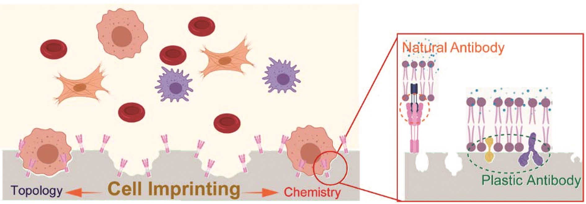
The mechanism behind is the synergy of “lock and key” topological and molecular interactions at the cell-biomaterials interface. Using target CTCs as an imprint template, they created a substrate mimicking their topologies and specific immunoaffinity. By leveraging soft lithography, hierarchical micro/nano-structures of CTCs could be recapitulated on an elastomer substrate (PDMS) to modulate cell adhesion behaviour: cell cytoskeleton staining showed that MCF-7 cells captured on the imprinted surface exhibited abundant filopodia and lamellipodia structures, while they showed an approximately spherical structure on flat substrate, indicating weak topographic interaction. Native proteins originating from CTC’s extracellular matrix (ECM) was anchored in the imprinted substrates providing artificial recognition receptors for selective CTC capture. This is supported by the significantly higher MCF-7 cells capture efficiency on MCF-7 imprinted substrates comparing to HeLa-cell (a human-cervical-cancer cell line) imprinted ones. Anti-EpCAM, a natural antibody, was also introduced to accelerate the CTC-substrate interactions. These interactions turned out to play a decisive role in cell capture, followed by that with plastic receptors.

By recapitulating the topological and chemical microenvironment of CTCs, this biomimetic cell-imprinted substrates demonstrate potential for rare cancer cell capture. Considering the significant roles of tissue ECM stiffness and viscoelasticity in metastasis, and the strong integrin-mediated focal adhesion at the CTCs-biomaterial interface, it might be interesting to incorporate mechanical signals into the interface design in the future for enhanced CTCs capture.

Read the full article for FREE until the 9th October!

Cell-imprinted biomimetic interface for intelligent recognition and efficient capture of CTCs, by Su Gao, Shuangshuang Chen and Qinghua Lu

About the web writer

Zhenwei Ma is currently a Ph.D. candidate in Mechanical Engineering at McGill University. He holds a M.E. degree in Chemical Engineering at McGill University and a B.E. degree in Chemical Engineering from Sichuan University. Find out more about him here.

Digg This
Reddit This
Stumble Now!
Share on Facebook
Bookmark this on Delicious
Share on LinkedIn
Bookmark this on Technorati
Post on Twitter
Google Buzz (aka. Google Reader)

Nominations open for the 2020 Biomaterials Science Lectureship

Do you know an early-career researcher who deserves recognition for their contribution to the biomaterials field?

Now is your chance to put them forward for the accolade they deserve.

Biomaterials Science is pleased to announce that nominations are now being accepted for the 2020 Biomaterials Science Lectureship award. This annual award was established in 2014 to honour an early-stage career scientist who has made a significant contribution to the biomaterials field.

The recipient of the award will be asked to present a lecture at the 11th World Biomaterials Congress in Glasgow in May 2020, where they will also be presented with the award. The Biomaterials Science Editorial Office will provide financial support to the recipient for travel and accommodation costs.

The recipient will also be asked to contribute a lead article to the journal and will have their work showcased free of charge on the front cover of the issue in which their article is published.

Professor April Kloxin

2019 winner Professor April Kloxin

Previous winners

2019 – April Kloxin, University of Delaware, USA

2018 – Zhen Gu, University of North Carolina at Chapel Hill & North Carolina State University, USA

2017 – Zhuang Liu, Soochow University, China

2016 – Fan Yang, Stanford University, USA

2015 – Joel Collier, Duke University, USA

2014 – Suzie Pun, University of Washington, USA

Eligibility

To be eligible for the lectureship, candidates should meet the following criteria:

  • Be an independent researcher, having completed PhD and postdoctoral studies
  • Be actively pursuing research within the biomaterials field, and have made a significant contribution to the field
  • Be at an early stage of their independent career (this should be within 12 years of attaining their doctorate or equivalent degree, but appropriate consideration will be given to those who have taken a career break, for example for childcare leave, or followed an alternative study path)

Although the Biomaterials Science Lectureship doesn’t explicitly reward support of or contributions to the journal, candidates with a history of either publishing in or refereeing for the journal would be viewed favourably.

Selection

  • Eligible nominated candidates will be notified of their nomination, and will be asked to provide 3 recent articles that they feel represent their current research.
  • All eligible nominated candidates will be assessed by a shortlisting panel, made up of members of the Biomaterials Science Advisory Board and a previous lectureship winner.
  • The shortlisting panel will consider the articles provided by the candidates as well as their CVs and letters of nomination.
  • Shortlisted candidates will be further assessed by the Biomaterials Science Editorial Board, and a winner will be selected based on an anonymous poll.
  • Selection is not based simply on quantitative measures. Consideration will be given to all information provided in the letter of recommendation and candidate CV, including research achievements and originality, contributions to the biomaterials community, innovation, collaborations and teamwork, publication history, and engagement with Biomaterials Science.

Nominations

  • Nominations must be made via email to biomaterialsscience-rsc@rsc.org, and should include a short CV (3 page maximum length) and a brief letter of nomination (1 page maximum length).
  • Self-nomination is not permitted
  • Nominators do not need to be senior researchers, and we encourage nominations from people at all career levels
  • As part of the Royal Society of Chemistry, we believe we have a responsibility to promote inclusivity and accessibility in order to improve diversity. Where possible, we encourage each nominator to consider nominating candidates of all genders, races, and backgrounds.
  • Candidates outside of the stated eligibility criteria may still be considered

Nominations should be submitted no later than 30th November 2019.

Digg This
Reddit This
Stumble Now!
Share on Facebook
Bookmark this on Delicious
Share on LinkedIn
Bookmark this on Technorati
Post on Twitter
Google Buzz (aka. Google Reader)

2019 Biomaterials Science Lectureship awarded to April Kloxin

Professor April Kloxin This year we are delighted to award the Lectureship to Professor April Kloxin (University of Delaware). She will present her Biomaterials Science lecture and receive her award at the European Society for Biomaterials Annual Meeting in Dresden, Germany in September 2019.

April Kloxin obtained her B.S. (Summa Cum Laude) and M.S. in Chemical Engineering from North Carolina State University and Ph.D. in Chemical Engineering from the University of Colorado, Boulder, as a NASA Graduate Student Research Program Fellow.  She trained as a Howard Hughes Medical Institute postdoctoral research associate at the University of Colorado before joining the faculty at the University of Delaware in 2011.

Her group aims to create unique materials with multiscale property control for addressing outstanding problems in human health. Her research currently focuses on the design of responsive biomaterials and development of controlled, dynamic models of disease and regeneration.  Her honors include the ACS PMSE Arthur K. Doolittle Award, a Susan G. Komen Foundation Career Catalyst Research award, a NSF CAREER award, and a Pew Scholars in Biomedical Sciences award.

To learn more about April’s research, have a look at her recent publications in Biomaterials Science and our sister journals:

Designing well-defined photopolymerized synthetic matrices for three-dimensional culture and differentiation of induced pluripotent stem cells
Elisa M. Ovadia,  David W. Colby  and  April M. Kloxin
Biomater. Sci., 2018,6, 1358-1370

Design of functionalized cyclic peptides through orthogonal click reactions for cell culture and targeting applications (Part of the 2018 ChemComm Emerging Investgators Issue)
 Paige J. LeValley,  Elisa M. Ovadia,  Christopher A. Bresette,  Lisa A. Sawicki,  Emanual Maverakis,  Shi Bai  and  April M. Kloxin 
Chem. Commun., 2018,54, 6923-6926

Design of thiol- and light-sensitive degradable hydrogels using Michael-type addition reactions (Part of the 2015 Polymer Chemistry Emerging Investgators Issue)
Prathamesh M. Kharkar,  Kristi L. Kiick  and  April M. Kloxin
Polym. Chem., 2015,6, 5565-5574

Design of thiol–ene photoclick hydrogels using facile techniques for cell culture applications
Lisa A. Sawicki  and  April M. Kloxin
Biomater. Sci., 2014,2, 1612-1626

Hydrogel scaffolds as in vitro models to study fibroblast activation in wound healing and disease
Megan E. Smithmyer,  Lisa A. Sawicki  and  April M. Kloxin 
Biomater. Sci., 2014,2, 634-650


Please join us in congratulating April on her award!


Digg This
Reddit This
Stumble Now!
Share on Facebook
Bookmark this on Delicious
Share on LinkedIn
Bookmark this on Technorati
Post on Twitter
Google Buzz (aka. Google Reader)

Biomaterials Science Prizes awarded at 3rd Biomaterials Discovery Workshop

The Third Annual Biomaterials Discovery Workshop took place on Wednesday 16th January at the University of Nottingham. The event was attended by Biomaterials Science Editorial Board member Professor Morgan Alexander and Biomaterials Science was pleased to sponsor two prizes.


The prize winners were

1st Place – Alessandra Travanut

2nd Place – Jennifer Ashworth

Congratulations to both winners!


Biomaterials Discovery

Digg This
Reddit This
Stumble Now!
Share on Facebook
Bookmark this on Delicious
Share on LinkedIn
Bookmark this on Technorati
Post on Twitter
Google Buzz (aka. Google Reader)

Paper of the month: Magnetic delivery of Fe3O4@polydopamine nanoparticle-loaded natural killer cells suggest a promising anticancer treatment

Natural killer (NK) cells have the intrinsic ability to recognise and eliminate cancer cells along with the potential to inhibit metastasis. NK cells utilize a variety of ways to kill tumor cells, such as stimulating cytokine release, direct cytotoxicity and activating targeted cells apoptosis. Non-small cell lung cancer (NSCLC) has shown significant response to NK cell based immunotherapy in clinical settings. Recently, researchers have begun investigating ways to augment the recruitment and infiltration of NK cells into tumors for improved theranostics. Hence, it is vital to develop non-invasive methods for in vivo control and for the monitoring of the administered NK cells with tissue targeting ability. FDA approved superparamagnetic iron oxide nanoparticles (SPIONs) based magnetic resonance imaging (MRI) contrast agents have been proven biocompatible delivery vehicles and imaging probes for NK cells.

c8bm00588e

In the present work Jiang et al have synthesized nanoparticles (NPs) composed of a Fe3O4 core and polydopamine (PDA) shell, for tumor theranostics. The aim of this study was to develop magnetic NPs for an immune-cell delivery system to target NSCLC cells. The system stimulated the accumulation of NK cells at the tumor site via the placement of a tiny external magnetic device inside animals. The NK cells actively took up the Fe3O4@PDA NPs due to its physiological stability, while the biology of NK cells was not affected, owing to its biocompatible nature. In vivo studies demonstrated the reduced expression of Ki-67 and the elevated apoptosis of A549 cancer cells upon treatment with Fe3O4@PDA NP-labeled NK cells. Though there are some limitations associated with the invasive approach, the magnetic delivery of NP-NKs can be of promising value in clinical applications.

Tips/comments from the authors:

  • Coating of magnetic NPs with PDA played a vital role in its cellular uptake, as surface modification is an essential factor in the determination of the biocompatibility and the cellular absorption of magnetic NPs.
  • Due to the biocompatible nature of magnetic NPs, even high concentrations (100 μg/mL) did not induce apoptosis of NK cells.
  • With improved retention over time the delivery of Fe3O4@PDA NP-labeled NK cells can be expedited to the tumor via application of local magnetic field.
  • The position of the tumor and the implanted magnetic field should be close enough, while sufficient time should be given to the magnetic field for the achievement of potent therapeutic effect.
  • The non-invasive nature of three-dimensional (3-D) rotating magnetic fields or high gradient magnetic fields can enhance the magnetic strength in a central point for the effective accumulation of NPs.


Read the full article here: Magnetic delivery of Fe3O4@polydopamine nanoparticle-loaded natural killer cells suggest a promising anticancer treatment Biomater. Sci., 2018,6, 2714-2725


About the web writer

Muhammad OvaisMuhammad Ovais is a Web Writer for Biomaterials Science. Currently, he is a PhD candidate in Prof. Chunying Chen Lab at CAS Center for Excellence in Nanoscience, CAS Key Laboratory for Biomedical Effects of Nanomaterials and Nanosafety, National Center for Nanoscience and Technology (NCNST), Beijing. His research interest lies in the development of novel nano-delivery systems for cancer immunotherapy. He has published a total of ~30 research/review articles. You can find or contact him on ResearchGate, LinkedIn and Chunying Chen’s lab

Contact Email: movais@bs.qau.edu.pk

Twitter: https://twitter.com/OVAISBiotec

Digg This
Reddit This
Stumble Now!
Share on Facebook
Bookmark this on Delicious
Share on LinkedIn
Bookmark this on Technorati
Post on Twitter
Google Buzz (aka. Google Reader)

Nominations open for the 2019 Biomaterials Science Lectureship

Do you know an early-career researcher who deserves recognition for their contribution to the biomaterials field?

Now is your chance to put them forward for the accolade they deserve!

Biomaterials Science is pleased to announce that nominations are now being accepted for its 2019 Lectureship award. This annual award was established in 2014 to honour an early-stage career scientist who has made a significant contribution to the biomaterials field.

The recipient of the award will be asked to present a lecture at the 2019 European Society for Biomaterials Annual Meeting, where they will also be presented with the award. The Biomaterials Science Editorial Office will provide financial support to the recipient for travel and accommodation costs.

The recipient will also be asked to contribute a lead article to the journal and will have their work showcased free of charge on the front cover of the issue in which their article is published.

Prof Zhen Gu (University of North Carolina at Chapel Hill and North Carolina State University)

Zhen Gu, winner of the 2018 Biomaterials Science Lectureship, receives his certificate from Executive Editor Neil Hammond


Previous winners

2018 – Zhen Gu, University of North Carolina at Chapel Hill & North Carolina State University, USA

2017 – Zhuang Liu, Soochow University, China

2016 – Fan Yang, Stanford University, USA

2015 – Joel Collier, Duke University, USA

2014 – Suzie Pun, University of Washington, USA

Eligibility

To be eligible for the lectureship, candidates should meet the following criteria:

  • Be an independent researcher, having completed PhD and postdoctoral studies
  • Be actively pursuing research within the biomaterials field, and have made a significant contribution to the field
  • Be at an early stage of their independent career (this should be within 12 years of attaining their doctorate or equivalent degree, but appropriate consideration will be given to those who have taken a career break, for example for childcare leave, or followed an alternative study path)

Although the Biomaterials Science Lectureship doesn’t explicitly reward support of or contributions to the journal, candidates with no history of either publishing in or refereeing for the journal would typically not be considered.

Selection

  • Eligible nominated candidates will be notified of their nomination, and will be asked to provide 3 recent articles that they feel represent their current research.
  • All eligible nominated candidates will be assessed by a shortlisting panel, made up of members of the Biomaterials Science Advisory Board and a previous lectureship winner.
  • The shortlisting panel will consider the articles provided by the candidates as well as their CVs and letters of nomination.
  • Shortlisted candidates will be further assessed by the Biomaterials Science Editorial Board, and a winner will be selected based on an anonymous poll.
  • Selection is not based simply on quantitative measures. Consideration will be given to all information provided in the letter of recommendation and candidate CV, including research achievements and originality, contributions to the biomaterials community, innovation, collaborations and teamwork, publication history, and engagement with Biomaterials Science.

Nominations

  • Nominations must be made via email to biomaterialsscience-rsc@rsc.org, and should include a short CV and a brief letter of nomination
  • Self-nomination is not permitted
  • Nominators do not need to be senior researchers, and we encourage nominations from people at all career levels
  • As part of the Royal Society of Chemistry, we believe we have a responsibility to promote inclusivity and accessibility in order to improve diversity. Where possible, we encourage each nominator to consider nominating candidates of all genders, races, and backgrounds.
  • Candidates outside of the stated eligibility criteria may still be considered
  • Nomination letters should be up to 1 page in length. They should particularly highlight contributions that the nominee has made to the field as an independent researcher, and any career breaks or alternative career paths that should be taken into consideration by the judging panel. Nomination of one candidate by multiple people in the same letter is accepted


Nominations should be submitted no later than 19th December 2018.


Digg This
Reddit This
Stumble Now!
Share on Facebook
Bookmark this on Delicious
Share on LinkedIn
Bookmark this on Technorati
Post on Twitter
Google Buzz (aka. Google Reader)

2018 Biomaterials Science Lectureship

Professor Zhen GuThe Biomaterials Science Lectureship is an annual award that honours an early-career researcher for their significant contribution to the biomaterials field. The recipient is selected by the Biomaterials Science Editorial Board from a list of candidates nominated by the community.

This year we are delighted to award the Lectureship to Professor Zhen Gu (University of North Carolina at Chapel Hill and North Carolina State University). He will present the Biomaterials Science lecture and receive his award at the European Society for Biomaterials Annual Meeting in Maastricht in September 2018.

Prof. Zhen Gu received his B.S. degree in Chemistry and M.S. degree in Polymer Chemistry and Physics from Nanjing University. In 2010, he obtained Ph.D. at the University of California, Los Angeles, under the guidance of Prof. Yi Tang in the Department of Chemical and Biomolecular Engineering. He was a Postdoctoral Associate working with Profs. Robert Langer and Daniel Anderson at MIT and Harvard Medical School during 2010 to 2012.

Prof. Zhen Gu is the recipient of the Young Investigator Award of the Controlled Release Society (CRS, 2017), Sloan Research Fellowship (2016), Pathway Award of the American Diabetes Association (ADA, 2015) and Young Innovator Award in Cellular and Molecular Engineering of the Biomedical Engineering Society (BMES, 2015). MIT Technology Review listed him in 2015 as one of the global top innovators under the age of 35 (TR35).

His group studies controlled drug delivery, bio-inspired materials and nanobiotechnology, especially for cancer and diabetes treatment.

To learn more about Zhen’s research, have a look at his recent publications in Biomaterials Science and our sister journals:

Engineering DNA scaffolds for delivery of anticancer therapeutics
Wujin Sun  and  Zhen Gu
Biomater. Sci., 2015,3, 1018-1024, Minireview

Advances in liquid metals for biomedical applications
Junjie Yan,  Yue Lu,  Guojun Chen,  Min Yang  and  Zhen Gu
Chem. Soc. Rev., 2018,47, 2518-2533, Tutorial Review

Investigation and intervention of autophagy to guide cancer treatment with nanogels
Xudong Zhang,  Xin Liang,  Jianjun Gu,  Danfeng Chang,b  Jinxie Zhang,  Zhaowei Chen,  Yanqi Ye,  Chao Wang,  Wei Tao,  Xiaowei Zeng,  Gan Liu,  Yongjun Zhang,  Lin Mei  and  Zhen Gu
Nanoscale, 2017,9, 150-163, Paper

Internalized compartments encapsulated nanogels for targeted drug delivery
Jicheng Yu,  Yuqi Zhang,  Wujin Sun,  Chao Wang,  Davis Ranson,  Yanqi Ye,  Yuyan Weng  and  Zhen Gu
Nanoscale, 2016,8, 9178-9184, Paper

Self-folded redox/acid dual-responsive nanocarriers for anticancer drug delivery
Yue Lu,  Ran Mo,  Wanyi Tai,  Wujin Sun,  Dennis B. Pacardo,  Chenggen Qian,  Qundong Shen,  Frances S. Ligler  and  Zhen Gu
Chem. Commun., 2014,50, 15105-15108, Communication

Please join us in congratulating Zhen on his award!


Digg This
Reddit This
Stumble Now!
Share on Facebook
Bookmark this on Delicious
Share on LinkedIn
Bookmark this on Technorati
Post on Twitter
Google Buzz (aka. Google Reader)

Congratulations to Biomaterials Science Award Winners at ICBZM2017

Biomaterials Science was proud to sponsor ICBZM2017, which took place this year in Tokyo, from the 18th to the 20th October. During the conference two Biomaterials Science Poster prizes were awarded.

Winners of the Biomaterials Science poster prize were;

Sarah Ward, (University of Massachusetts), for her poster presentation on ‘Polymer Zwitterion Prodrugs as Chemotherapeutics’.

Sarah Ward

Sarah Ward with Professor Todd Emrick

Erik Liu, (University of Washington), for his poster presentation on ‘Expression of EK Fusion Proteins to Enhance Protein Kinetics and Stability’.

Erik Liu

Erik Liu with Professor Shaoyi Jiang


Congratulations to both Sarah and Erik!

Digg This
Reddit This
Stumble Now!
Share on Facebook
Bookmark this on Delicious
Share on LinkedIn
Bookmark this on Technorati
Post on Twitter
Google Buzz (aka. Google Reader)

Repurposing Drug Action with Targeted Nanomedicine

In cancers, rapid tumour growth is attributed to overexpression of anti-apoptotic proteins, inhibition or functional inefficiency of apoptotic proteases like caspases. Among caspases, caspase-8 signalling cascade is vital and interesting because of its ability to induce cell death by involving both mitochondria mediated intrinsic as well as death receptor (DR) – mediated extrinsic pathways. Notably, among ovarian cancer patients, tumors with low levels of caspase 8 are inherently resistant to chemotherapies. Incidentally, aggressive melanoma cells have functional expression of both Folate receptor (FR) on cell membrane and Estrogen receptor (ER) in cytoplasm. Stitching these basic facts one can deliver anti-cancer drugs, possibly targeting ER, using a liposomal system which will carry FR-targeting ligand to treat the aggressive melanoma cells.

In this present work, the Banerjee group used a hydrophobic drug molecule called NME2 (a recently developed ER-targeted anticancer drug for the treatment in breast cancer). Using a special FR-targeted liposome, the drug was successfully delivered to FR-moderately expressing melanoma cells.

Melanoma Regression in Mice

The efficient targeting to FR-moderately expressed melanoma cells was accomplished by a new robust, cationic folate ligand named FA8. This efficiency of delivery is in stark contrast to other available FR-targeted liposomes which target only FR-over expressing cancer cells. The concoction of NME2 in FA8-associated liposome selectively induced caspase-8 expression-mediated apoptotic cell death in melanoma cancer cells (in vitro and in vivo). However, the drug in pristine state or in non-targeted liposome could not induce caspase-8 mediated apoptosis. Preliminarily, docetaxel, another potent anticancer drug, showed a similar result upon FA8-mediated delivery. Clearly, the given FR-targeted, liposomal delivery methodology indicated a change in mechanism of anticancer action of drug cargo and hence exemplified an interesting possibility to elude impending drug resistance (if any) against the given drug.

Tips from the authors:

1) In MDR cancers repurposing drug’s mechanistic pathway is essential, as acquired drug resistance is one of the major obstacles in fruitful cancer treatment.

2) The given FR-targeted formulation affected the change of mechanism of action of drug cargo (here, NME2) from non-caspase 8 to caspase-8 mediated apoptosis, thereby repurposing the apoptotic pathway of encapsulated drug.

3) The unique cationic lipid-conjugated folic acid based-ligand facilitated a) targeting to FR-moderately expressed melanoma cells; b) modification of mechanistic action of drug-cargo.

4) The liposomal delivery system with an FR-targeting ligand instigated an independent cell death pathway through the up-regulation of caspase-8 with subsequent cleavage of pro-survival factor RIP-1.

Article Link:

Cationic folate-mediated liposomal delivery of bis-arylidene oxindole induces efficient melanoma tumor regression Biomater. Sci., 2017, 5, 1898-1909

About the Webwriter:

Dr. Sudip Mukherjee Dr. Sudip Mukherjee is a Webwriter for Biomaterials Science. He is currently a Postdoctoral Research Associate working alongside Dr. Omid Veiseh at the Department of Bioengineering at the Rice University. His research is involved in the development of advanced nanomaterials for drug/gene delivery in cancer theranostics, immunomodulatory applications & angiogenesis. He published a total of ~30 research articles/patents. He serves as International Advisory Board Member for ‘Materials Research Express‘, IOP Sciences. He is an associate member (AMRSC) of The Royal Society of Chemistry, UK. He serves as reviewer for several international journals like Chem Comm, J Mater Chem A, J Mater Chem B, Journal of Biomedical Nanotechnology, RSC Advances, IOP Nanotechnology etc.

Digg This
Reddit This
Stumble Now!
Share on Facebook
Bookmark this on Delicious
Share on LinkedIn
Bookmark this on Technorati
Post on Twitter
Google Buzz (aka. Google Reader)

Mapping Oxygen Gradients in 3D Cell Cultures

Microenvironmental oxygen levels and gradients within three-dimensional (3D) tissue cultures directly influence cellular behavior and function, dictating the mode of proliferation, metabolism and interaction of cells with each other and their environment. While advances and prevalence of in vitro generated 3D cultures have spurred new techniques and systems for biological interrogation, it is necessary to develop and implement parallel systems to monitor and characterize the oxygen microenvironment within the tissue cultures and around them in the vessel used for the cultures. Conventional oxygen evaluation platforms can be ill-suited for continuous oxygen evaluation in custom tissue cultures. The Takayama group was able to robustly evaluate multiple 3D culture platforms by combining the use of phase-fluorimetry and lab-fabricated dispersible oxygen responsive microparticles. Oxygen microsensors were used to evaluate two spheroid culture vessels, hanging-drop and low-adhesion microwell plates, to highlight the variations in the oxygen levels peripheral to the spheroids in the two culture techniques. Dramatic differences can be seen in the steady state oxygen levels between the two culture techniques because of the difference in distance between the spheroids and the air-liquid interface in these two vessel types. These results highlighted the importance of minding the gas exchange location as compared to the cell culture to ensure appropriate tissue culture microenvironments.

Figure 1

Furthermore, these microsensors were used to map radial oxygen distribution across a circular, cell-patterned hydrogel by dispersing the microsensors within the culture. Coupling the spatial oxygen mapping to computational models of oxygen diffusion, the authors were able to estimate oxygen uptake behavior of the tissue culture. While 3D tissue culture platforms leverage the in vitro tissue architecture to produce more physiologically similar phenomena, integrated design and analysis of these 3D cell cultures from both biomaterial and oxygen supply aspects will be paramount in enabling researchers to effectively recreate some of the complexities present within both healthy and diseased tissues.

Tips from the authors:

  1. When fabricating oxygen microsensing beads, infusion with Dichloromethane enabled large amount of Ruthenium caging within the PDMS microspheres, while leaving them oxygen sensitive. While other solvents swell PDMS more readily and enabled higher efficiency infusion of ruthenium, these solvents resulted in oxygen unresponsive ruthenium loaded PDMS beads.
  2. Microsensors cannot be effectively integrated in the multicellular spheroids we tried with HEK293T, HS-5 and MDA-MB-231 cells; as the spheroids contract microsensors are ejected out of the spheroids.
  3. The only limitation of phase-fluorimetry for the oxygen measurements is sufficient signal output that it can be detected by the photodiode, or other detection system. This was generally not a problem with beads greater than 80 microns assuming the culture systems was less than 1-mm thick. However, we were unable to effectively infuse beads under 80 microns with enough ruthenium to have enough output signals from the microsensors to get robust readings with cultures > 1 mm.

Article Link:

Dispersible oxygen microsensors map oxygen gradients in three-dimensional cell cultures Biomater. Sci., 2017,5, 2106-2113

About the WDr. Sudip Mukherjee ebwriter:

Dr. Sudip Mukherjee is a Web Writer for Biomaterials Science. He is currently a Postdoctoral Research Associate working alongside Dr. Omid Veiseh at the Department of Bioengineering at the Rice University. His research is involved in the development of advanced nanomaterials for drug/gene delivery in cancer theranostics, immunomodulatory applications & angiogenesis. He published a total of ~30 research articles/patents. He serves as International Advisory Board Member for ‘Materials Research Express’, IOP Sciences. He is an associate member (AMRSC) of RSC, UK. He serves as reviewer for several international journals like Chem Comm, J Mater Chem A, J Mater Chem B, Journal of Biomedical Nanotechnology, RSC Advances, IOP Nanotechnology etc.

Digg This
Reddit This
Stumble Now!
Share on Facebook
Bookmark this on Delicious
Share on LinkedIn
Bookmark this on Technorati
Post on Twitter
Google Buzz (aka. Google Reader)