Archive for the ‘Hot Article’ Category

Decreasing Tumor Growth with Magnetic Nanohydrogels

Cancer is the second leading cause of death worldwide, and continues to be a challenging disease to treat therapeutically. One strategy to treat cancer patients is to surgically remove the primary tumor and attempt to prevent the spread of cancer cells to other organs in the body. Unfortunately, surgical resection of tumors may be difficult in sensitive organs (e.g. the brain), and tumors may need to be treated directly with chemotherapy drugs to inhibit growth.

Recently, there has been an increased interest in utilizing biomaterials to deliver chemotherapy drugs directly to the primary tumor. Current research at the Indian Institute of Technology explores the use of nanohydrogels supplemented with iron oxide magnetic nanoparticles as a vehicle to deliver chemotherapy drugs to the primary tumor. Interestingly, these thermo-responsive nanohydrogels could release their anticancer drug cargo when heated with a magnetic field. Although this study did not use chemotherapy drugs, the magnetic hydrogels were used to heat the tumor locally to reduce tumor size – upon stimulation with a magnetic field. This study focused on characterizing the material properties of the magnetic nanohydrogels, in addition to analyzing the effects of the nanohydrogel on inhibiting tumor growth.

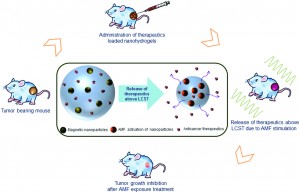
The magnetic nanohydrogel is composed of iron oxide nanoparticles, chitosan, and poly-N-isopropylacrylamide (NIPAAm).  The chitosan-poly-(NIPAAm) nanohydrogels are hydrophilic below the lower critical solution temperature (LCST) of 42oC. When stimulated with the appropriate magnetic field, the magnetic nanoparticles heat the hydrogel above the LCST. When the temperature is increased above the LCST, the polymers in the hydrogel change from “expanded coils” to “compact globules,” and cause the release of the aqueous contents of the hydrogel. The hydrophilic to hydrophobic transition is the main mechanism by which the anticancer drug cargo could be released from the hydrogel.

The biocompatibility and efficacy of the hydrogel in reducing tumor growth in vivo were analyzed. Using a mouse model of fibrosarcoma, the magnetic nanohydrogels were delivered via injection to the primary tumor site, and then stimulated with a magnetic field. Various biocompatibility studies were conducted, showing that serum protein concentrations and blood cell counts were not affected. The biodistribution of the magnetic hydrogel was also quantified, with minimal accumulation of the nanohydrogel in various organs (14 days post-delivery). Additionally, the magnetic nanohydrogel decelerated the growth of the primary tumor. After magnetic stimulation, the magnetic particles in the nanohydrogel successfully heated the tumor locally with minimal damage to the surrounding tissues. Despite not containing chemotherapy drugs, the magnetic nanohydrogels were still efficient in inhibiting the primary tumor growth.

This study describes the thermo-responsive properties of magnetic nanohydrogels, and demonstrates the ability to reduce tumor size using nanohydrogels heated with a magnetic field. Future studies are needed to show efficacy in delivering chemotherapy drugs using the magnetic nanohydrogels as a delivery vehicle. Still, the magnetic nanohydrogels are a promising platform for a minimally invasive method of decreasing tumor growth in vivo.

Biocompatibility, biodistribution and efficacy of magnetic nanohydrogels in inhibiting growth of tumors in experimental mice models
Manish K. Jaiswal, Manashjit Gogoi, Haladhar Dev Sarma, Rinti Banerjee and D. Bahadur
Biomater. Sci., 2014, Advance Article DOI: 10.1039/C3BM60225G

Brian Aguado is currently a Ph.D. Candidate and NSF Fellow in the Biomedical Engineering department at Northwestern University. He holds a B.S. degree in Biomechanical Engineering from Stanford University and a M.S. degree in Biomedical Engineering from Northwestern University. When he’s not in the lab, Brian enjoys traveling, cooking, swimming, and spending time with family and friends. Read more about Brian’s research publications here.

To keep up-to-date with all the latest research, sign-up to our RSS feed or Table of contents

Digg This
Reddit This
Stumble Now!
Share on Facebook
Bookmark this on Delicious
Share on LinkedIn
Bookmark this on Technorati
Post on Twitter
Google Buzz (aka. Google Reader)

Achieving improved separation of output signals in ‘sense and treat’ biosensors via an enzymatic filter

The next generation of diagnostic devices is currently being realized in the form of ‘smart’ biosensors.  These sensors use logic gates to digitalize biochemical signals and produce a binary output that either indicates the presence or absence of a disease.  The biochemical signals, which are often the product of enzymatic cascades, are also capable of triggering drug release from stimuli-responsive materials, thus creating a device that is capable of both sensing and treating a particular condition.

However, these integrated sense and treat biosensors have a major obstacle to overcome if they are to be successfully implemented.  Many of the enzymatic cascades used for input will produce low, but non-zero concentrations of the sensing analytes under normal, physiological conditions.  Over time, the accumulation of these background signals can rise to a level that is high enough to trigger drug release, resulting in the dosage of a perfectly healthy patient which often has adverse consequences.  In this work, a team from Clarkson University and Oak Ridge National Laboratory propose a novel solution to this problem which involves the incorporation of an enzymatic filter into these devices.

The group created a system that was designed to detect and treat liver injury using input from two known biomarkers of liver disease: alanine transaminase (ALT) and aspartate transaminase, type I (AST).  When both ALT and AST are present in elevated concentrations, they will produce high levels of molecules which can be used to synthesize citric acid and another enzyme known as CoA-SH.  While CoA-SH can be readily detected and used to generate a positive signal indicating liver injury, citric acid can be used to trigger drug release by dissolving the iron-cross-linked alginate beads which were used in this study to simulate drug release capsules.  The group then introduced an enzymatic filter, malate dehydrogenase (MDH), into the system.  MDH is able to regulate the production of citric acid, thus controlling the degradation of the alginate beads and subsequent drug release.  In the test cases listed, the modified system generated the correct diagnostic result 100% of the time.  However, the filter was not able to completely eliminate bead dissolution and drug release under conditions associated with a normal, healthy environment.

To tackle this problem, the group decided to try improving the stability of the beads themselves.  By alternately depositing layers of poly-L-lysine with additional layers of alginate, PLA bi-layers were formed on the surface of the alginate cores.   The group found that, as more layers were added, the speed of bead dissolution and the amount of drug released were reduced.  The combination of the filter and the PLA coatings showed enhanced suppression of the negative signals while continuing to have a minimal effect on the positive signal.

Although the group was unable to completely eliminate drug release under normal conditions due to long-term noise accumulation, they were able to reduce it significantly.  Their approach marks a promising step forwards in addressing one of the major obstacles faced by smart biosensor systems.

Enzymatic filter for improved separation of output signals in enzyme logic systems towards ‘sense and treat’ medicine
Shay Mailloux, Oleksandr Zavalov, Nataliia Guz, Evgeny Katz and Vera Bocharova  
Biomater. Sci., 2014, Advance Article DOI: 10.1039/C3BM60197H

Ellen Tworkoski is a guest web-writer for Biomaterials Science.  She is currently a second year Ph.D. student in the biomedical engineering department at Northwestern University (Evanston, IL, USA).

Follow the latest journal news on Twitter @BioMaterSci or go to our Facebook page.

To keep up-to-date with all the latest research, sign-up to our RSS feed or Table of contents alert.

Digg This
Reddit This
Stumble Now!
Share on Facebook
Bookmark this on Delicious
Share on LinkedIn
Bookmark this on Technorati
Post on Twitter
Google Buzz (aka. Google Reader)

Sulfated hyaluronic acid hydrogels for protein delivery

Hyaluronic acid (HA) is a biological polymer found ubiquitously in the extracellular matrix (ECM) of several tissues. Given the biocompatibility of HA, there is great interest in utilizing HA polymers to generate hydrogels for protein/drug delivery and tissue engineering applications.

The ability for a hydrogel to bind proteins (such as growth factors or cytokines) is an important design parameter to consider for protein-mediated tissue remodeling. For instance, incorporating sulfate groups in hydrogels increases the binding of proteins to a hydrogel through electrostatic interactions between sulfates and amino acids. Unfortunately, HA does not readily bind protein because the polymer does not have sulfate groups.

To address this limitation of HA, recent research in the Burdick Lab at the University of Pennsylvania focuses on designing HA polymers with sulfate groups to create robust hydrogels capable of protein binding and release. HA has an abundance of carboxylic acid groups along the polymer chain, making HA amenable to a variety of chemical modifications – including the addition of sulfate groups.

Researchers took advantage of carboxylic acid residues on HA to generate modified HA polymers with hydroxyethyl methacrylate (HEMA-HA). The methacrylate groups on HEMA-HA allow for free radical cross-linking of the polymers. The ester groups present in the cross-links also make the gel susceptible to hydrolytic degradation, which is an important feature for temporal release of bound protein from the hydrogel. Additionally, HEMA-HA polymers were allowed to react with SO3 to form sulfated HEMA-HA (HEMA-SHA). The resulting HEMA-HA and HEMA-SHA polymer products were characterized extensively with NMR and molecular weight/polydispersity measurements. 

The binding affinities of HEMA-HA and HEMA-SHA were characterized using protein-binding assays of stromal cell-derived factor-1α (SDF-1α). Interestingly, SDF-1α binding to sulfated HA was comparable to heparin controls, and virtually no SDF-1α was bound to non-sulfated HA.  The enhanced binding to HEMA-SHA is likely due to the increased negative charge from the sulfate groups on the HEMA-SHA (confirmed with zeta potential measurements).

After characterizing the binding efficiency of the polymers, HEMA-SHA and HEMA-HA were used to fabricate hydrogels. Using a 90%-10% blend of HEMA-HA and HEMA-SHA and a 100% solution HEMA-HA as a control, the polymer solutions were cross-linked into hydrogels using a redox initiator system for free radical cross-linking. Rheometry data show that the presence of sulfate groups in the hydrogel does not alter gel cross-linking kinetics and gel stiffness. Additionally, the sulfated HA hydrogel showed prolonged release of SDF-1α over time compared to the non-sulfated hydrogel control. These results show the ability to generate a robust sulfated HA hydrogel capable of controlled release of encapsulated protein.

This work describes a novel method to easily incorporate sulfate groups into HA hydrogels for enhanced binding and prolonged release of protein. The described technique is poised for use in future HA hydrogel designs for a wide array of drug delivery and tissue engineering applications. 

Incorporation of sulfated hyaluronic acid macromers into degradable hydrogel scaffolds for sustained molecule delivery
Brendan P. Purcell, Iris L. Kim, Vanessa Chuo, Theodore Guenin, Shauna M. Dorsey and Jason A. Burdick  
Biomater. Sci., 2014, Advance Article DOI: 10.1039/C3BM60227C

Brian Aguado is currently a Ph.D. Candidate and NSF Fellow in the Biomedical Engineering department at Northwestern University. He holds a B.S. degree in Biomechanical Engineering from Stanford University and a M.S. degree in Biomedical Engineering from Northwestern University. When he’s not in the lab, Brian enjoys traveling, cooking, swimming, and spending time with family and friends. Read more about Brian’s research publications here.

To keep up-to-date with all the latest research, sign-up to our RSS feed or Table of contents alert.

Digg This
Reddit This
Stumble Now!
Share on Facebook
Bookmark this on Delicious
Share on LinkedIn
Bookmark this on Technorati
Post on Twitter
Google Buzz (aka. Google Reader)

Patterned 3D cornea replacements

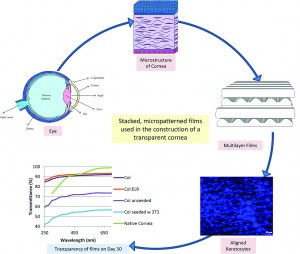
Hasirci and colleagues have developed a new type of engineered corneal construct using a combination of cells derived from the cornea as well as organized elastin-based biomaterials.  

The stroma is a layer in our cornea that consists of highly organized layers of collagen fibers and keratocytes, a special type of corneal cell.  The cells and the collagen layers are aligned with one another and follow a specific pattern of organization.   This organized structure allows the cornea to be transparent and to have the mechanical strength necessary to function correctly.

This study used specific biomaterials that draw inspiration from nature.  Specifically, the researchers used elastin, a commonly found biomaterial, as a template, and created a biomaterial with a similar protein composition. They modified a traditional elastin-based sequence to include the amino acid chain YIGSR, which is known to help cells attach to biomaterial structures.  They blended their elastin-based biomaterial with collagen, and used a silicon template to mold these biomaterials into sheets with surfaces that contain micron-scale grooves or micropatterns that cells can sense and align to. 

The biomaterial sheets were dehydrated completely under vacuum, causing the films to form internal bonds or crosslinks.  The crosslinking confers mechanical stability to the sheets, ensuring that the sheets do not degrade too quickly in response to enzymes secreted by keratocytes.  To test this idea, the biomaterial sheets were then subjected to a collagen-degrading enzyme, collagenase II.  The crosslinked sheets were better able to resist degradation for 24 hours as compared to their non-crosslinked counterparts.  This suggested that the keratocytes could form organized layers on the crosslinked biomaterials before degrading away the original structures. To further test these materials, the biomaterial sheets were then immersed in a saline solution for 4 weeks to study non-enzyme based degradation, and the crosslinked sheets again resisted degradation better than the non-crosslinked sheets. The crosslinked sheets were also able to transmit light, which is crucial for their application as corneal replacements.

Finally, the researchers tested these biomaterial sheets with human corneal keratocytes. The cells were able to survive on the biomaterial and grew well on the micropatterned biomaterial sheets. The cells also followed the alignment of the micropatterns on the surface of the biomaterials. The researchers then placed multiple layers of the micropatterned scaffolds together such that each successive patterned layer was perpendicular to the previous layer, and ‘glued’ the scaffolds together using droplets of collagen.  Cells were then seeded onto each layer using a syringe. They found that the keratocytes remained aligned for a period of 21 days in culture. The material was transparent even with the presence of cells for up to 4 weeks. In fact, the presence of keratocytes contributed to the construct’s overall transparency.

This work is an elegant demonstration of how biomaterials and cells can be combined to produce a viable tissue-engineered construct that may one day be used as corneal replacement therapy.

A collagen-based corneal stroma substitute with micro-designed architecture
Cemile Kilic, Alessandra Girotti, J. Carlos Rodriguez-Cabello and Vasif Hasirci  
Biomater. Sci., 2014, Advance Article DOI: 10.1039/C3BM60194C

Debanti Sengupta recently completed her PhD in Chemistry from Stanford University.  She is currently a Siebel postdoctoral scholar at the University of California, Berkeley.  

Follow the latest journal news on Twitter @BioMaterSci or go to our Facebook page.

To keep up-to-date with all the latest research, sign-up to our RSS feed or Table of contents alert.

Digg This
Reddit This
Stumble Now!
Share on Facebook
Bookmark this on Delicious
Share on LinkedIn
Bookmark this on Technorati
Post on Twitter
Google Buzz (aka. Google Reader)

Honeycomb surfaces for stem cell differentiation

The ability for a stem cell to differentiate into a specific cell type depends on the cell’s surrounding microenvironment. Variables such as substrate stiffness, presence of certain growth factors, fluid flow, and extracellular matrix composition in the cell culture environment all play a role in the cell’s ability to differentiate. Additionally, there is great interest in studying the effects of surface topology on stem cell differentiation in vitro to more accurately mimic the in vivo microenvironment. Using various micro-fabrication technologies, 2D and 3D substrates with varying topographic features including grooves, pits, pores, and posts can be fabricated and tested with cells to evaluate cell response.

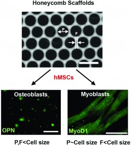
Researchers at Tohoku University in Japan have fabricated “honeycomb” topological substrates to regulate the differentiation of mesenchymal stem cells. The substrates are manufactured using a chloroform solution of polystyrene and an amphiphilic co-polymer, poly(N-dodecyl acrylamide-co-6-acrylamidehexanoic acid), or dioleoylphosphatidylethanolamine (DOPE). The solutions were cast on a glass slide and exposed to a flow of humid air to generate polystyrene films that resemble honeycomb from a beehive. The honeycomb substrates had varying pore size and strut width depending on the chloroform evaporation time.

After obtaining four different honeycomb films with varying pore size and strut width, human mesenchymal stem cells (hMSCs) were seeded on the surfaces to evaluate cell differentiation into osteo-specific (bone) and myo-specific (muscle) cells. To determine the cell differentiation state, cells were stained with osteoblast and myoblast markers, osteopontin and MyoD1, respectively.

Interestingly, when both the pore size and strut width were smaller than hMSC size, cells showed globular morphologies and increased expression of osteopotin (consistent with osteo-specific differentiation). On the other hand, when the pore size was the same size as hMSCs, the cells expressed MyoD1 and had elongated morphologies (consistent with myo-specific differentiation). Given the promising morphological and staining results, further studies are required to fully characterize the molecular mechanisms of the differentiation process.

This study demonstrates that geometries of honeycomb topographical substrates may have a direct influence on stem cell differentiation. Surface topology of biomaterial surfaces must be taken into account for future designs of cell culture environments for stem cell differentiation studies.

Honeycomb-shaped surface topography induces differentiation of human mesenchymal stem cells (hMSCs): uniform porous polymer scaffolds prepared by the breath figure technique
Takahito Kawano, Madoka Sato, Hiroshi Yabu and Masatsugu Shimomura 
Biomater. Sci., 2014, Advance Article DOI: 10.1039/C3BM60195A

Brian Aguado is currently a Ph.D. Candidate and NSF Fellow in the Biomedical Engineering department at Northwestern University. He holds a B.S. degree in Biomechanical Engineering from Stanford University and a M.S. degree in Biomedical Engineering from Northwestern University. When he’s not in the lab, Brian enjoys traveling, cooking, swimming, and spending time with family and friends. Read more about Brian’s research publications here.

To keep up-to-date with all the latest research, sign-up to our RSS feed or Table of contents alert.
Digg This
Reddit This
Stumble Now!
Share on Facebook
Bookmark this on Delicious
Share on LinkedIn
Bookmark this on Technorati
Post on Twitter
Google Buzz (aka. Google Reader)

Bioactive hydrogel beads for iPS cell expansion


Induced pluripotent stem (iPS) cells continue to hold great promise for cell transplantation therapies for numerous debilitating diseases. For instance, a patient’s skin cells can be “re-programmed” into iPS cells and converted into heart, liver, or bone cells (to name a few). These cells can then be used to regenerate specific tissues, and would theoretically bypass the patient’s immune response since the original cells come directly from the patient.

However, the scalability and growth of iPS cell in vitro continues to be a significant challenge. To address this problem, researchers at the University of Tokyo and École Polytechnique Fédérale de Lausanne have engineered bioactive hydrogel beads to significantly increase the expansion of iPS cells in vitro while maintaining their pluripotency.

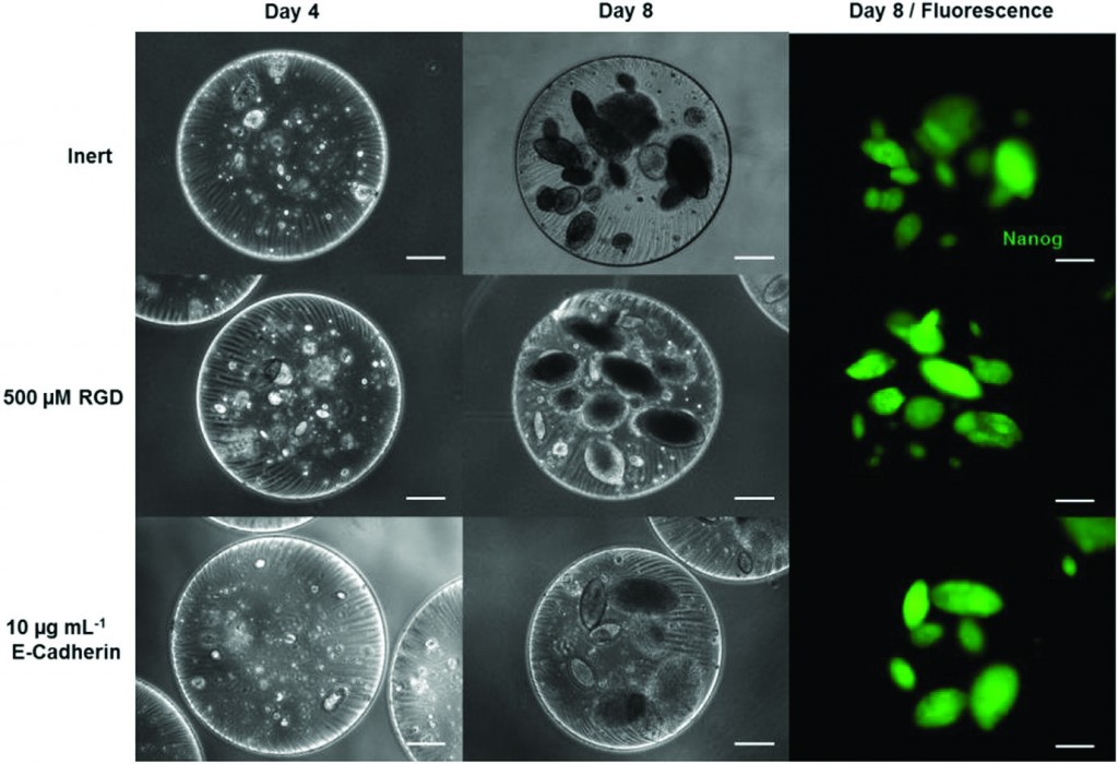
Mouse iPS cells were readily encapsulated and expanded in alginate/polyethylene glycol (PEG) gel beads using an effective three-step protocol.  First, iPS cells were added to a precursor solution of functionalized PEG and alginate. The precursor solution was loaded into a linear extrusion vessel and allowed to drip into a calcium chloride bath to crosslink the alginate and form gel beads. Second, the maleimide-functionalized 4-armed PEG in the beads was further crosslinked with di-thiolated linear PEG using a Michael addition reaction. Finally, the fully-crosslinked beads were coated with poly(L-lysine) to prevent volumetric swelling of the beads, since swelling imparts harmful mechanical stress to the encapsulated cells during the culture process.

Interestingly, the encapsulated iPS cells were pluripotent after 8 days in culture (confirmed with Nanog expression and colony morphology). To make the beads “bioactive,” the PEG networks were functionalized with cell adhesion components (RGD peptide or E-cadherin) to enhance iPS cell expansion. Overall, increasing the concentration of RGD binding sites in the bead offers up to an 80% increase in iPS cell proliferation compared to non-bioactive bead controls.

Additional experiments demonstrate that the expression of pluripotency markers and primitive endoderm markers is a function of bead design. Notably, the pluripotency marker Rex1 was significantly more expressed in beads containing RGD or E-Cadherin compared to non-bioactive beads. Furthermore, endoderm markers such as Gata4 and HNF4 were down regulated in beads containing specific concentrations of RGD. These results support the conclusion that the design of the bioactive bead has direct effects on the pluripotency of encapsulated iPS cells.

Ultimately, bioactive hydrogel beads are a promising solution for increasing iPS cell expansion in vitro, and are well suited for use in future regenerative medicine applications.

Development of bioactive hydrogel capsules for the 3D expansion of pluripotent stem cells in bioreactors
Yoji Tabata, Ikki Horiguchi, Matthias P. Lutolf and Yasuyuki Sakai  
Biomater. Sci., 2014, Advance Article DOI: 10.1039/C3BM60183H

Brian Aguado is currently a Ph.D. Candidate and NSF Fellow in the Biomedical Engineering department at Northwestern University. He holds a B.S. degree in Biomechanical Engineering from Stanford University and a M.S. degree in Biomedical Engineering from Northwestern University. When he’s not in the lab, Brian enjoys traveling, cooking, swimming, and spending time with family and friends. Read more about Brian’s research publications here.

To keep up-to-date with all the latest research, sign-up to our RSS feed or Table of contents alert.

Digg This
Reddit This
Stumble Now!
Share on Facebook
Bookmark this on Delicious
Share on LinkedIn
Bookmark this on Technorati
Post on Twitter
Google Buzz (aka. Google Reader)

Creating functional muscle in a dish

Hickman et al. have developed methods to successfully differentiate human muscle progenitor cells into functional, multinucleated myofibers. 

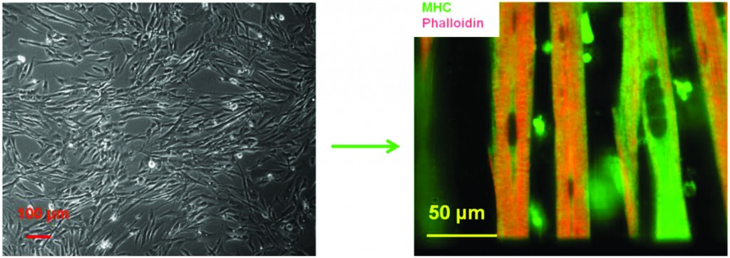
The first step towards replacing muscle lost due to diseases such as muscular dystrophy is to build functional model systems of healthy muscle tissue.  In order to do this with human cells, human muscle progenitor cells must be removed from the body, cultured in a dish, and treated with appropriate signaling molecules in order to appropriately differentiate into functional muscle fibers, known as myotubes or myofibers.  Hickman et al. have done precisely this – the authors demonstrate that it is possible to differentiate muscle progenitor cells into human myofibers outside of the body.  These myofibers are also shown to contract both in response to external electrical stimulation as well as spontaneously.

The researchers developed a special media cocktail that allowed these myofibers to grow in culture over a period of more than two weeks.  Importantly, the media cocktail did not contain serum, an undefined extract from animal blood that is often used to keep grow cells in culture outside of the body.  This has important implications for eventual clinical translation.  The researchers found that their differentiation medium produced myofibers that contained organized sarcomeres (the internal cell machinery that allows fibers to contract and do work). Muscle contraction generally occurs through the uptake of calcium by the cells from their surroundings.  Calcium uptake requires the presence of two different types of calcium channels, DHPR and RyR.  The researchers generated myofibers that demonstrated the presence of both of these channels.  Finally, these myofibers also contracted on their own, as well as in response to an applied electrical potential. 

This research marks an important step forward in muscle development research, and can be found here.


Spontaneous contraction of human myotubes in culture

In vitro differentiation of functional human skeletal myotubes in a defined system
Xiufang Guo, Keshel Greene, Nesar Akanda, Alec S. T. Smith, Maria Stancescu, Stephen Lambert, Herman Vandenburgh and James J. Hickman  
Biomater. Sci., 2013, Advance Article  DOI: 10.1039/C3BM60166H

Debanti Sengupta recently completed her PhD in Chemistry from Stanford University.  She is currently a Siebel postdoctoral scholar at the University of California, Berkeley.  

Follow the latest journal news on Twitter @BioMaterSci or go to our Facebook page.

To keep up-to-date with all the latest research, sign-up to our RSS feed or Table of contents alert.

Digg This
Reddit This
Stumble Now!
Share on Facebook
Bookmark this on Delicious
Share on LinkedIn
Bookmark this on Technorati
Post on Twitter
Google Buzz (aka. Google Reader)

Better bioceramics for bone generation: the importance of crystal structure

Bioactive ceramics are commonly used for the repair and replacement of damaged bone tissues.  However, there is still a limited understanding of how the inherent material properties of these ceramics can be used to predict their osteoinductive properties.  The development of models which relate these quantities could lead to the more efficient and intelligent design of materials that promote bone formation.

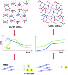
In a study that pushes this goal one step closer to reality, researchers from the University of Wisconsin, Madison, the University of Akron and the University of Michigan investigate the role which crystal structure plays in controlling the attachment and osteogenic differentiation of human mesenchymal stem cells (hMSCs).  The group studied hMSC viability and differentiation on two CaSiO3 polymorphs: pseudowollastonite (psw) and wollastonite (wol).  As polymorphs, these materials have identical chemical compositions and stoichiometries, and can be fabricated to have nearly equivalent surface roughness.  The only significant difference between these two materials is their crystal structure.  While the wol polymorph has a stable, open silicate chain structure, the psw polymorph has a highly strained silicate ring structure. 

Interestingly, significant differences in hMSC growth and differentiation were observed, with the psw polymorph exhibiting superior osteoinductive properties.  Although the hMSCs cultured on psw initially exhibited poor viability, they were able to recover and, after 20 days, exhibited a statistically higher cell count than the hMSCs cultured on wol.  Fine-grained calcium phosphate precipitate developed on the surfaces of both polymorphs but only the psw polymorph exhibited polycrystalline calcite aggregates as well as nodular amorphous precipitates which were identified as CaP, or bone nodules.

The group attributed these differences in hMSC behavior to the different soluble factor release rates exhibited by the two materials.  Psw has a much higher initial dissolution rate than wol, presumably because its highly strained structure makes it more susceptible to hydrolysis.  This resulted in a large initial release of silicon atoms, which caused the initial low cell viability, and calcium atoms, which caused increased calcite and calcium phosphate formation.

Overall, the study proved that psw is more osteoinductive than wol, thereby showing that crystal structure can play an enormous role in determining the osteoconductive and osteoinductive properties of a bioceramic.  The collected data mark a promising step forward in the creation of a model that can successfully predict osteoinductivity based on a bioceramic’s inherent material properties.

Crystal structures of CaSiO3 polymorphs control growth and osteogenic differentiation of human mesenchymal stem cells on bioceramic surfaces
Nianli Zhang, James A. Molenda, Steven Mankoci, Xianfeng Zhou, William L. Murphy and Nita Sahai  
Biomater. Sci., 2013, 1, 1101-1110 DOI: 10.1039/C3BM60034C

Ellen Tworkoski is a guest web-writer for Biomaterials Science.  She is currently a second year Ph.D. student in the biomedical engineering department at Northwestern University (Evanston, IL, USA).

Follow the latest journal news on Twitter @BioMaterSci or go to our Facebook page.

To keep up-to-date with all the latest research, sign-up to our RSS feed or Table of contents alert.

Digg This
Reddit This
Stumble Now!
Share on Facebook
Bookmark this on Delicious
Share on LinkedIn
Bookmark this on Technorati
Post on Twitter
Google Buzz (aka. Google Reader)

Multi-layer collagen sheets for soft tissue engineering

Collagen has been widely used in scaffold fabrication protocols in the tissue-engineering field for decades, since the material is biocompatible and biodegradable. However, the structure of collagen molecules is often disrupted during various processing techniques (e.g. decellularization or electrospinning), which decreases the mechanical stiffness of the material. There is an urgent need to generate mechanically robust materials that integrate with the host tissue for soft tissue engineering applications.

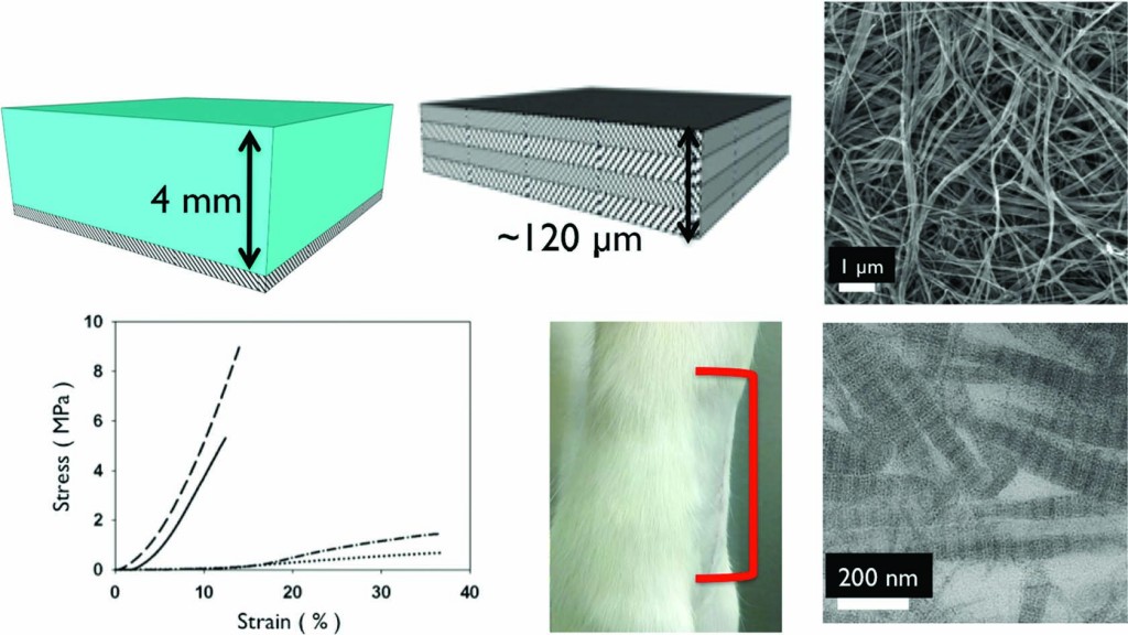
To address this issue, Elliot Chaikof’s group at Harvard Medical School has taken a “bottom-up approach” to fabricate thick collagen mats without damaging the structure of collagen molecules. Collagen gels were cast in a rectangular mold and dried to generate a sheet of collagen approximately 15-40 microns thick. Using a layer-by-layer fabrication strategy, subsequent collagen layers were cast on top of the previous layer and dried to form 120 μm thick sheets. The casting method successfully preserves the collagen molecule microstructure, and allows for tunable mechanical properties over several orders of magnitude as a function of concentration, thickness, and number of layers.

After elegantly characterizing the material properties, the collagen mats were used in an in vivo model of ventral hernia repair. The mats were compared to commercially available PermacolTM collagen sheets currently used for hernia repair. After surgically forming a hernia in the abdomen of rats, the collagen mats were sutured on the abdominal wall. After 3 months, the collagen mats showed significantly enhanced integration at the implant site over the PermacolTM controls.

Vivek Kumar, lead author on the study, explains that the collagen mats were 65% thinner than the 1 mm thick PermacolTM matrices – potentially accounting for the enhanced integration of the mat at the implant site. Even though the collagen mats were thinner than the commercially available collagen sheets, they were able to prevent re-herniation in all the rats tested due to the comparable mechanical properties of the collagen mat to the PermacolTM matrices.

Additionally, the layer-by-layer stacking method could be used to incorporate specific drugs in between the layers (much like making a sandwich) to reduce the risk of re-herniation and stimulate new tissue formation. Kumar also claims that these collagen mats can be used for a wide array of applications, such as vascular tissue replacements, artificial skin, and dura mater replacements.

 Interestingly, Kumar also suggests the method he developed is scalable. “We have cast much larger gels and much smaller gels successfully,” Kumar says. “The fabrication scheme is amenable to a variety of other geometries such as tubes and other intricate mold designs. The simplicity of our approach suggests good potential for use in industry – where often times a simpler fabrication method is better.”

This innovative research shows a simple method to create mechanically robust collagen mats that integrate exceptionally well with the surrounding tissue, and competes with current commercially available materials.

Collagen-based substrates with tunable strength for soft tissue engineering
Vivek A. Kumar, Jeffrey M. Caves, Carolyn A. Haller, Erbin Dai, Liying Liu, Stephanie Grainger and Elliot L. Chaikof 
Biomater. Sci., 2013, 1, 1193-1202 DOI: 10.1039/C3BM60129C

Brian Aguado is currently a Ph.D. Candidate and NSF Fellow in the Biomedical Engineering department at Northwestern University. He holds a B.S. degree in Biomechanical Engineering from Stanford University and a M.S. degree in Biomedical Engineering from Northwestern University. When he’s not in the lab, Brian enjoys traveling, cooking, swimming, and spending time with family and friends. Read more about Brian’s research publications here.

To keep up-to-date with all the latest research, sign-up to our RSS feed or Table of contents alert.

Digg This
Reddit This
Stumble Now!
Share on Facebook
Bookmark this on Delicious
Share on LinkedIn
Bookmark this on Technorati
Post on Twitter
Google Buzz (aka. Google Reader)

Using structural changes in carbon nanostructures to modify cellular responses

Carbon nanostructures have rapidly become subjects of increasing interest for use in biomedical applications ranging from drug delivery to bioimaging.  This is primarily due to their exceptionally high surface areas as well as their unique optical and electronic properties.  In this study, researchers from the Piramal Group in collaboration with the University of Florida investigated how the surface composition and structure of these materials affects their biological impact.

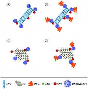
Specifically, the group investigated the protein binding capabilities, degree of cellular internalization, and cytotoxicity of carbon nanotubes (CNT) and graphene (G) sheets as well as CNT and G structures which had been conjugated with PAMAM dendrimers (G4) and polyethylene glycol (PEG).  These two polymers were chosen as conjugates as they are both known to improve the dispersibility and stability of various materials in solution.

Initial protein binding studies showed that the CNT and G conjugates exhibited weaker interactions with a model protein, bovine serum albumin (BSA), when compared to their non-conjugated counterparts.  These weaker interactions, which were thought to be primarily electrostatic in nature, could be extremely beneficial in limiting any possible functional interference which could arise from protein agglomeration on the nanostructure’s surface.  The group also studied the degree to which the different nanostructures were internalized within cells.  CNT conjugates were consistently found to be internalized in higher quantities than the G conjugates, presumably because their tubular shape promoted increased cell membrane penetration.  Finally, cytotoxicity studies illustrated that the conjugated nanostructures typically exhibited reduced toxicity levels compared to unmodified CNT or G, particularly at higher concentrations of the nanomaterial.

Overall, the study demonstrated that carbon nanostructures can have different biological activities depending on their shape and surface composition.  It also suggests that conjugation of carbon materials with polymers such as PEG and G4 might be an effective method for limiting protein binding and reducing cytotoxicity.


Structure effect of carbon nanovectors in regulation of cellular responses
Shashwat S. Banerjee, Archana Jalota-Badhwar, Prateek Wate, Sneha Asai, Khushbu R. Zope, Russel Mascarenhas, Dimple Bhatia and Jayant Khandare 
Biomater. Sci., 2013, Advance Article DOI: 10.1039/C3BM60082C

Ellen Tworkoski is a guest web-writer for Biomaterials Science.  She is currently a second year Ph.D. student in the biomedical engineering department at Northwestern University (Evanston, IL, USA).

Follow the latest journal news on Twitter @BioMaterSci or go to our Facebook page.

To keep up-to-date with all the latest research, sign-up to our RSS feed or Table of contents alert.

Digg This
Reddit This
Stumble Now!
Share on Facebook
Bookmark this on Delicious
Share on LinkedIn
Bookmark this on Technorati
Post on Twitter
Google Buzz (aka. Google Reader)