Author Archive

Facile Synthesis of Antibacterial Graphene Films Doped with Silver Nanoparticles

The development of antibacterial materials that can guard against microbial infections during medical procedures has been a topic of increasing interest. In this paper, a team of researchers from Beijing University of Chemical Technology and the University of Bremen describe a facile synthesis method for a hybrid nanomaterial that has previously demonstrated excellent antibacterial activity: reduced graphene oxide films decorated with silver nanoparticles. The efficient, dimensionally scalable, and environmentally friendly nature of this method make it a potentially valuable tool for other groups interested in fabricating these hybrid films for applications ranging from medical biomaterials, to cell culture scaffolds, to drug delivery platforms, to environmental remediation.

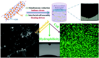
Simultaneous reduction and thermal evaporation-driven self assembly fabrication method used to create RGO/AgNP hybrid film
Schematic model for fabricating RGO/AgNP hybrid film

Zhang et al. created these hybrid films by using sodium citrate to simultaneously reduce aqueous graphene oxide (GO) and aqueous silver nitrate to reduced graphene oxide (RGO) and silver nanoparticles (AgNPs), respectively. This was followed by a prolonged incubation at 80°C to induce thermal evaporation-driven self-assembly of the hybrid RGO/AgNP films. The mass ratio of silver nitrate to GO in the initial solution could be adjusted to produce films with varying densities of AgNPs. In addition, this mass ratio as well the thermal evaporation duration provided some control over the thickness of the resulting hybrid film. The film’s size could be readily changed via adjustments to the dimensions of the reaction container and the film could be later transferred to other substrates without any additional post processing steps.

Subsequent characterisation of the film confirmed the successful reduction of GO as well as the formation of primarily spherical AgNPs between 12 and 30 nm in size on the surface of the film. The resulting material was found to be very hydrophilic and supported the adhesion and proliferation of mouse osteoblasts. An antibacterial assay measuring E. coli adhesion on the surface of silicon wafers coated with these RGO/AgNP films found that the few bacteria that did manage to adhere to the film’s surface exhibited damaged cell walls. A subsequent E. coli colony viability assay proved that no living bacterial colonies were present on the surface of the hybrid film, a result that could not be obtained when using either an uncoated silicon wafer or a pure RGO film.

The biocompatibility, hydrophilicity, durability, and antibacterial activity of these RGO/AgNP hybrid films make them intriguing candidates for a number of medical and environmental applications. The simple, dimension-scalable, and environmental friendly synthesis method presented by Zhang et al. in this paper opens up new avenues for the creation and widespread use of these versatile hybrid films.

Check out the full article:
Graphene film doped with silver nanoparticles: self-assembly formation, structural characterizations, antibacterial ability, and biocompatibility by Panpan Zhang, Haixia Wang, Xiaoyuan Zhang, Wei Xu, Yang Li, Qing Li, Gang Wei, and Zhiqiang Su

Ellen Tworkoski is a Web Writer for Biomaterials Science and is currently a graduate student in the biomedical engineering department at Northwestern University (Evanston, IL, US).

Follow the latest journal news on Twitter @BioMaterSci or go to our Facebook page.

Digg This
Reddit This
Stumble Now!
Share on Facebook
Bookmark this on Delicious
Share on LinkedIn
Bookmark this on Technorati
Post on Twitter
Google Buzz (aka. Google Reader)

Size of Internalized Calcium Phosphate Particles Plays a Critical Role in Cell Fate

Calcium phosphate (CaP) based materials have long been popular choices for a range of medical applications including bone replacement and drug delivery.  However, recent studies indicate a need for a closer look at how cells react to small, degraded CaP particles that find their way into the cell’s interior.  In a recent study, a research team from the University of Birmingham demonstrated that when CaP particles with a diameter larger than 1.5μm penetrated the interior of the cell but were not sequestered by the cell’s lysosomes, a series of events eventually leading to cell death could be observed.

Within the past few years, an increased focus on the end results of CaP degradation have shown that the eventual cytotoxicity of these materials is heavily dependent on the total volume of internalized material.  A study by Motskin et al. in 2009 illustrated that many of these degraded particles are localized to the lysosomes, cellular structures whose acidic environments are responsible for the degradation of waste products which, in the case of CaP, results in the generation of calcium ions.  It was suggested that the generation of a surplus of these ions could interfere with the cell’s signalling pathways, potentially triggering apoptosis.

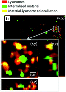
Intracellular Distribution of CaP showing co-localization with lysosomes

In a study published recently in Biomaterials Science, Williams et al. quantified the volume and size distribution of CaP material within cells by chemically grafting a fluorescent probe to the surface of silicon-substituted hydroxyapatite (SiHA), and then exposing a mouse osteoblast precursor cell line to the labelled particles.  Following exposure, the group observed changes in cell behaviour that were indicative of the onset of cell death including alterations in cell morphology, an increase in the number of lysosomes, and cellular detachment from the underlying substrate.  As the cells transitioned from the early stages of cell death (morphology changes) to later stages (detachment from the substrate), there was a marked increase in the amount of SiHA within the cytoplasm but outside of the lysosomes.  Moreover, the onset of cell death was correlated with SiHA aggregates within the cytoplasm that were 1.5μm in diameter or larger.

The group hypothesized that this may have been due to a destabilization of the lysosome membrane which prevented undissolved CaP from remaining within the lysosomes or, alternatively, the destabilization of endosomes which were responsible for delivering particles to the lysosomes.  Regardless, this study proves a need for additional research into the size-dependent effects of CaP particles on cell health and suggests a new design concern for CaP based medical materials.

Check out the full article here:
Quantification of volume and size distribution of internalised calcium phosphate particles and their influence on cell fate
by Richard L. Williams, Isaac Vizcaíno-Castón, and Liam M. Grover


Ellen Tworkoski is a Web Writer for Biomaterials Science and a graduate student in the department of Biomedical Engineering at Northwestern University.

Digg This
Reddit This
Stumble Now!
Share on Facebook
Bookmark this on Delicious
Share on LinkedIn
Bookmark this on Technorati
Post on Twitter
Google Buzz (aka. Google Reader)

Controlling neuronal behavior with nano-topography

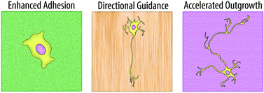
Morphological and cellular changes in neurons in response to different nanotopographies

The ability to control neuronal behavior and growth is highly sought among researchers interested in neuro-regenerative medicine.  Over the past two decades, it has become widely accepted that modifications in a substrate’s physical surface topography can influence the growth patterns of seeded neurites.  However, the advent of techniques that are capable of fabricating nano-textured surfaces has revealed that the magnitude of this influence may be much greater than originally thought.  In this paper, a group of scientists from KAIST review a number of recent advances in the development of nano-topographies which can influence neuronal behavior.

Many of these advances revolve around improving neural adhesion, providing directional guidance for axonal growth, and accelerating the speed of neurite outgrowth.  Numerous groups have become interested in using nanowire arrays to improve neuron adhesion, eliminating the need for any additional surface coating.  In a 2010 Nano Letters publication, Xie et al. were able to demonstrate the creation of a silicon nanopillar array that could selectively pin cortical neurons to desired locations.  Any cells that were seeded onto, or later came into contact with the nanopillars became immobilized, enabling a long-term observational study of their electrical activity.  Other groups have found that nanofiber bundles make excellent scaffolding materials for neurons and can be used to direct and align neurite growth.  In addition, when compared to planar surfaces, nanofiber-based substrates were shown to increase the speed at which initial neurite formation occurred.

Although the benefits of nano-textured substrates are clear, the mechanism by which neurons translate these physical cues into biological signals is still something of a mystery.  However, many of the reviewed papers suggest that actin filaments and focal adhesions (FA) play a major role.  One group found that f-actin often forms networks which resemble the shape of the underlying surface topography and that the impairment of f-actin polymerization completely erases any ability neurons have to respond to nanotopographical cues.  Other groups have demonstrated that the size of FA is proportional to the degree of neuronal alignment with underlying surface structures.  An increase in alignment leads to a larger FA which ultimately results in a more stable neurite attachment.

Regardless of the mechanism, it has become clear in recent years that nanotopography is an important parameter for the controlled manipulation of neuronal behavior.  In this mini-review, Kim et al. are able to provide an interesting overview on the progress that has already been made within this area.

Neurons on nanometric topographies: insights into neuronal behaviors in vitro
M. Kim, M. Park, K. Kang, and I.S. Choi.
Biomater. Sci., 2014, 2, 148-155 DOI: 10.1039/C3BM60255A

Ellen Tworkoski is a guest web-writer for Biomaterials Science.  She is currently a graduate student in the biomedical engineering department at Northwestern University (Evanston, IL, USA).

Follow the latest journal news on Twitter @BioMaterSci or go to our Facebook page.

To keep up-to-date with all the latest research, sign-up to our RSS feed or Table of contents alert.

Digg This
Reddit This
Stumble Now!
Share on Facebook
Bookmark this on Delicious
Share on LinkedIn
Bookmark this on Technorati
Post on Twitter
Google Buzz (aka. Google Reader)

Inducing biomimetic mineralization using a graphene oxide-chitosan hybrid material

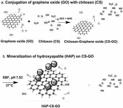
Chemical conjugation of graphene oxide with chitosan and subsequent mineralization of hydroxyapatite

One of the primary goals within the field of hard tissue engineering is to create biomaterials that can both promote osteoblast proliferation and support the natural bone mineralization process.  In this study, a trio of researchers at the University of Louisiana at Lafayette has developed a promising, layered hybrid system that contains hydroxyapatite (HAP), chitosan (CS), and graphene oxide (GO).  The GO base provides mechanical strength and biocompatibility while the surface layer which is composed of HAP, a natural constituent of bone, has a high bioactivity.  The intermediate chitosan layer further strengthens the hybrid biomaterial and, perhaps more importantly, has been shown to promote more homogeneous mineralization that mimics the natural biomineralization process.

In order to test the bone forming capabilities of this hybrid material the group soaked it, along with two control materials (HAP-GO and pure GO), in simulated body fluid for three weeks.  Each substrate was then incubated with MC3T3-E1 pre-osteoblast cells.  Among the three biomaterials, the HAP-CS-GO hybrid produced the highest levels of cell proliferation and expansion.   It also induced the greatest expression levels of vinculin, actin, and fibronectin, proteins that are essential for cell adhesion, cytoskeletal organization, and osteoblast morphogenesis and mineralization.  The hybrid material also demonstrated a highly significant increase in mineralized area and bone nodule formation compared to either of the two controls.  This was primarily attributed to the strong electrostatic interactions between the functional groups in the CS-GO base and the calcium ions present in solution.

Overall the hybrid HAP-CS-GO material was able to favorably modulate cellular activity in a way that was conducive to bone formation, making it a promising candidate for future bone tissue engineering applications.   However, as the authors have highlighted, there is room for improvement.  A reoccurring issue that many bone tissue engineering materials, including this novel hybrid material, have faced is an inability to attain the proper ratio of mineral deposit to cellular matrix in the regenerated tissue.  This mineral to matrix ratio has been shown to be relatively high in healthy, intact bone (~4.35-5) but was observed in a significantly lower amount (1.76) in the HAP-CS-GO material.  Nonetheless, the hybrid biomaterial reported in this study represents a promising direction forward in the field of bone tissue engineering.

The synergistic effect of a hybrid graphene oxide-chitosan system and biomimetic mineralization on osteoblast functions
D. Depan, T. C. Pesacreta and R. D. K. Misra
Biomater. Sci., 2014, 2, 264-274 DOI: 10.1039/C3BM60192G

Ellen Tworkoski is a guest web-writer for Biomaterials Science.  She is currently a graduate student in the biomedical engineering department at Northwestern University (Evanston, IL, USA).

Follow the latest journal news on Twitter @BioMaterSci or go to our Facebook page.
To keep up-to-date with all the latest research, sign-up to our RSS feed or Table of contents alert.

Digg This
Reddit This
Stumble Now!
Share on Facebook
Bookmark this on Delicious
Share on LinkedIn
Bookmark this on Technorati
Post on Twitter
Google Buzz (aka. Google Reader)