Author Archive

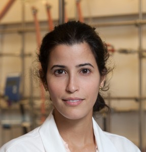
2014 Biomaterials Science Lectureship Awarded to Suzie Pun

It is with great pleasure that we announce Professor Suzie Pun (University of Washington) as the recipient of the inaugural Biomaterials Science Lectureship.

This award, which will be an annual event for the journal,  honours a younger scientist who has made a significant contribution to the biomaterials field. The recipient is selected by the Biomaterials Science Editorial Board from a list of candidates nominated by the community.

More about Suzie…

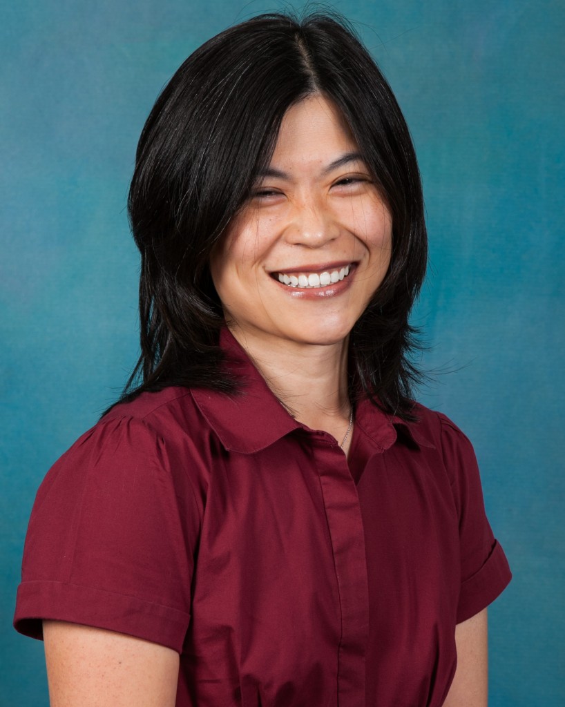
Suzie H. Pun received her Chemical Engineering Ph.D. degree in 2000 from the California Institute of Technology.  She then worked as a senior scientist at Insert Therapeutics/Calando Pharmaceuticals for 3 years developing polymeric drug delivery systems before joining the Department of Bioengineering at University of Washington (UW).  She is currently the Robert J Rushmer Associate Professor of Bioengineering, an Adjunct Associate Professor of Chemical Engineering, and a member of the Molecular Engineering and Sciences Institute at UW.  Her research focus area is in drug and gene delivery systems and she has published over 70 research articles in this area.  Current application areas for her group include biologics delivery to the central nervous system and cancer.  For this work, she was recognized with a Presidential Early Career Award for Scientists and Engineers in 2006. 

Take a look at this paper for an example of Suzie’s recent research:

Comparative study of guanidine-based and lysine-based brush copolymers for plasmid delivery Peter M. Carlson, Joan G. Schellinger, Joshuel A. Pahang, Russell N. Johnson and Suzie H. Pun  
Biomater. Sci., 2013, 1, 736-744 DOI: 10.1039/C3BM60079C 

We would like to thank everybody who nominated a candidate for the Lectureship- we received many excellent nominations, and the Editorial Board had a difficult task in choosing between some outstanding candidates.

We invite you to join us in congratulating Suzie in the comments below.

Digg This
Reddit This
Stumble Now!
Share on Facebook
Bookmark this on Delicious
Share on LinkedIn
Bookmark this on Technorati
Post on Twitter
Google Buzz (aka. Google Reader)

Cancer therapy with nanoparticles

Researchers have demonstrated that controlling nanoparticle shape and packing can impact how cancer therapeutics interact with cells.

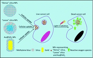
Photodynamic therapy is an innovative emerging therapy for cancer.  This therapeutic modality consists of introducing light-activated molecules known as ‘photosensitizers’ into cancer cells.  When activated with light, these photosensitizers are able to produce toxic molecules within the cells (also known as reactive oxygen species or ROS) that can eventually cause cancer cell death.  Photosensitizers are often introduced into the cells using nanoparticle carriers. In order for effective cell death to occur, ROS must be formed within the nanoparticles and then diffuse out of the nanoparticles into the cells.

In this article, Chu et al. have designed silica-based nanocarriers that can control how photosensitizers interact with cells. They previously found that when photosensitizer-loaded nanoparticles were packed more densely, the amount of ROS that was released decreased.  In contrast, more loosely packed nanoparticles allowed for greater release of ROS into cells. In this study, the researchers designed a third type of nanoparticle consisting of a gold nanorod coated with dense silica. They found that this third type of nanoparticle released photosensitizers much more efficiently than the either densely packed or loosely packed silica-only particles. They also discovered that the nanorod-based particles produced a different distribution of ROS as compared to the loose or dense silica-only particles. When cells were treated with these nanoparticles, loosely-packed silica particles and the nanorod-based particles demonstrated dramatic decreases in cell-viability due to the action of the photosensitizers. Further, while the silica-based loose and dense particles caused cells to die via a programmed cell death (apoptotic) route, the nanorod-based particles caused cell death via cell injury resulting in premature cell death (necrosis).

These experiments suggest that controlling the composition and shape of the nanoparticles that carry photosensitizers to cancer cells can alter both the number of cancer cells killed as well as the mechanism by which the cells die. This work has implications for future photodynamic therapeutic strategies.

Zhiqin Chu, Silu Zhang, Chun Yin, Ge Lin and Quan Li
Biomater. Sci., 2014, Advance Article DOI: 10.1039/C4BM00024B

Debanti Sengupta recently completed her PhD in Chemistry from Stanford University.  She is currently a Siebel postdoctoral scholar at the University of California, Berkeley.

Follow the latest journal news on Twitter @BioMaterSci or go to our Facebook page.

Digg This
Reddit This
Stumble Now!
Share on Facebook
Bookmark this on Delicious
Share on LinkedIn
Bookmark this on Technorati
Post on Twitter
Google Buzz (aka. Google Reader)

Emerging Investigators themed issue now published

We are pleased to announce that the Biomaterials Science 2014 Emerging Investigators themed issue is now available to read online.

Edited by Phillip Messersmith and Norio Nakatsuji, co-Editors in Chief of Biomaterials Science, this issue highlights the exciting and important work being carried out by some of the most talented up-and-coming researchers in the field.  Read more about the issue in the Editorial.

Here is a sample of the reviews, communications and papers that feature in the Emerging Investigators themed issue:

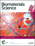
On the cover

Fabrication of zeolite–polymer composite nanofibers for removal of uremic toxins from kidney failure patients Koki Namekawa, Makoto Tokoro Schreiber, Takao Aoyagi and Mitsuhiro Ebara 

 
Review
Smart hydrogels as functional biomimetic systems Han L. Lim, Yongsung Hwang, Mrityunjoy Kar and Shyni Varghese 
 
Minireviews
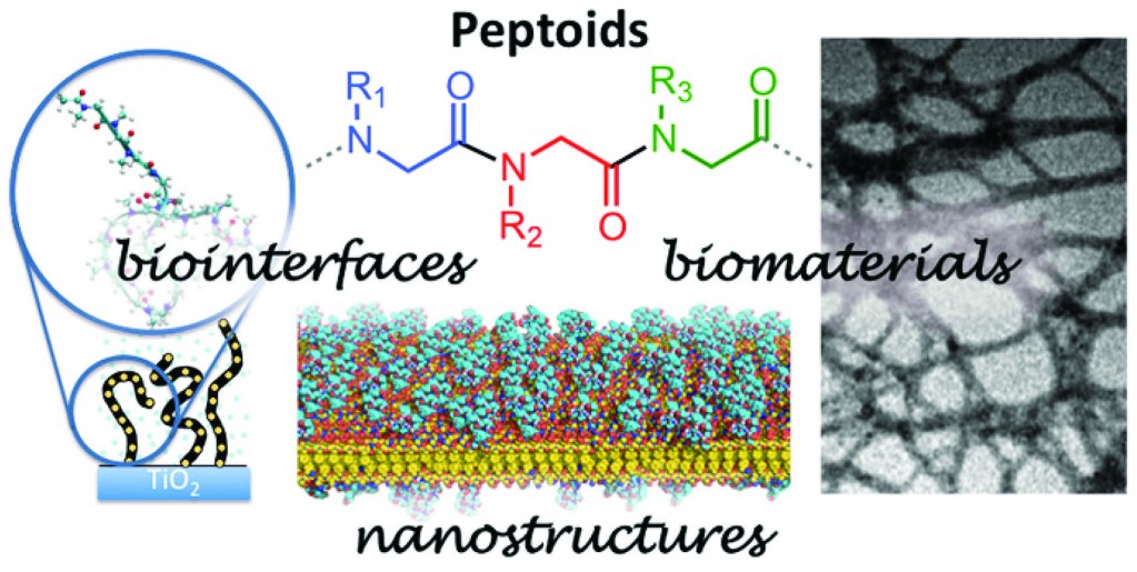
Peptoids for biomaterials science King Hang Aaron Lau 
 
Communications
 
Hyaluronic acid hydrogel stiffness and oxygen tension affect cancer cell fate and endothelial sprouting Yu-I Shen, Hasan E. Abaci, Yoni Krupski, Lien-Chun Weng, Jason A. Burdick and Sharon Gerecht
 
Papers
Translocation of flexible polymersomes across pores at the nanoscale Carla Pegoraro, Denis Cecchin, Jeppe Madsen, Nicholas Warren, Steven P. Armes, Sheila MacNeil, Andrew Lewis and Giuseppe Battaglia
 
Structural reinforcement of cell-laden hydrogels with microfabricated three dimensional scaffolds Chaenyung Cha, Pranav Soman, Wei Zhu, Mehdi Nikkhah, Gulden Camci-Unal, Shaochen Chen and Ali Khademhosseini
 
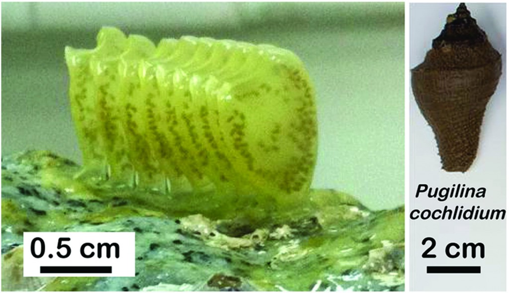
Integrative and comparative analysis of coiled-coil based marine snail egg cases – a model for biomimetic elastomers Paul A. Guerette, Gavin Z. Tay, Shawn Hoon, Jun Jie Loke, Arif F. Hermawan, Clemens N. Z. Schmitt, Matthew J. Harrington, Admir Masic, Angelo Karunaratne, Himadri S. Gupta, Koh Siang Tan, Andreas Schwaighofer, Christoph Nowak and Ali Miserez
 
 
Molecular farming of fluorescent virus-based nanoparticles for optical imaging in plants, human cells and mouse models S. Shukla, C. Dickmeis, A. S. Nagarajan, R. Fischer, U. Commandeur and N. F. Steinmetz 
 
More papers from the themed issue can be downloaded here.
 
Digg This
Reddit This
Stumble Now!
Share on Facebook
Bookmark this on Delicious
Share on LinkedIn
Bookmark this on Technorati
Post on Twitter
Google Buzz (aka. Google Reader)

NanoBio Australia 2014

Incorporating the 5th International NanoBio Conference & 3rd International Conference on BioNano Innovation (ICBNI), NanoBio Australia 2014 will take place on 6-10th July 2014 in Brisbane, Australia.

The intersection of Biology with Nanoscience and Nanotechnology currently represents one of the most exciting wellsprings of scientific innovation, and is also a major stimulus for a plethora of high value technologies and industries. By combining the two largest conferences in the world which focus on this scientific frontier, NanoBio Australia 2014 will feature a diverse array of multi-disciplinary science designed to connect world-leading scientists, engineers and entrepreneurs working in this space.

International Plenary Speakers:

Please see the conference website to register your attendance or submit an abstract.

Digg This
Reddit This
Stumble Now!
Share on Facebook
Bookmark this on Delicious
Share on LinkedIn
Bookmark this on Technorati
Post on Twitter
Google Buzz (aka. Google Reader)

Lectureship nominations close on Friday 7th March!

Time is running out to make your nominations for the Biomaterials Science Lectureship! Please submit your nominations by Friday 7th March 2014.

A reminder of the details…

Qualification

To be eligible for the Biomaterials Science Lectureship, the candidate should be in the earlier stages of their scientific career, typically within 15 years of attaining their doctorate or equivalent degree, and will have made a significant contribution to the field.

Description

The recipient of the award will be asked to present a lecture three times, one of which will be located in the home country of the recipient. The Biomaterials Science Editorial Office will provide the sum of £1000 to the recipient for travel and accommodation costs.

The award recipient will be presented with the award at one of the three award lectures. They will also be asked to contribute a lead article to the journal and will have their work showcased on the back cover of the issue in which their article is published.

Selection

The recipient of the award will be selected and endorsed by the Biomaterials Science Editorial Board.

Nominations

Those wishing to make a nomination should send details of the nominee, including a brief C.V. (no longer than 2 pages A4) together with a letter (no longer than 2 pages A4) supporting the nomination, to the Biomaterials Science Editorial Office (biomaterialsscience-rsc@rsc.org ) by 7th March 2014.  Self-nomination is not permitted.

We look forward to receiving your nominations!

Digg This
Reddit This
Stumble Now!
Share on Facebook
Bookmark this on Delicious
Share on LinkedIn
Bookmark this on Technorati
Post on Twitter
Google Buzz (aka. Google Reader)

Introducing new Editorial Board Member Adah Almutairi

We are very pleased to welcome Professor Adah Almutairi to the Biomaterials Science Editorial Board.

Adah Almutairi is co-director of the joint KACST-UC San Diego Center for Excellence in Nanomedicine and Engineering and an associate professor in the Skaggs School of Pharmacy and Pharmaceutical Sciences at UC San Diego, with secondary appointments in NanoEngineering and Materials Science. Her own research group, the Laboratory for Bioresponsive Materials, creates novel smart materials for on-demand drug delivery, regeneration of damaged tissue, and safe image-based diagnosis. She came to UC San Diego in 2008 from UC Berkeley, where she worked with Professor Jean Fréchet to develop several nanoprobes for in vivo imaging of pH and angiogenesis.  Prof. Almutairi is the recipient of an NIH New Innovator Award and has been recognized as a rising star in the field of polymeric materials by Chemical Communications and the ACS Division of Polymeric Materials Science and Engineering.

Adah’s recent papers include:

Increasing materials’ response to two-photon NIR light via self-immolative dendritic scaffolds
Nadezda Fomina, Cathryn L. McFearin and Adah Almutairi  
Chem. Commun., 2012, 48, 9138-9140 DOI: 10.1039/C2CC00072E

Metal chelating crosslinkers form nanogels with high chelation stability
Jacques Lux, Minnie Chan, Luce Vander Elst, Eric Schopf, Enas Mahmoud, Sophie Laurent and Adah Almutairi  
J. Mater. Chem. B, 2013, 1, 6359-6364 DOI: 10.1039/C3TB21104E, Paper

Antigen-loaded pH-sensitive hydrogel microparticles are taken up by dendritic cells with no requirement for targeting antibodies
Laura E. Ruff, Enas A. Mahmoud, Jagadis Sankaranarayanan, José M. Morachis, Carol D. Katayama, Maripat Corr, Stephen M. Hedrick and Adah Almutairi  
Integr. Biol., 2013, 5, 195-203 DOI: 10.1039/C2IB20109G

Digg This
Reddit This
Stumble Now!
Share on Facebook
Bookmark this on Delicious
Share on LinkedIn
Bookmark this on Technorati
Post on Twitter
Google Buzz (aka. Google Reader)

Announcing new members of the Biomaterials Science Advisory Board

We are very pleased to introduce the new members of the Biomaterials Science Advisory Board:

Jianwu Dai is currently a Professor at the Intitute of Genetics and Developmental Biology at the Chinese Academy of Sciences. His research is focused on stem cells and regerative medicine.

Professor Dai obtained a B.Sc. in Cell Biology at Wuhan University, China, before completing an M.Sc. in Biophysics at Beijing Medical University. He received his Ph.D. from Duke University Medical Center (USA) in 1998, before joining Harvard Medical School as a Postdoctoral trainee, working on animal genetics and stem cells.


Ali Khademhosseini is an Associate Professor at Harvard-MIT Division of Health Sciences and Technology, Brigham and Women’s Hospital and Harvard Medical School as well as an Associate Faculty at the Wyss Institute for Biologically Inspired Engineering and a Junior PI at Japan’s World Premier International-Advanced Institute for Materials Research at Tohoku University where he directs a satellite laboratory. He has authored more than 300 papers and 50 book chapters.   He has engineered a range of hydrogels for tissue engineering and utilized various micro- and nanoengineering approaches to further modify the hydrogel properties / architecture.

Dr. Khademhosseini’s interdisciplinary research has been recognized by over 30 major national and international awards.  He has received early career awards from three major engineering discipline societies: electrical (IEEE Engineering in Medicine and Biology Society award and IEEE Nanotechnology award), chemical (Colburn award from the AIChE) and mechanical engineering (Y.C. Fung award from the ASME).  He is also a recipient of the Presidential Early Career Award for Scientists and Engineers, the highest honour given by the US government for early career investigators. He is a fellow of the American Institute of Medical and Biological Engineering (AIMBE) and the American Association for the Advancement of Science (AAAS).   He received his Ph.D. in bioengineering from MIT (2005), and MASc (2001) and BASc (1999) degrees from University of Toronto, both in chemical engineering.
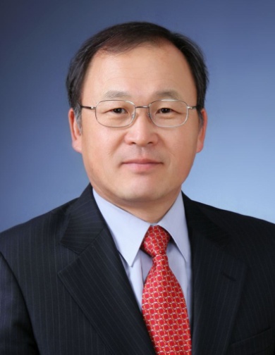

Doo Sung Lee received his B.S. degree in Chemical Engineering from the Seoul National University in 1978 and his M.S. and Ph.D. in Chemical Engineering from the Korea Advanced Institute of Science and Technology (KAIST). Since 1984 he has been a Professor of  the School of Chemical Engineering at the Sungkyunkwan University, where he served as the Dean of the College of Engineering from 2005 to 2007.

Doo Sung Lee was elected as a member of the Korean Academy of Science and Technology in 2011 and was made a member of the National Academy of Engineering of Korea in 2012. He was a president of the Polymer Society of Korea in 2013. Since 2010, he has been a director of Theranostic Macromolecules Research Center funded by National Research Foundation of Korea  His research group studies on the development of functionalized & biodegradable injectable hydrogels and micelles for controlled drug and protein delivery and molecular imaging.
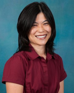

Suzie H. Pun received her Chemical Engineering Ph.D. degree in 2000 from the California Institute of Technology.  She then worked as a senior scientist at Insert Therapeutics for 3 years before joining the Department of Bioengineering at University of Washington (UW).  She is currently the Robert J Rushmer Associate Professor of Bioengineering, an Adjunct Associate Professor of Chemical Engineering, and a member of the Molecular Engineering and Sciences Institute at UW.  Her research focus area is in drug and gene delivery systems and she has published over 75 research articles in this area.  For this work, she was recognized with a Presidential Early Career Award for Scientists and Engineers in 2006.
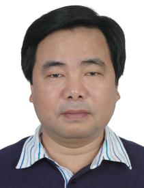

Xintao Shuai received his Ph. D. degree in 1996 from Beijing Institute of Technology (China). After working for some years as a visiting scholar or postdoc at North Carolina State University, Philipps-University Marburg and Case Western Reserve University, he joined Sun Yat-sen University, China in 2005 as a professor of polymer science in the School of Chemistry and Chemical Engineering and professor by courtesy of biomedical engineering in the School of Medicine. Dr. Shuai’s research interests include polymeric nano-biomaterials for drug delivery and MRI-visible theranostic systems for disease diagnosis and treatment. He has published over 80 peer reviewed journal articles.
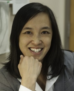

Joyce Wong is a Professor in Biomedical Engineering and Materials Science & Engineering at Boston University. She directs the Biomimetic Materials Engineering Laboratory which is focused on developing biomaterial systems that mimic physiological and pathophysiological environments to study fundamental cellular processes at the biointerface. Current research includes vascular tissue engineering, theranostics, and engineering biomimetic systems to study restenosis and cancer metastasis.


We are delighted to welcome these six distinguished scientists to the Biomaterials Science team. For a full list of Biomaterials Science Editorial and Advisory board members, please see the website.
Digg This
Reddit This
Stumble Now!
Share on Facebook
Bookmark this on Delicious
Share on LinkedIn
Bookmark this on Technorati
Post on Twitter
Google Buzz (aka. Google Reader)

3D micropatterns to grow cells

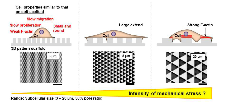
This paper by Sunami et al. studies the impact of 3D patterning on cells, and shows that cells grown on 3D micropatterns respond by changing their migration speed, their proliferation rate, and their expression of actin.

It is standard laboratory practice to grow cells outside the body on flat tissue culture plates.  However, cells inside the body are surrounded by other cells and connective tissue organized in three dimensions.  It is therefore very important to develop culture platforms for cells that more accurately capture a cell’s microenvironment inside the body. 

In this paper, the authors developed a three-dimensional culture platform that could be modified to study the properties of fibroblasts (skin cells) in culture. The authors changed only one factor – the area of the three-dimensional triangles that the cells were cultured on – to study the impact of 3D micropatterned areas on fibroblasts. The resultant surfaces that they used were about as adhesive as a smooth glass surface.  Interestingly, they found that fibroblast spreading was very different on surfaces with different areas, with maximum cell spreading observed on a surface with an intermediate pattern area.  The cells primarily adhered to the upper surface of the 3D micropatterns. Further, the density of the cells was also dependent on the micropattern area – as micropattern area increased, the density of the cells correspondingly increased as well. The cells also proliferated faster on surfaces with larger micropattern areas, while cell proliferation slowed on smaller triangles. The authors then used timelapse microscopy to study how these cells migrated over time, and found that while cells migrated more slowly on all of the micropatterned surfaces as compared to a completely flat surface, migration was slowest on the micropatterns with the smallest areas. Similarly, the authors found that f-actin expression was increased on the patterns with the largest areas. The authors hypothesize that this may mean that cells experience less mechanical stress as pattern size decreases.

Influence of the pattern size of micropatterned scaffolds on cell morphology, proliferation, migration and F-actin expression
Hiroshi Sunami, Ikuko Yokota and Yasuyuki Igarashi
Biomater. Sci., 2014, Advance Article DOI: 10.1039/C3BM60237K

Debanti Sengupta recently completed her PhD in Chemistry from Stanford University.  She is currently a Siebel postdoctoral scholar at the University of California, Berkeley.  

Follow the latest journal news on Twitter @BioMaterSci or go to our Facebook page.

Digg This
Reddit This
Stumble Now!
Share on Facebook
Bookmark this on Delicious
Share on LinkedIn
Bookmark this on Technorati
Post on Twitter
Google Buzz (aka. Google Reader)

Editor-in-Chief Phillip Messersmith interviewed in Chemistry World

Phillip Messersmith, Biomaterials Science co-Editor-in-Chief, has been interviewed in Chemistry World about his work on biological adhesives to develop new biomaterials for the repair, replacement, or augmentation of human tissue.

Here are some highlights from the interview:

…What are the main applications for your synthetic polymers, are they just biomedical?
Not exclusively, but the funding sources right now are primarily in health related areas. We have a lot of funding from the National Institutes of Health in the US and obviously their main interest at the end of the day is to contribute to basic understanding as well as the applications of new materials, new devices and new therapies. So we work through the government funding as well as some corporate and institutional funding towards applications. The application you mentioned before, fetal surgery, has been a great passion for me over the last few years. I only really became involved in this three–four years ago but it’s become really important to me.
It’s the kind of medical problem that has too small a market to interest big companies and so the surgeons work in this area and do wonderful things without having all the tools they would like. One example of a tool they need is for sealing ruptures in the fetal membrane that occur spontaneously or after an interventional procedure. The ruptures can lead to leakage of amniotic fluid and when that happens you have two major problems. First, is the risk of infection and second is the premature induction of labour. Either way you have a very serious medical problem for the mother and the fetus and there aren’t many ways to treat this apart from bed rest.
There’s a small community of fetal surgeons that have trained for many years to try and avoid these ruptures but if it happens there’s not a lot they can do. So we’re developing materials to try and seal the membranes after rupture. Here obviously the tissue is wet and there’s a large volume of high ionic strength fluid. This is not very different from the conditions encountered by mussels- thus providing a great argument for learning how mussels and other marine organisms can accomplish wet adhesion.

How easy is it to make these materials biocompatible?
That’s a great question and something we spend a lot of time thinking about. Biocompatibility is an all-encompassing word: but it’s all about context. All we can say is we try to develop systems based on biocompatible polymers and DOPA and then formulate them in a way that doesn’t induce a severe inflammatory response. But any synthetic material has some level of that response. There’s an interesting give and take between in vitro results and in vivo results. A positive in vitro result won’t necessarily translate to a positive in vivo result. One of the interesting things is that the opposite is also true. Sometimes in vivo cell toxicity assays give a borderline response but in vivo we see really good results. We choose the polymers and how we go about the functionalisation and purification very carefully and then we do in vitro and in vivo tests.

Going back slightly, what made you get into bioadhesion?
The guy I mentioned earlier, Herbert Waite. When I was a young faculty member I used to block off one full day a month and just go to the library and look at all the new journal issues that had come in. And I used to try and make a point of trying to read out of my comfort zone, in areas I really wasn’t trained in. And one of those times I encountered one of his papers which described these proteins, the mussel adhesive proteins. And I said, wow, this is really interesting. Then I started looking for more of his papers and it just struck me, as a materials scientist, as an interesting translation opportunity, which it’s turned out to be. To this day I often tell my students that story because I don’t think they really appreciate how important it is not just to read the literature, but to read the literature outside of what you happen to be looking for that day, that hour…

Read the full interview with Laura Howes here

Follow the latest journal news on Twitter @BioMaterSci or go to our Facebook page.

To keep up-to-date with all the latest research, sign-up to our RSS feed or Table of contents

Digg This
Reddit This
Stumble Now!
Share on Facebook
Bookmark this on Delicious
Share on LinkedIn
Bookmark this on Technorati
Post on Twitter
Google Buzz (aka. Google Reader)

Decreasing Tumor Growth with Magnetic Nanohydrogels

Cancer is the second leading cause of death worldwide, and continues to be a challenging disease to treat therapeutically. One strategy to treat cancer patients is to surgically remove the primary tumor and attempt to prevent the spread of cancer cells to other organs in the body. Unfortunately, surgical resection of tumors may be difficult in sensitive organs (e.g. the brain), and tumors may need to be treated directly with chemotherapy drugs to inhibit growth.

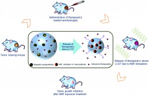
Recently, there has been an increased interest in utilizing biomaterials to deliver chemotherapy drugs directly to the primary tumor. Current research at the Indian Institute of Technology explores the use of nanohydrogels supplemented with iron oxide magnetic nanoparticles as a vehicle to deliver chemotherapy drugs to the primary tumor. Interestingly, these thermo-responsive nanohydrogels could release their anticancer drug cargo when heated with a magnetic field. Although this study did not use chemotherapy drugs, the magnetic hydrogels were used to heat the tumor locally to reduce tumor size – upon stimulation with a magnetic field. This study focused on characterizing the material properties of the magnetic nanohydrogels, in addition to analyzing the effects of the nanohydrogel on inhibiting tumor growth.

The magnetic nanohydrogel is composed of iron oxide nanoparticles, chitosan, and poly-N-isopropylacrylamide (NIPAAm).  The chitosan-poly-(NIPAAm) nanohydrogels are hydrophilic below the lower critical solution temperature (LCST) of 42oC. When stimulated with the appropriate magnetic field, the magnetic nanoparticles heat the hydrogel above the LCST. When the temperature is increased above the LCST, the polymers in the hydrogel change from “expanded coils” to “compact globules,” and cause the release of the aqueous contents of the hydrogel. The hydrophilic to hydrophobic transition is the main mechanism by which the anticancer drug cargo could be released from the hydrogel.

The biocompatibility and efficacy of the hydrogel in reducing tumor growth in vivo were analyzed. Using a mouse model of fibrosarcoma, the magnetic nanohydrogels were delivered via injection to the primary tumor site, and then stimulated with a magnetic field. Various biocompatibility studies were conducted, showing that serum protein concentrations and blood cell counts were not affected. The biodistribution of the magnetic hydrogel was also quantified, with minimal accumulation of the nanohydrogel in various organs (14 days post-delivery). Additionally, the magnetic nanohydrogel decelerated the growth of the primary tumor. After magnetic stimulation, the magnetic particles in the nanohydrogel successfully heated the tumor locally with minimal damage to the surrounding tissues. Despite not containing chemotherapy drugs, the magnetic nanohydrogels were still efficient in inhibiting the primary tumor growth.

This study describes the thermo-responsive properties of magnetic nanohydrogels, and demonstrates the ability to reduce tumor size using nanohydrogels heated with a magnetic field. Future studies are needed to show efficacy in delivering chemotherapy drugs using the magnetic nanohydrogels as a delivery vehicle. Still, the magnetic nanohydrogels are a promising platform for a minimally invasive method of decreasing tumor growth in vivo.

Biocompatibility, biodistribution and efficacy of magnetic nanohydrogels in inhibiting growth of tumors in experimental mice models
Manish K. Jaiswal, Manashjit Gogoi, Haladhar Dev Sarma, Rinti Banerjee and D. Bahadur
Biomater. Sci., 2014, Advance Article DOI: 10.1039/C3BM60225G

Brian Aguado is currently a Ph.D. Candidate and NSF Fellow in the Biomedical Engineering department at Northwestern University. He holds a B.S. degree in Biomechanical Engineering from Stanford University and a M.S. degree in Biomedical Engineering from Northwestern University. When he’s not in the lab, Brian enjoys traveling, cooking, swimming, and spending time with family and friends. Read more about Brian’s research publications here.

To keep up-to-date with all the latest research, sign-up to our RSS feed or Table of contents

Digg This
Reddit This
Stumble Now!
Share on Facebook
Bookmark this on Delicious
Share on LinkedIn
Bookmark this on Technorati
Post on Twitter
Google Buzz (aka. Google Reader)