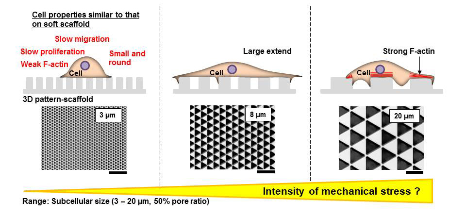
This paper by Sunami et al. studies the impact of 3D patterning on cells, and shows that cells grown on 3D micropatterns respond by changing their migration speed, their proliferation rate, and their expression of actin.
It is standard laboratory practice to grow cells outside the body on flat tissue culture plates. However, cells inside the body are surrounded by other cells and connective tissue organized in three dimensions. It is therefore very important to develop culture platforms for cells that more accurately capture a cell’s microenvironment inside the body.
In this paper, the authors developed a three-dimensional culture platform that could be modified to study the properties of fibroblasts (skin cells) in culture. The authors changed only one factor – the area of the three-dimensional triangles that the cells were cultured on – to study the impact of 3D micropatterned areas on fibroblasts. The resultant surfaces that they used were about as adhesive as a smooth glass surface. Interestingly, they found that fibroblast spreading was very different on surfaces with different areas, with maximum cell spreading observed on a surface with an intermediate pattern area. The cells primarily adhered to the upper surface of the 3D micropatterns. Further, the density of the cells was also dependent on the micropattern area – as micropattern area increased, the density of the cells correspondingly increased as well. The cells also proliferated faster on surfaces with larger micropattern areas, while cell proliferation slowed on smaller triangles. The authors then used timelapse microscopy to study how these cells migrated over time, and found that while cells migrated more slowly on all of the micropatterned surfaces as compared to a completely flat surface, migration was slowest on the micropatterns with the smallest areas. Similarly, the authors found that f-actin expression was increased on the patterns with the largest areas. The authors hypothesize that this may mean that cells experience less mechanical stress as pattern size decreases.
Influence of the pattern size of micropatterned scaffolds on cell morphology, proliferation, migration and F-actin expression
Hiroshi Sunami, Ikuko Yokota and Yasuyuki Igarashi
Biomater. Sci., 2014, Advance Article DOI: 10.1039/C3BM60237K
Debanti Sengupta recently completed her PhD in Chemistry from Stanford University. She is currently a Siebel postdoctoral scholar at the University of California, Berkeley.
Follow the latest journal news on Twitter @BioMaterSci or go to our Facebook page.


