Author Archive

Shining light on lung alveoli using photodegradable PEG hydrogels

When I was in elementary school, I remember having lots of fun making papier-mâché piñatas.  To form a spherical, hollow structure, I would inflate a balloon and layer papier-mâché on top. Once the papier-mâché dried, I popped the balloon with great satisfaction to leave behind a hollow sphere, which I then painted and filled with candy to complete my piñata. The best part of the whole process was enjoying the candy.

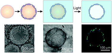
The Anseth Lab at the University of Colorado in Boulder has developed a clever biomaterials technique that reminds me of my favorite arts & crafts activity. In their Biomaterials Science paper Katherine Lewis et al. describe how they used photodegradable PEG microspheres (analogous to balloons) coated with lung epithelial cells (analogous to papier-mâché) to generate cyst structures that mimic lung alveoli in vitro. In alveoli, lung epithelial cells form tight junctions to create a barrier between the airway and blood vessels. To appropriately model this barrier in vitro, the photodegradable microspheres were functionalized with the adhesive peptide CRGDS to allow epithelial cell attachment to the surface of the microsphere. Subsequently, cell-coated microspheres were encapsulated in a PEG hydrogel with stiffness similar to lung tissue, which was functionalized with CRGDS and enzymatically-cleavable peptide cross-linkers. Finally, the photodegradable microspheres were degraded away with cell-compatible light to form cysts, similar to popping the balloon when making a piñata. The cells forming the spherical cysts retained their tight junctions because they also adhered to the surrounding encapsulating hydrogel. The morphology and cell–cell junctions of the cysts were elegantly characterized with confocal microscopy and immuno-staining to demonstrate barrier formation. These 3D models of alveolar cysts demonstrate yet another unique application of photodegradable PEG hydrogels. These cysts may be used to develop models of diseases including pulmonary fibrosis for in vitro screening of potential therapeutics. Discovering treatments to lung-associated diseases using this technology in the future would certainly be a sweeter success than enjoying candy from a piñata.

Check out the June cover article here: In vitro model alveoli from photodegradable microsphere templates by Katherine J.R. Lewis, Mark W. Tibbitt, Yi Zhao, Kelsey Branchfield, Xin Sun, Vivek Balasubramaniam, and Kristi S. Anseth


Brian AguadoDr. Brian Aguado (@BrianAguado) completed his Ph.D. in Biomedical Engineering from Northwestern University as an NSF fellow in 2015. He holds a B.S. degree in Biomechanical Engineering from Stanford University and a M.S. degree in Biomedical Engineering from Northwestern University. Read more about Brian’s research publications here.


Follow the latest journal news on Twitter @BioMaterSci or go to our Facebook page.

Digg This
Reddit This
Stumble Now!
Share on Facebook
Bookmark this on Delicious
Share on LinkedIn
Bookmark this on Technorati
Post on Twitter
Google Buzz (aka. Google Reader)

Making it personal – Engineered tissues for neural regeneration

The “holy grail” of neural tissue engineering is to develop functional tissue constructs in an effort to reconnect nerves that have been previously disconnected from disease or injury (i.e. spinal cord injury).  One technique for developing personalized regenerative cell therapies is to use adult somatic cells from a patient (such as skin cells) and re-program them to an induced pluripotent stem cell (iPSC) state. After isolating iPSCs, patient-specific neurons may be generated and transplanted back into the patient, with the hope that the newly formed neurons will form connections in the tissue and restore function.

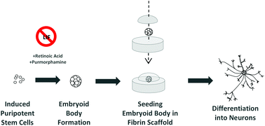
At the University of Victoria, Montgomery et al. have developed a method to differentiate mouse iPSCs into neurons within a fibrin gel. Fibrin has been regarded as a traditional biomaterial to effectively differentiate embryonic stem cells into neurons. In this study, fibrin was utilized to evaluate the efficiency of two iPS differentiation protocols. The first protocol tested was the traditional “4-/4+” method, involving no treatment to iPS embryoid bodies for 4 days of culture, then adding retinoic acid (RA) for the subsequent 4 days of culture. The second protocol tested was the newer “2-/4+” method, involving no treatment to iPS embryoid bodies for 2 days of culture, then adding RA and purmorphamine for the subsequent 4 days of culture. Individual embryoid bodies were then isolated and placed within a 3D fibrin gel to observe differentiation of neurons.


Interestingly, the overall differentiation efficiency was increased using the 6 day 2-/4+ method compared to using the traditional 4-/4+ method after seeding the treated iPS cells in 3D fibrin gels. Phenotypes were characterized using immunofluorescence staining and RT-PCR, with increases in expression of the neuronal markers TUJ1 and nestin at Day 14 of culture using the 2-/4+ method compared to the 4-/4+ method. The SOX2 pluripotent marker remained low at Day 14, indicating cells were differentiating to a neuronal state.

Taken together, this study demonstrates the ability to differentiate neurons from mouse iPSCs using simple differentiation and encapsulation protocols. Future work will need to be conducted to see if these protocols can be implemented to achieve neuronal differentiation using human iPSCs.

Check out the full article:
Engineering personalized neural tissue by combining induced pluripotent stem cells with fibrin scaffolds, by Amy Montgomery, Alix Wong, Nicole Gabers and Stephanie M. Willerth


Brian Aguado

Brian Aguado (@BrianAguado) is currently a Ph.D. Candidate and NSF Fellow in the Biomedical Engineering department at Northwestern University. He holds a B.S. degree in Biomechanical Engineering from Stanford University and a M.S. degree in Biomedical Engineering from Northwestern University. Read more about Brian’s research publications here.


Follow the latest journal news on Twitter @BioMaterSci or go to our Facebook page.

Digg This
Reddit This
Stumble Now!
Share on Facebook
Bookmark this on Delicious
Share on LinkedIn
Bookmark this on Technorati
Post on Twitter
Google Buzz (aka. Google Reader)

Nanovehicles for targeted bone metastasis chemotherapy

The cover story for the July 2014 issue of Biomaterials Science highlights the use of nanovehicle particles for the targeted delivery of chemotherapy drugs to tumor cells in bone.

Bone metastasis, or the spreading of tumor cells from the initial tumor site to bone tissue, marks the stage of cancer progression where the diseased is deemed incurable. Currently, the main method to treat bone metastasis is to utilize aggressive chemotherapy drugs and destroy metastatic tumor cells.  Unfortunately, untargeted chemotherapy treatments result in death of both cancerous and healthy tissue. Treatments could be significantly improved with the technology to specifically target the destruction of tumor cells without affecting the surrounding bone tissue.

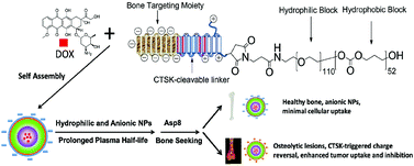
Researchers at the University of Utah, University of Iowa, and Wuhan University in China are making this desired technology a reality. Wang et al. have developed a nanovehicle capable of targeting tumor cells within bone tissue.  Nanovehicles (NVs) are essentially nano-sized cargo containers capable of delivering drugs to a desired destination. The cleverly designed NVs are designed to target diseased bone tissue to deliver chemotherapy drugs at a specific site.

The NVs developed in this study have three design features:

(1) The NVs core is made of hydrophilic and hydrophobic block co-polymers able to form a shell-like structure known as a micelle. The micelle serves as the cargo vessel that contains the chemotherapy drug doxorubicin.

(2) The outermost segments of the NVs are negatively charged bone-targeting peptides. The peptide is responsible for the accumulation of NVs in skeletal tissue, and the negative charge prevents cellular uptake of the NVs into healthy tissue.

(3) For uptake into cancer cells, the particle must be positively charged. Therefore, a positively charged peptide linker was added in between the micelle core and the negatively charged bone-targeting region. The peptide linker is cleavable by cathepsin K enzyme (CTSK), which is overexpressed in bone metastatic lesions. When the NV enters a metastatic lesion, the CTSK cleaves off the negatively charged peptides and exposes the positively charged peptides. This charge reversal allows for tumor cells present in the metastatic lesion to uptake the NV.

After extensively characterizing the nanoparticles with NMR and zeta potential measurements, the NVs were used to observe decreased tumor cell viability under CTSK-rich conditions in vitro. More strikingly, in vivo tests of the NVs in a mouse model of bone metastasis showed significant increases in mouse survival and decreases in overall tumor burden.

Collectively, these ingeniously designed nanovehicles show great promise in providing an effective targeted therapy for bone metastasis.

Peptide decoration of nanovehicles to achieve active targeting and pathology-responsive cellular uptake for bone metastasis chemotherapy
X. Wang, Y. Yang, H. Jia, W. Jia, S. Miller, B. Bowman, J. Feng, and F. Zhan
Biomater. Sci., 2014, Advance Article DOI: 10.1039/C4BM00020J

Brian Aguado is currently a Ph.D. Candidate and NSF Fellow in the Biomedical Engineering department at Northwestern University. He holds a B.S. degree in Biomechanical Engineering from Stanford University and a M.S. degree in Biomedical Engineering from Northwestern University. Read more about Brian’s research publications here.

Follow the latest journal news on Twitter @BioMaterSci or go to our Facebook page.

Digg This
Reddit This
Stumble Now!
Share on Facebook
Bookmark this on Delicious
Share on LinkedIn
Bookmark this on Technorati
Post on Twitter
Google Buzz (aka. Google Reader)

High throughput evaluation of surfaces for stem cell culture

Polymer microarrays for stem cell adhesion studies

This study identifies new surfaces optimized for human pluripotent stem cell expansion using a high-throughput polymer microarray chip.

The biomaterials community is moving toward using high-throughput tools to evaluate cell-material interactions at an unprecedented rate. For instance, polymer microarrays have become popular for identifying candidate surfaces that elicit a desired biological response. Using a single microarray chip, low volumes of polymer solution are deposited in a grid-like format using robotic ink-jet or contact printing. The chip is polymerized to manufacture a polymer microarray. After fabricating the microarray, cells are deposited directly onto the spots of the chip and cultured to determine materials that support a specific cell phenotype.

At the Laboratory of Biophysics and Surface Analysis at the University of Nottingham, Celiz et al. evaluated human pluripotent stem cell growth on a polymer microarray containing 141 varieties of (meth)acrylate and (meth)acrylamide photo-curable polymers. To maximize the diversity of the microarray, monomers containing a variety of nitrogen, fluorine, oxygen, aromatic, and aliphatic side chains were utilized.  Monomers were selected on their ability to be photo-polymerized by UV irradiation. The surface chemistry after polymerization was assessed using ToF-SIMS, and water contact angle measurements determined the surface wettability of each spot.

Partial least squares analysis (PLS) was utilized to correlate surface chemistry and wettability to identify surfaces that would yield high human pluripotent stem cell (hPSC) adhesion. Of the 141 surfaces screened in the array, 47 polymers supported hPSC attachment. Interestingly, no relationship was observed between surface wettability and cell adhesion, indicating wettability of a surface is not sufficient to predict hPSC adhesion. PLS analysis subsequently identified correlations between polymer surface chemistry and experimental hPSC adhesion. Additional experiments confirmed that increased protein adsorption on specific polymer spots was a contributor to cell adhesion to the polymer surface.

Collectively, the polymer array developed in this study was able to operate as a high-throughput tool to identify surfaces amenable to hPSC adhesion. Moving forward, polymer microarrays have the potential to identify a broad library of surfaces capable of supporting sustained hPSC growth and pluripotency.

Chemically diverse polymer microarrays and high throughput surface characterization: a method for discovery of materials for stem cell culture
A. D. Celiz, J. G. W. Smith, A. K. Patel, R. Langer, D. G. Anderson, D. A. Barrett, L. E. Young, M. C. Davies, C. Denning and M. R. Alexander
Biomater. Sci., 2014, Advance Article DOI: 10.1039/C4BM00054D

Brian Aguado is currently a Ph.D. Candidate and NSF Fellow in the Biomedical Engineering department at Northwestern University. He holds a B.S. degree in Biomechanical Engineering from Stanford University and a M.S. degree in Biomedical Engineering from Northwestern University. Read more about Brian’s research publications here.

Follow the latest journal news on Twitter @BioMaterSci or go to our Facebook page.

Digg This
Reddit This
Stumble Now!
Share on Facebook
Bookmark this on Delicious
Share on LinkedIn
Bookmark this on Technorati
Post on Twitter
Google Buzz (aka. Google Reader)

Cerebellar neuron development on hybrid matrix constructs

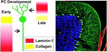
The cerebellum is a region in the brain responsible for regulating motor control and cognitive functions such as attention and language. During development of the cerebellum (and of other tissues), cells interact with the surrounding microenvironment known as the extracellular matrix (ECM). These neuron-ECM interactions regulate neuronal differentiation, growth, formation of synapses, and neurite outgrowth. Specifically, ECM components including collagen and laminin-1 (lam-1) are known to regulate the alignment, migration, and neurite outgrowth of Purkinje cells (PCs). However, the dual signaling roles of collagen and laminin-1 during cerebellar tissue development have not been fully explored.

Dr. Shantanu Sur, a postdoctoral fellow in Prof. Samuel Stupp’s laboratory at Northwestern University, has collaborated with Dr. Thomas Launey at the RIKEN Brain Science Institute in Japan to explore cerebellar tissue development using biomaterial hydrogels. Sur has developed an artificial matrix consisting of collagen and synthetic peptide amphiphile (PA) molecules presenting IKVAV, the peptide on laminin-1 responsible for cell adhesion and neurite outgrowth. Sur et al. evaluated the spatiotemporal expression of lam-1 and collagen in rat cerebellums during PC development (embryonic to post-natal) using histology and immunostaining. Given the changes in the ratio of lam-1 to collagen during PC formation and growth, these results suggest the dynamic involvement of these ECM proteins in forming the neural architecture of the cerebellum.

Using a biomimetic approach to mimic the critical lam-1 to collagen ratio, Sur proceeded to model the dynamic nature of the ECM during PC development with a synthetic collagen-PA hydrogel. Collagen (types I-V) and IKVAV-PA molecules were mixed together in solution at varying concentrations and gelled using ammonia vapor. This simple method allowed Sur to evaluate the density of PC growth, axon guidance, and dendrite morphology in gels using a wide array of collagen and IKVAV-PA concentrations. Strikingly, effects on PC phenotype were observed as a function of the collagen:IKVAV-PA ratio and not the absolute concentrations of each ECM component within the matrix.

Sur comments that the hybrid matrix provides an easily tunable environment to enable the in vitro testing of the role of ECM signals on neuronal maturation. “Our study shows that the optimal ECM-derived cues for neurons change at specific stages of development,” Sur says. “This observation will drive us to work on the design of a dynamic matrix where the extracellular signals delivered to the neuron can be tuned spatiotemporally.” Additionally, the collagen/IKVAV-PA gels may be used to identify cell-ECM interactions during the development of other tissue types, given the simplicity of the technique.

Collectively, this study demonstrates the exciting use of engineered matrices to evaluate spatiotemporal cell-ECM interactions, with the hopes to further elucidate mechanisms of tissue development.

Synergistic regulation of cerebellar Purkinje neuron development by laminin epitopes and collagen on an artificial hybrid matrix construct
Shantanu Sur, Mustafa O. Guler, Matthew J. Webber, Eugene T. Pashuck, Masao Ito, Samuel I. Stupp, and Thomas Launey
Biomater. Sci., 2014, Advance Article, DOI: 10.1039/C3BM60228A

Brian Aguado is currently a Ph.D. Candidate and NSF Fellow in the Biomedical Engineering department at Northwestern University. He holds a B.S. degree in Biomechanical Engineering from Stanford University and a M.S. degree in Biomedical Engineering from Northwestern University. Read more about Brian’s research publications here.

To keep up-to-date with all the latest research, sign-up to our RSS feed or Table of contents

Digg This
Reddit This
Stumble Now!
Share on Facebook
Bookmark this on Delicious
Share on LinkedIn
Bookmark this on Technorati
Post on Twitter
Google Buzz (aka. Google Reader)

Cells as Legos: Using cells for gelation of hydrophobically-modified polymers

Engineering tissue mimics in vitro using cells and materials is one of the core goals of biomaterials science. To develop a tailored cell culture environment, natural and/or synthetic hydrogels are used to mimic the extracellular matrix. Cells are embedded in a hydrogel matrix in a process known as “encapsulation.” During encapsulation, polymers in solution are chemically or physically cross-linked to immobilize cells in a mesh-like structure. The cell-loaded hydrogels are subsequently used for various regenerative medicine applications.

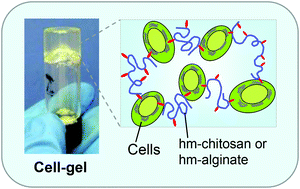
Researchers at the University of Maryland have developed a novel encapsulation technique for embedding cells in hydrogel materials. Instead of passively encapsulating cells in the polymer mesh, cells serve as “active structural elements” and are connected to the polymer chains. Using hydrophobically-modified (hm) alginate and chitosan, hydrophobic regions along the polymer chains are physically embedded in the hydrophobic cell membrane. The cells serve as struts to link together the polymer mesh. Rheological studies were performed to confirm the sol-gel transition when hm-polymers were combined to form gels (unmodified polymer controls did not form gels). To show the versatility of the system, various cell types were used to form gels with hm-alginate and hm-chitosan, including human umbilical vein endothelial cells (HUVECs), MCF7 breast cancer cells, and blood cells.

Analogous to building block toys such as Legos, the non-covalent hm-polymer and cell interactions are reversible with the addition of excess hydrophobic binding “pockets.” Using α-cyclodextrin, the gels are immediately transformed into free-flowing solutions (confirmed using rheology). The α-cyclodextrin serves to sequester the hydrophobic tails of the hm-polymer and gently release the cells from the mesh. Interestingly, α-cyclodextrin does not affect cell viability because the molecule is not large enough to bind two-tailed lipids of the cell membrane. Viability of the cells was confirmed before and after the gentle release from the gel, indicating that cell membranes remained unharmed during the gel reversal process.

Taken together, hm-polymers could serve as a unique technique to embed and release cells from a hydrogel matrix. Using cells as building blocks is highly desirable for several applications including 3D cell culture models and injectable cell therapies.

Reversible gelation of cells using self-assembling hydrophobically-modified biopolymers: toward self-assembly of tissue
Vishal Javvaji, Matthew B. Dowling, Hyuntaek Oh, Ian M. White and Srinivasa R. Raghavan
Biomater. Sci., 2014, Advance Article, DOI: 10.1039/C4BM00017J

Brian Aguado is currently a Ph.D. Candidate and NSF Fellow in the Biomedical Engineering department at Northwestern University. He holds a B.S. degree in Biomechanical Engineering from Stanford University and a M.S. degree in Biomedical Engineering from Northwestern University. Read more about Brian’s research publications here.

To keep up-to-date with all the latest research, sign-up to our RSS feed or Table of contents

Digg This
Reddit This
Stumble Now!
Share on Facebook
Bookmark this on Delicious
Share on LinkedIn
Bookmark this on Technorati
Post on Twitter
Google Buzz (aka. Google Reader)

Mending a broken heart: Myocardial matrix hydrogels for cardiac tissue engineering

According to the World Health Organization, cardiovascular disease causes 17.3 million deaths worldwide, with projections reaching 23.3 million deaths by the year 2030. Unfortunately, heart attack patients still have limited therapeutic options, commonly relying on left ventricular assist devices (LVADs) and heart transplantation. To provide more modern therapies, physicians have turned to tissue engineers to develop biomaterials that enable local regeneration of the heart to restore function and ideally improve the quality of life for the patient.

Professor Karen Christman’s lab at the University of California – San Diego (UCSD) is exploring methods to fabricate injectable hydrogels for cardiac repair. In the present study, the lab has developed human myocardial matrix (HMM), and compared the material properties of HMM to previously fabricated porcine myocardial matrix (PMM). The materials are made directly from the decellularized extracellular matrix (ECM) from human and porcine hearts. The decellularized matrices are composed of the natural structural proteins found in the heart, which make the ideal environment needed to promote cardiac cell growth and maturation.

To produce HMM, seven human hearts with a patient age range of 41-69 years were decellularized using sodium dodecyl sulfate (SDS) and a series of lyophilization, milling, and digestion steps.  Due to the dramatic patient-to-patient variability between hearts, over 50% of the HMM solutions were not able to self-assemble into hydrogels at physiological conditions, unlike self-assembling PMM hydrogels. The irreproducibility of HMM hydrogel fabrication is likely due to differences in protein composition between HMM and PMM. Using mass spectrometry to identify the proteins present in the decellularized matrices, the authors showed that porcine and human hearts have inherent differences in their matrix composition.

Although HMM did not produce hydrogels reproducibly, the matrix was still useful for in vitro cell culture protocols. Using PMM and HMM as coatings for cell culture plates, increased proliferation of rat aortic smooth muscle cells (RASMCs) and human coronary artery endothelial cells (HCAECs) was observed on HMM coated plates compared to PMM coated plates. Additionally, cell cultured on both HMM and PMM matrices showed increased expression of early cardiac transcription factor markers in human fetal cardiomyocyte progenitor cells (hCMPCs). This result indicates that biochemical cues from the HMM and PMM proteins may enhance early stages of cardiomyocyte differentiation.

Even though HMM was shown to not be a likely candidate for clinical translation due to large variability between samples, PMM injectable hydrogels are still a promising alternative for improving cardiac repair in vivo. Additionally, HMM materials may be used for future in vitro cell culture and cardiomyocyte differentiation protocols.

Human versus porcine tissue sourcing for an injectable myocardial matrix hydrogel
Todd D. Johnson, Jessica A. DeQuach, Roberto Gaetani, Jessica Ungerleider, Dean Elhag, Vishal Nigam, Atta Behfar, and Karen L. Christman
Biomater. Sci., 2014, Advance Article, DOI: 10.1039/C3BM60283D

Brian Aguado is currently a Ph.D. Candidate and NSF Fellow in the Biomedical Engineering department at Northwestern University. He holds a B.S. degree in Biomechanical Engineering from Stanford University and a M.S. degree in Biomedical Engineering from Northwestern University. Read more about Brian’s research publications here.

To keep up-to-date with all the latest research, sign-up to our RSS feed or Table of contents

Digg This
Reddit This
Stumble Now!
Share on Facebook
Bookmark this on Delicious
Share on LinkedIn
Bookmark this on Technorati
Post on Twitter
Google Buzz (aka. Google Reader)

Nanoscale semiconductor devices as new biomaterials

Most biological organisms depend on electrical signals to function properly. For instance, neurons are electrically active cells that transmit signals to other neurons using electrical signals known as action potentials. Other electrically active cells such as cardiomyocytes in the heart spontaneously generate electrical signals that regulate normal heart rhythm. Additionally, electrical signals are also known to mediate certain processes such as cell migration and cell division. Knowing that biological organisms rely on electrical signals to function, biomaterials scientists are searching for opportunities on how to use innovative electrically conductive materials to deepen our understanding of fundamental biological processes.

Professor Bozhi Tian and his team at the University of Chicago have recently written a mini-review discussing nanoscale semiconductor devices and how they can revolutionize our understanding of cellular electrophysiology. Semiconductor devices such as silicon nanowire field effect transistors (NWFETs) operate at a sufficient resolution to detect the slightest changes in chemical and electrical signals (down to the pico- and femto- unit scales). These devices could theoretically be used as a low-cost strategy for disease marker detection in the clinic.

Ramya Parameswaran, an MD/PhD student at the University of Chicago and second author on the mini-review, explains that the use of nanoscale semiconductor devices in the clinic is not far away. The authors discuss the development of “multiplex sensing” semiconductor devices seek to replace enzyme-linked immunosorbent assays (ELISA) in the future, with the ability to detect concentrations of proteins, nucleic acids, and other ions within cells simultaneously with one device. “Multiplexing nanomaterials can hopefully soon be used to detect biomarkers such as cancer antigens or other important players involved in disease processes in a rapid fashion,” says Parameswaran. “While clinical trials for devices such as these have not been performed as of yet, the proof of concept exists.” These devices would ultimately accelerate patient diagnosis, and would ideally improve patient treatments for a variety of diseases.

The application of nanoscale semiconductor devices in the tissue-engineering field is also emerging. Nanoscale semiconductors embedded in tissue-engineered implants could be used to record electrical information for monitoring and ensuring proper functionality of the implant. Interestingly, nanoelectronic scaffolds can be used for a variety of tissue targets. “Free-standing nanowire nanoelectronic scaffolds can be interfaced with neurons, smooth muscle cells, and cardiomyocytes to monitor local electrical activity within the hybrid scaffolds,” says Parameswaran. Using these nanoelectronic scaffolds, tissue responses to drug treatments could also be “monitored meticulously,” which is another advantage for accelerating the translation of these devices to the clinic.

This mini-review describes the current status of nanoscale semiconductor devices and how they can advance our understanding of cell electrophysiology. Additionally, these devices could have a transformative role in the design of medical devices and tissue engineering scaffolds for clinical applications.

Nanoscale semiconductor devices as new biomaterials
John Zimmerman, Ramya Parmeswaran, and Bozhi Tian
Biomaterial Sci., Advance Article, DOI: 10.1039/C3BM60280J

Brian Aguado is currently a Ph.D. Candidate and NSF Fellow in the Biomedical Engineering department at Northwestern University. He holds a B.S. degree in Biomechanical Engineering from Stanford University and a M.S. degree in Biomedical Engineering from Northwestern University. Read more about Brian’s research publications here.

To keep up-to-date with all the latest research, sign-up to our RSS feed or Table of contents

Digg This
Reddit This
Stumble Now!
Share on Facebook
Bookmark this on Delicious
Share on LinkedIn
Bookmark this on Technorati
Post on Twitter
Google Buzz (aka. Google Reader)

Dextran polyampholyte hydrogels for cell cryopreservation

One of the goals of tissue engineers is to manufacture “spare body parts” made of cells and biomaterials to repair damaged or diseased tissues. A current challenge for commercializing cell-biomaterial constructs is maintaining their viability during shipping and/or storage. The storage of cells in liquid suspension is relatively simple with the use of common cryoprotectants (such as dimethyl sulfoxide), but current preservation methods for large cell-biomaterial constructs yield low cell viability in comparison.  More effective preservation methods for cell-biomaterial constructs are needed to produce “off the shelf” tissue engineered products and accelerate the translation of tissue engineered products to the clinic.

In a current study conducted at the Japan Advanced Institute of Science and Technology (JAIST), researchers developed a dextran-based polyampholyte (charged polymer) hydrogel with cryoprotective properties. First, dextran polymers were functionalized with azide groups (azide-Dex) to provide sites for crosslinking. Then, positively charged amine groups were introduced on the dextran using poly-L-lysine to form azide-amino-Dex. To add negatively charged carboxyl groups, succinic anhydride was added at various concentrations to azide-amino-Dex to convert amine groups to carboxyl groups. This newly formed dextran polyampholyte (azide-Dex-PA) polymer was utilized for cryopreservation studies. After encapsulating and freezing cells with various azide-Dex-PA solutions, cell viability post-cryopreservation was shown to increase with higher concentrations of azide-Dex-PA. Viability was also dependent on the ratio of amine and carboxyl groups on the polymer, indicating that the cryoprotective properties of the polyampholyte are likely due to polymer charge.

The azide groups on the azide-Dex-PA were utilized to crosslink the dextran polyampholytes to form hydrogels. After synthesizing alkyne-functionalized dextran with dibenzylcyclooctyne (DBCO-Dex), the two components were mixed with cells to form hydrogels with azide-alkyne click chemistry. Encapsulated cells in the hydrogel were cryopreserved, thawed, and evaluated for viability. Compared to the 0% viability observed with collagen hydrogel and non-polyampholyte dextran hydrogel controls, the dextran polyampholyte hydrogels substantially increased cell viability to 93-94%. The increased viability post-cryopreservation may be due to the polyampholyte polymer adsorbing onto the cell membrane, providing a polymer “shell” of protection during thawing. Images of cells coated with FITC labeled azide-Dex-PA provide evidence to support this hypothesis.

All in all, dextran polyampholyte hydrogels have cryoprotective properties, and are able to preserve cell viability during the cryopreservation process. The dramatic increase in cell viability post-cryopreservation justifies future studies for using these hydrogels for preserving larger cell-biomaterial constructs for long term storage.

Hydrogelation of dextran-based polyampholytes with cryoprotective properties via click chemistry
Minkle Jain, Robin Rajan, Suong-Hyu Hyon, and Kazuaki Matsumura
Biomaterials Science, 2014, Advance Article DOI: 10.1039/C3BM60261C

Brian Aguado is currently a Ph.D. Candidate and NSF Fellow in the Biomedical Engineering department at Northwestern University. He holds a B.S. degree in Biomechanical Engineering from Stanford University and a M.S. degree in Biomedical Engineering from Northwestern University. Read more about Brian’s research publications here.

To keep up-to-date with all the latest research, sign-up to our RSS feed or Table of contents

Digg This
Reddit This
Stumble Now!
Share on Facebook
Bookmark this on Delicious
Share on LinkedIn
Bookmark this on Technorati
Post on Twitter
Google Buzz (aka. Google Reader)