Archive for January, 2014

Nanoscale semiconductor devices as new biomaterials

Most biological organisms depend on electrical signals to function properly. For instance, neurons are electrically active cells that transmit signals to other neurons using electrical signals known as action potentials. Other electrically active cells such as cardiomyocytes in the heart spontaneously generate electrical signals that regulate normal heart rhythm. Additionally, electrical signals are also known to mediate certain processes such as cell migration and cell division. Knowing that biological organisms rely on electrical signals to function, biomaterials scientists are searching for opportunities on how to use innovative electrically conductive materials to deepen our understanding of fundamental biological processes.

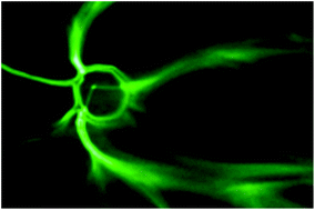
Professor Bozhi Tian and his team at the University of Chicago have recently written a mini-review discussing nanoscale semiconductor devices and how they can revolutionize our understanding of cellular electrophysiology. Semiconductor devices such as silicon nanowire field effect transistors (NWFETs) operate at a sufficient resolution to detect the slightest changes in chemical and electrical signals (down to the pico- and femto- unit scales). These devices could theoretically be used as a low-cost strategy for disease marker detection in the clinic.

Ramya Parameswaran, an MD/PhD student at the University of Chicago and second author on the mini-review, explains that the use of nanoscale semiconductor devices in the clinic is not far away. The authors discuss the development of “multiplex sensing” semiconductor devices seek to replace enzyme-linked immunosorbent assays (ELISA) in the future, with the ability to detect concentrations of proteins, nucleic acids, and other ions within cells simultaneously with one device. “Multiplexing nanomaterials can hopefully soon be used to detect biomarkers such as cancer antigens or other important players involved in disease processes in a rapid fashion,” says Parameswaran. “While clinical trials for devices such as these have not been performed as of yet, the proof of concept exists.” These devices would ultimately accelerate patient diagnosis, and would ideally improve patient treatments for a variety of diseases.

The application of nanoscale semiconductor devices in the tissue-engineering field is also emerging. Nanoscale semiconductors embedded in tissue-engineered implants could be used to record electrical information for monitoring and ensuring proper functionality of the implant. Interestingly, nanoelectronic scaffolds can be used for a variety of tissue targets. “Free-standing nanowire nanoelectronic scaffolds can be interfaced with neurons, smooth muscle cells, and cardiomyocytes to monitor local electrical activity within the hybrid scaffolds,” says Parameswaran. Using these nanoelectronic scaffolds, tissue responses to drug treatments could also be “monitored meticulously,” which is another advantage for accelerating the translation of these devices to the clinic.

This mini-review describes the current status of nanoscale semiconductor devices and how they can advance our understanding of cell electrophysiology. Additionally, these devices could have a transformative role in the design of medical devices and tissue engineering scaffolds for clinical applications.

Nanoscale semiconductor devices as new biomaterials
John Zimmerman, Ramya Parmeswaran, and Bozhi Tian
Biomaterial Sci., Advance Article, DOI: 10.1039/C3BM60280J

Brian Aguado is currently a Ph.D. Candidate and NSF Fellow in the Biomedical Engineering department at Northwestern University. He holds a B.S. degree in Biomechanical Engineering from Stanford University and a M.S. degree in Biomedical Engineering from Northwestern University. Read more about Brian’s research publications here.

To keep up-to-date with all the latest research, sign-up to our RSS feed or Table of contents

Digg This
Reddit This
Stumble Now!
Share on Facebook
Bookmark this on Delicious
Share on LinkedIn
Bookmark this on Technorati
Post on Twitter
Google Buzz (aka. Google Reader)

Inducing biomimetic mineralization using a graphene oxide-chitosan hybrid material

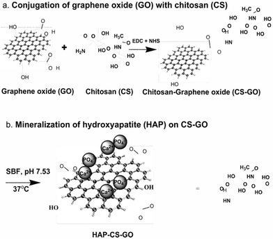
Chemical conjugation of graphene oxide with chitosan and subsequent mineralization of hydroxyapatite

One of the primary goals within the field of hard tissue engineering is to create biomaterials that can both promote osteoblast proliferation and support the natural bone mineralization process.  In this study, a trio of researchers at the University of Louisiana at Lafayette has developed a promising, layered hybrid system that contains hydroxyapatite (HAP), chitosan (CS), and graphene oxide (GO).  The GO base provides mechanical strength and biocompatibility while the surface layer which is composed of HAP, a natural constituent of bone, has a high bioactivity.  The intermediate chitosan layer further strengthens the hybrid biomaterial and, perhaps more importantly, has been shown to promote more homogeneous mineralization that mimics the natural biomineralization process.

In order to test the bone forming capabilities of this hybrid material the group soaked it, along with two control materials (HAP-GO and pure GO), in simulated body fluid for three weeks.  Each substrate was then incubated with MC3T3-E1 pre-osteoblast cells.  Among the three biomaterials, the HAP-CS-GO hybrid produced the highest levels of cell proliferation and expansion.   It also induced the greatest expression levels of vinculin, actin, and fibronectin, proteins that are essential for cell adhesion, cytoskeletal organization, and osteoblast morphogenesis and mineralization.  The hybrid material also demonstrated a highly significant increase in mineralized area and bone nodule formation compared to either of the two controls.  This was primarily attributed to the strong electrostatic interactions between the functional groups in the CS-GO base and the calcium ions present in solution.

Overall the hybrid HAP-CS-GO material was able to favorably modulate cellular activity in a way that was conducive to bone formation, making it a promising candidate for future bone tissue engineering applications.   However, as the authors have highlighted, there is room for improvement.  A reoccurring issue that many bone tissue engineering materials, including this novel hybrid material, have faced is an inability to attain the proper ratio of mineral deposit to cellular matrix in the regenerated tissue.  This mineral to matrix ratio has been shown to be relatively high in healthy, intact bone (~4.35-5) but was observed in a significantly lower amount (1.76) in the HAP-CS-GO material.  Nonetheless, the hybrid biomaterial reported in this study represents a promising direction forward in the field of bone tissue engineering.

The synergistic effect of a hybrid graphene oxide-chitosan system and biomimetic mineralization on osteoblast functions
D. Depan, T. C. Pesacreta and R. D. K. Misra
Biomater. Sci., 2014, 2, 264-274 DOI: 10.1039/C3BM60192G

Ellen Tworkoski is a guest web-writer for Biomaterials Science.  She is currently a graduate student in the biomedical engineering department at Northwestern University (Evanston, IL, USA).

Follow the latest journal news on Twitter @BioMaterSci or go to our Facebook page.

To keep up-to-date with all the latest research, sign-up to our RSS feed or Table of contents alert.

Digg This
Reddit This
Stumble Now!
Share on Facebook
Bookmark this on Delicious
Share on LinkedIn
Bookmark this on Technorati
Post on Twitter
Google Buzz (aka. Google Reader)

Inaugural Biomaterials Science Lectureship: Nominations now open

Do you know someone who deserves recognition for their contribution to the biomaterials field? 

Now is your chance to propose they receive the accolade they deserve. 

Biomaterials Science is pleased to announce that nominations are now being accepted for its Biomaterials Science Lectureship 2014.  New in 2014, this award will be run annually by the journal to honour a younger scientist who has made a significant contribution to the biomaterials field. 

Qualification 

To be eligible for the Biomaterials Science Lectureship, the candidate should be in the earlier stages of their scientific career, typically within 15 years of attaining their doctorate or equivalent degree, and will have made a significant contribution to the field. 

Description 

The recipient of the award will be asked to present a lecture three times, one of which will be located in the home country of the recipient. The Biomaterials Science Editorial Office will provide the sum of £1000 to the recipient for travel and accommodation costs. 

The award recipient will be presented with the award at one of the three award lectures. They will also be asked to contribute a lead article to the journal and will have their work showcased on the back cover of the issue in which their article is published. 

Selection 

The recipient of the award will be selected and endorsed by the Biomaterials Science Editorial Board. 

Nominations 

Those wishing to make a nomination should send details of the nominee, including a brief C.V. (no longer than 2 pages A4) together with a letter (no longer than 2 pages A4) supporting the nomination, to the Biomaterials Science Editorial Office (biomaterialsscience-rsc@rsc.org ) by 7th March 2014.  Self-nomination is not permitted.

Digg This
Reddit This
Stumble Now!
Share on Facebook
Bookmark this on Delicious
Share on LinkedIn
Bookmark this on Technorati
Post on Twitter
Google Buzz (aka. Google Reader)

3D micropatterns to grow cells

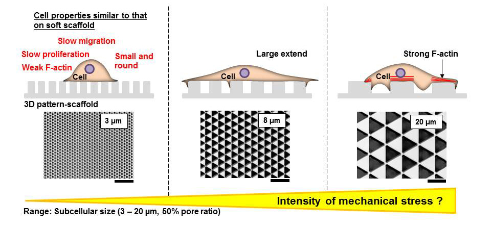
This paper by Sunami et al. studies the impact of 3D patterning on cells, and shows that cells grown on 3D micropatterns respond by changing their migration speed, their proliferation rate, and their expression of actin.

It is standard laboratory practice to grow cells outside the body on flat tissue culture plates.  However, cells inside the body are surrounded by other cells and connective tissue organized in three dimensions.  It is therefore very important to develop culture platforms for cells that more accurately capture a cell’s microenvironment inside the body. 

In this paper, the authors developed a three-dimensional culture platform that could be modified to study the properties of fibroblasts (skin cells) in culture. The authors changed only one factor – the area of the three-dimensional triangles that the cells were cultured on – to study the impact of 3D micropatterned areas on fibroblasts. The resultant surfaces that they used were about as adhesive as a smooth glass surface.  Interestingly, they found that fibroblast spreading was very different on surfaces with different areas, with maximum cell spreading observed on a surface with an intermediate pattern area.  The cells primarily adhered to the upper surface of the 3D micropatterns. Further, the density of the cells was also dependent on the micropattern area – as micropattern area increased, the density of the cells correspondingly increased as well. The cells also proliferated faster on surfaces with larger micropattern areas, while cell proliferation slowed on smaller triangles. The authors then used timelapse microscopy to study how these cells migrated over time, and found that while cells migrated more slowly on all of the micropatterned surfaces as compared to a completely flat surface, migration was slowest on the micropatterns with the smallest areas. Similarly, the authors found that f-actin expression was increased on the patterns with the largest areas. The authors hypothesize that this may mean that cells experience less mechanical stress as pattern size decreases.

Influence of the pattern size of micropatterned scaffolds on cell morphology, proliferation, migration and F-actin expression
Hiroshi Sunami, Ikuko Yokota and Yasuyuki Igarashi
Biomater. Sci., 2014, Advance Article DOI: 10.1039/C3BM60237K

Debanti Sengupta recently completed her PhD in Chemistry from Stanford University.  She is currently a Siebel postdoctoral scholar at the University of California, Berkeley.  

Follow the latest journal news on Twitter @BioMaterSci or go to our Facebook page.

Digg This
Reddit This
Stumble Now!
Share on Facebook
Bookmark this on Delicious
Share on LinkedIn
Bookmark this on Technorati
Post on Twitter
Google Buzz (aka. Google Reader)