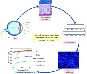
 Hasirci and colleagues have developed a new type of engineered corneal construct using a combination of cells derived from the cornea as well as organized elastin-based biomaterials.
The stroma is a layer in our cornea that consists of highly organized layers of collagen fibers and keratocytes, a special type of corneal cell. The cells and the collagen layers are aligned with one another and follow a specific pattern of organization. This organized structure allows the cornea to be transparent and to have the mechanical strength necessary to function correctly.
This study used specific biomaterials that draw inspiration from nature. Specifically, the researchers used elastin, a commonly found biomaterial, as a template, and created a biomaterial with a similar protein composition. They modified a traditional elastin-based sequence to include the amino acid chain YIGSR, which is known to help cells attach to biomaterial structures. They blended their elastin-based biomaterial with collagen, and used a silicon template to mold these biomaterials into sheets with surfaces that contain micron-scale grooves or micropatterns that cells can sense and align to.
The biomaterial sheets were dehydrated completely under vacuum, causing the films to form internal bonds or crosslinks. The crosslinking confers mechanical stability to the sheets, ensuring that the sheets do not degrade too quickly in response to enzymes secreted by keratocytes. To test this idea, the biomaterial sheets were then subjected to a collagen-degrading enzyme, collagenase II. The crosslinked sheets were better able to resist degradation for 24 hours as compared to their non-crosslinked counterparts. This suggested that the keratocytes could form organized layers on the crosslinked biomaterials before degrading away the original structures. To further test these materials, the biomaterial sheets were then immersed in a saline solution for 4 weeks to study non-enzyme based degradation, and the crosslinked sheets again resisted degradation better than the non-crosslinked sheets. The crosslinked sheets were also able to transmit light, which is crucial for their application as corneal replacements.
Finally, the researchers tested these biomaterial sheets with human corneal keratocytes. The cells were able to survive on the biomaterial and grew well on the micropatterned biomaterial sheets. The cells also followed the alignment of the micropatterns on the surface of the biomaterials. The researchers then placed multiple layers of the micropatterned scaffolds together such that each successive patterned layer was perpendicular to the previous layer, and ‘glued’ the scaffolds together using droplets of collagen. Cells were then seeded onto each layer using a syringe. They found that the keratocytes remained aligned for a period of 21 days in culture. The material was transparent even with the presence of cells for up to 4 weeks. In fact, the presence of keratocytes contributed to the construct’s overall transparency.
This work is an elegant demonstration of how biomaterials and cells can be combined to produce a viable tissue-engineered construct that may one day be used as corneal replacement therapy.
A collagen-based corneal stroma substitute with micro-designed architecture
Cemile Kilic, Alessandra Girotti, J. Carlos Rodriguez-Cabello and Vasif Hasirci
Biomater. Sci., 2014, Advance Article DOI: 10.1039/C3BM60194C
Debanti Sengupta recently completed her PhD in Chemistry from Stanford University. She is currently a Siebel postdoctoral scholar at the University of California, Berkeley.
Follow the latest journal news on Twitter @BioMaterSci or go to our Facebook page.
To keep up-to-date with all the latest research, sign-up to our RSS feed or Table of contents alert.


