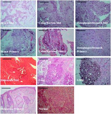
According to the Brain Research UK, brain tumours are diagnosed in ~13000 people in Britain each year (of which 60 % are metastatic), 20 % of all cancer patients will develop secondary metastatic tumours in the brain and brain tumours are the most common solid tumour in children. Dr. Matt Baker and colleagues from the University of Central Lancaster, Lancashire Teaching Hospital and Dublin Institute of Technology have reported a study on the use of Raman spectroscopy to aid the diagnosis of brain cancer.
In this HOT new Analytical Methods paper, the team showed differences in spectra obtained by Raman scattering at the air-tissue interface and immersion Raman (where the sample is submerged in an appropriate medium, such as water) at the water-tissue interface. Immersion Raman was then used to identify differences between the tissue classes. Multivariate analysis was employed to differentiate between primary malignant tumours (glioblastoma multiforme), metastatic tumours and normal brain tissue with impressive sensitivity and specificity. To read more about this study download the full article below, which is free to access until March 7th.
1
Investigating the use of Raman and immersion Raman spectroscopy for spectral histopathology of metastatic brain cancer and primary sites of origin
Leanne M. Fullwood, Graeme Clemens, Dave Griffiths, Katherine Ashton, Timothy P. Dawson, Robert W. Lea, Charles Davis, Franck Bonnier, Hugh J. Byrne and Matthew J. Baker
Journal Article
Anal. Methods, 2014, Advance Article
DOI: 10.1039/C3AY42190B


