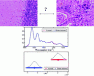
Scientists at Lancaster University and Lancashire Teaching Hospitals NHS Trust have used infrared and Raman spectroscopy for brain tumour diagnosis.
Professor Frank Martin and colleagues used the techniques combined with statistical analysis to discriminate between normal brain tissue and three different tumour types based on the unique spectral fingerprints of their biochemical composition.
Current diagnostic approaches lack the spatial resolution required by surgeons, and delineating the excision border is important in making sure the tumour is completely removed. Vibrational spectroscopy has potential for analysing brain tissue, and Raman in particular can be performed in vivo within seconds or minutes. The information obtained by this method can be combined with conventional methods, for example immunohistochemistry, to diagnose and grade brain tumours to allow for more accurate planning and execution of surgery and/or radiation therapy. This offers more potential for individualised treatment and better long-term survival.
Professor Martin says, “These are really exciting developments which could lead to significant improvements for individual patients diagnosed with brain tumours. We and other research teams are now working towards a sensor which can be used during brain surgery to give surgeons precise information about the tumour and tissue type that they are operating on.”
Read the article in full using the link below – it will be free to access until 8 October.
Diagnostic segregation of human brain tumours using Fourier-transform infrared and/or Raman spectroscopy coupled with discriminant analysis
Ketan Gajjar, Lara D. Heppenstall, Weiyi Pang, Katherine M. Ashton, Júlio Trevisan, Imran I. Patel, Valon Llabjani, Helen F. Stringfellow, Pierre L. Martin-Hirsch, Timothy Dawson and Francis L. Martin
Anal. Methods, 2012, Advance Article
DOI: 10.1039/C2AY25544H


