DNA Detection Using Novel Fiber-Optic Biosensing Strategy

Pui Sai Lau is a guest web writer for Analyst. She is currently a PhD student at McMaster University, Hamilton, ON, Canada.

DNA Detection Using Fiber Optic Biosensor

Overview of DNA Detection Using Fiber Optic Biosensor

Ming-jie Yin and others from The Hong Kong Polytechnic University in China have made the latest stride towards development of label-free fiber-optic biosensors for DNA detection.

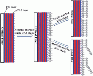
Their new design involves fabrication of a DNA sensor as part of a thin sensing film on the surface of a thin-core fiber modal interferometer (TCFMI) to measure differences in refractive-index upon detection of target DNA sequences. The research team makes use of a layer-by-layer self-assembly approach, which enables controlled tunability in preparation of the sensing film.

Unlike many other reported fiber-optic biosensing methods, this current strategy confers the advantages of high detection sensitivity, lower cost and ease in preparation.

Read more about the latest discovery by accessing the link below. This article will be free to read until March 6th.

Label-free, disposable fiber-optic biosensors for DNA hybridization detection
Ming-jie Yin ,  Chuang Wu ,  Li-yang Shao ,  Wing Kin Edward Chan ,  A. Ping Zhang ,  Chao Lu and Hwa-yaw Tam
Analyst, 2013, Advance Article
DOI: 10.1039/C3AN36791F

Digg This
Reddit This
Stumble Now!
Share on Facebook
Bookmark this on Delicious
Share on LinkedIn
Bookmark this on Technorati
Post on Twitter
Google Buzz (aka. Google Reader)

Rayleigh, Ramsay, Rutherford and Raman

In his recently published Editorial, Robin Clark talks about the key contributions of the four great Nobel Laureates – Lord Rayleigh, Sir William Ramsay, Lord Rutherford and Sir Chandrasekhara Raman – to the understanding of light scattering, to the identification and classification of the rare gases, and to the discovery of the Raman effect.

Rayleigh, Ramsay, Rutherford and Raman

Rayleigh, Ramsay, Rutherford and Raman – their connections with, and contributions to, the discovery of the Raman effect
Robin J. H. Clark
Analyst, 2013, 138, 729-734
DOI: 10.1039/C2AN90124B

To commemorate this wonderful editorial, we have gathered together a collection of papers from the last few year on Raman spectroscopy that have been published in Analyst. These papers will be free to read until March 22nd. Enjoy!

SERS-based sandwich immunoassay using antibody coated magnetic nanoparticles for Escherichia coli enumeration
Burcu Guven, Nese Basaran-Akgul, Erhan Temur, Ugur Tamer and İsmail Hakkı Boyacı
Analyst, 2011, 136, 740-748
DOI: 10.1039/C0AN00473A

Evaluation of tip-enhanced Raman spectroscopy for characterizing different virus strains
Peter Hermann, Antje Hermelink, Veronika Lausch, Gudrun Holland, Lars Möller, Norbert Bannert and Dieter Naumann
Analyst, 2011, 136, 1148-1152
DOI: 10.1039/C0AN00531B

Surface enhanced optical spectroscopies for bioanalysis
Iain A. Larmour and Duncan Graham
Analyst, 2011, 136, 3831-3853
DOI: 10.1039/C1AN15452D

Recent advancements in optical DNA biosensors: Exploiting the plasmonic effects of metal nanoparticles
Hsin-I Peng and Benjamin L. Miller
Analyst, 2011, 136, 436-447
DOI: 10.1039/C0AN00636J

Non-invasive analysis of turbid samples using deep Raman spectroscopy
Kevin Buckley and Pavel Matousek
Analyst, 2011, 136, 3039-3050
DOI: 10.1039/C0AN00723D

Non-invasive analysis of turbid samples using deep Raman spectroscopy

Buckley & Matousek, Analyst, 2011, 136, 3039

Subcellular localization of early biochemical transformations in cancer-activated fibroblasts using infrared spectroscopic imaging
Sarah E. Holton, Michael J. Walsh and Rohit Bhargava
Analyst, 2011, 136, 2953-2958
DOI: 10.1039/C1AN15112F

Poor quality drugs: grand challenges in high throughput detection, countrywide sampling, and forensics in developing countries
Facundo M. Fernandez, Dana Hostetler, Kristen Powell, Harparkash Kaur, Michael D. Green, Dallas C. Mildenhall and Paul N. Newton
Analyst, 2011, 136, 3073-3082
DOI: 10.1039/C0AN00627K

Rapid, sensitive DNT vapor detection with UV-assisted photo-chemically synthesized gold nanoparticle SERS substrates
Maung Kyaw Khaing Oo, Chia-Fang Chang, Yuze Sun and Xudong Fan
Analyst, 2011, 136, 2811-2817
DOI: 10.1039/C1AN15110J

Surface enhanced Raman scattering for multiplexed detection
Jennifer A. Dougan and Karen Faulds
Analyst, 2012, 137, 545-554
DOI: 10.1039/C2AN15979A

Direct SERS detection of contaminants in a complex mixture: rapid, single step screening for melamine in liquid infant formula
Jordan F. Betz, Yi Cheng and Gary W. Rubloff
Analyst, 2012, 137, 826-828
DOI: 10.1039/C2AN15846A

A quantitative solid-state Raman spectroscopic method for control of fungicides
Bojidarka Ivanova and Michael Spiteller
Analyst, 2012, 137, 3355-3364
DOI: 10.1039/C2AN35174A

Extracting biological information with computational analysis of Fourier-transform infrared (FTIR) biospectroscopy datasets: current practices to future perspectives
Júlio Trevisan, Plamen P. Angelov, Paul L. Carmichael, Andrew D. Scott and Francis L. Martin
Analyst, 2012, 137, 3202-3215
DOI: 10.1039/C2AN16300D

Differentiating intrinsic SERS spectra from a mixture by sampling induced composition gradient and independent component analysis
Justin L. Abell, Joonsang Lee, Qun Zhao, Harold Szu and Yiping Zhao
Analyst, 2012, 137, 73-76
DOI: 10.1039/C1AN15623C

Surface enhanced Raman spectroscopy for microfluidic pillar arrayed separation chips
Lisa C. Taylor, Teresa B. Kirchner, Nickolay V. Lavrik and Michael J. Sepaniak
Analyst, 2012, 137, 1005-1012
DOI: 10.1039/C2AN16239C

Total internal reflection Raman spectroscopy
David A. Woods and Colin D. Bain
Analyst, 2012, 137, 35-48
DOI: 10.1039/C1AN15722A

Understanding the molecular information contained in principal component analysis of vibrational spectra of biological systems
F. Bonnier and H. J. Byrne
Analyst, 2012, 137, 322-332
DOI: 10.1039/C1AN15821J

2p or not 2p: tuppence-based SERS for the detection of illicit materials

Goodacre et al., Analyst, 2013, 138, 118

Streptococcus suis II immunoassay based on thorny gold nanoparticles and surface enhanced Raman scattering

Kun Chen, Heyou Han and Zhihui Luo
Analyst, 2012, 137, 1259-1264
DOI: 10.1039/C2AN15997J

2p or not 2p: tuppence-based SERS for the detection of illicit materials
Samuel Mabbott, Alex Eckmann, Cinzia Casiraghi and Royston Goodacre
Analyst, 2013, 138, 118-122
DOI: 10.1039/C2AN35974J

Toward development of a surface-enhanced Raman scattering (SERS)-based cancer diagnostic immunoassay panel
Jennifer H. Granger, Michael C. Granger, Matthew A. Firpo, Sean J. Mulvihill and Marc D. Porter
Analyst, 2013, 138, 410-416
DOI: 10.1039/C2AN36128K

3D confocal Raman imaging of endothelial cells and vascular wall: perspectives in analytical spectroscopy of biomedical research
Katarzyna Majzner, Agnieszka Kaczor, Neli Kachamakova-Trojanowska, Andrzej Fedorowicz, Stefan Chlopicki and Malgorzata Baranska
Analyst, 2013, 138, 603-610
DOI: 10.1039/C2AN36222H

Digg This
Reddit This
Stumble Now!
Share on Facebook
Bookmark this on Delicious
Share on LinkedIn
Bookmark this on Technorati
Post on Twitter
Google Buzz (aka. Google Reader)

HOT Articles in Analyst!

In the latest issue of Analyst we have some wonderful HOT articles for you to read. They cover a wide variety of topics including iodine determination by optical emission spectrometry, detection of trinitrotoluene in seawater, and immunosensors for surface plasmon field-enhanced fluorescence spectroscopy in serum.

If you want to know more about the latest discoveries published in Analyst, take a look at the papers below, they will be free to read until February 28th.

Iodine excitation in a dielectric barrier discharge micro-plasma and its determination by optical emission spectrometry
Yong-Liang Yu ,  Shuai Dou ,  Ming-Li Chen and Jian-Hua Wang
Analyst, 2013, Advance Article
DOI: 10.1039/C2AN36780G

Graphenes prepared from multi-walled carbon nanotubes and stacked graphene nanofibers for detection of 2,4,6-trinitrotoluene (TNT) in seawater
Shu Min Tan ,  Chun Kiang Chua and Martin Pumera
Analyst, 2013, Advance Article
DOI: 10.1039/C3AN00089C

Graphene nanopfibers for detection of trinitrotoluene

A mixed alkanethiol based immunosensor for surface plasmon field-enhanced fluorescence spectroscopy in serum
Andreas Scholten ,  Bernhard Menges ,  Martin Juebner ,  Markus A. Rothschild and Katja Bender
Analyst, 2013, Advance Article
DOI: 10.1039/C3AN35657D

Facile formation of folic acid-modified dendrimer-stabilized gold–silver alloy nanoparticles for potential cellular computed tomography imaging applications
Hui Liu ,  Mingwu Shen ,  Jinglong Zhao ,  Jingyi Zhu ,  Tingting Xiao ,  Xueyan Cao ,  Guixiang Zhang and Xiangyang Shi
Analyst, 2013, Advance Article
DOI: 10.1039/C3AN36649A

In-process phase growth measurement technique in the fabrication of monolithic porous layer open tubular (monoPLOT) columns using capacitively coupled contactless conductivity
David A. Collins ,  Ekaterina P. Nesterenko ,  Dermot Brabazon and Brett Paull
Analyst, 2013, Advance Article
DOI: 10.1039/C3AN00133D

Digg This
Reddit This
Stumble Now!
Share on Facebook
Bookmark this on Delicious
Share on LinkedIn
Bookmark this on Technorati
Post on Twitter
Google Buzz (aka. Google Reader)

Showcase your own research at the Analytical Research Forum 2013 (ARF13)

Showcase your own research at the Analytical Research Forum 2013 (ARF13)

The deadline for submitting oral abstracts is fast approaching – 15 February 2013

This is the premier Analytical Science meeting of the RSC designed primarily for early-career analytical scientists (including PhD students and postdoctoral fellows) to present their latest results in the context of the wider analytical science community.

Analytical Research Forum 2013A major part of the programme will be oral and poster contributions from students and young researchers, medal winners and international leaders in the field – so make sure you don’t miss out! An added feature this year will be a tour of the GSK site in Stevenage, which is where the lectures and poster presentations will take place.

Make sure you don’t miss out – submit your abstract now!

Check out the event website to find out more – http://rsc.li/arf13

Digg This
Reddit This
Stumble Now!
Share on Facebook
Bookmark this on Delicious
Share on LinkedIn
Bookmark this on Technorati
Post on Twitter
Google Buzz (aka. Google Reader)

Diagnosing bacterial growth

Antibiotics are used regularly for treating bacterial infections, but there is currently no quick and simple test to determine the most effective type or dose of antibiotic for a specific patient infection. As a result, it’s estimated that around 30% of all antibiotic prescriptions are not the optimum choice. This can lead to the formation of drug-resistant bacteria, delayed recovery, and in some cases death from an infection.

Tests for the most appropriate antibiotic choice are performed for life-threatening patient infections. However, microbes have to be grown on agar plates from a very small patient sample which delays results for a few days. Hiroaki Suzuki et al have designed a microfluidic device that is able to determine the most effective antibiotic treatment in just 12 hours.


To read the full article please visit Chemistry World.

A microfluidic microbial culture device for rapid determination of the minimum inhibitory concentration of antibiotics
Rika Takagi ,  Junji Fukuda ,  Keiji Nagata ,  Yutaka Yawata ,  Nobuhiko Nomura and Hiroaki Suzuki
Analyst, 2013,138, 1000-1003
DOI: 10.1039/C2AN36323B

Digg This
Reddit This
Stumble Now!
Share on Facebook
Bookmark this on Delicious
Share on LinkedIn
Bookmark this on Technorati
Post on Twitter
Google Buzz (aka. Google Reader)

Diagnose and Deliver: Hybrid Nanoparticles to Detect and Treat Disease

Polly-Anna Ashford is a guest web writer for Analyst. She is currently a PhD student at the University of East Anglia, UK.

Using nanoparticles for diagnosis and drug release

A vast array of illnesses can be diagnosed by detecting specific proteins or fragments of DNA in the body. But is it possible to build a 2-in-1 system which both detects a disease and provides treatment?

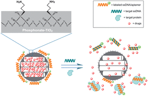
Researchers at Nankai University, China, have created the first functioning system for simultaneous diagnosis and controlled drug release, a so-called “diagnospy” carrier.   To demonstrate the concept, De-Ming Kong and co-workers constructed a mesoporous phosphonate-TiO2 nanoparticle scaffold loaded with ibuprofen. The amino groups on the phosphonates hold fluorescein-labelled single strands of DNA in place over the porous surface, trapping the drug molecules inside. When a complementary target strand is present, a displacement reaction uncaps the pores and the drug is released.

This mechanism relies on the highly specific affinity between complementary strands of DNA, or between proteins and their aptamers, resulting in a very sensitive detection and drug delivery system. The simultaneous sense-and-release approach is hoped to make treatments more efficient and convenient for patients in the future.

To find out more, please access the full article below. It will be free to read for the next two weeks.

Mesoporous phosphonate-TiO2 nanoparticles for simultaneous bioresponsive sensing and controlled drug release
Hui Li,  Tian-Yi Ma,  De-Ming Kong and Zhong-Yong Yuan
Analyst, 2013, 138, 1084-1090
DOI: 10.1039/C2AN36631B

Digg This
Reddit This
Stumble Now!
Share on Facebook
Bookmark this on Delicious
Share on LinkedIn
Bookmark this on Technorati
Post on Twitter
Google Buzz (aka. Google Reader)

Using Chemosensors to Monitor pH Dynamics

Pui Sai Lau is a guest web writer for Analyst. She is currently a PhD student at McMaster University, Hamilton, ON, Canada.

The cell effectively uses the mechanism of pH fluctuation as a form of regulatory control for diverse physiological functions. Over the years, optical based methods have been used to develop chemosensors to measure the dynamics of intracellular pH, some of which have demonstrated considerable potential for biological applications.

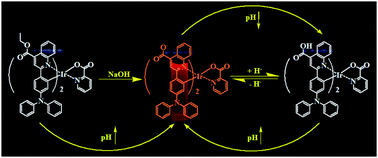
Wei Huang and colleagues from the Nanjing University of Posts and Telecommunications, China, have developed the latest optical pH probes by making use of cyclometalated iridium (III) complexes, a well known phosphorescent emitter. Huang’s group has expanded the repertoire of useful chemosensors by synthesizing and characterizing the photophysical properties of two iridium (III) complexes using a novel substituent, carboxylic-acid.

Find out more about their discovery by accessing the link below. This paper will be free to read for the next two weeks.

Effect of pH on the photophysical properties of two new carboxylic-substituted iridium(III) complexes
Jiena Weng ,  Qunbo Mei ,  Weiwei Jiang ,  Quli Fan ,  Bihai Tong ,  Qidan Ling and Wei Huang
Analyst, 2013, Advance Article
DOI: 10.1039/C2AN36298H

pH effect on carboxylic-substituted iridium(III) complex

pH effect on carboxylic-substituted iridium(III) complex

Digg This
Reddit This
Stumble Now!
Share on Facebook
Bookmark this on Delicious
Share on LinkedIn
Bookmark this on Technorati
Post on Twitter
Google Buzz (aka. Google Reader)

HOT articles in Analyst Issue 5

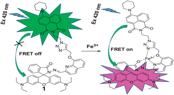
Along with Analyst Issue 5, here is a list of selected HOT articles for you to read all about. Topics vary from lipid imaging by mass spectrometry to determination of drugs by titanium oxide nanoparticles. Featured below are the work of Asit Baran Mandal and colleagues who present a new single molecular FRET-based sensor for determination of iron, and a study from the National Sun Yat-sen University, Taiwan, on direct monitoring of chemical transformations. All these papers will be free to read until February 28th. Have a read now!

Lipid imaging by mass spectrometry – a review
David Gode and Dietrich A. Volmer
Analyst, 2013,138, 1289-1315
DOI: 10.1039/C2AN36337B

Single molecular FRET-based sensor for detection of Fe2+

A highly selective and efficient single molecular FRET based sensor for ratiometric detection of Fe3+ ions
Narendra Reddy Chereddy ,  Sathiah Thennarasu and Asit Baran Mandal
Analyst, 2013,138, 1334-1337
DOI: 10.1039/C3AN36577H

Impact of charge state on gas-phase behaviors of noncovalent protein complexes in collision induced dissociation and surface induced dissociation
Mowei Zhou ,  Shai Dagan and Vicki H. Wysocki
Analyst, 2013,138, 1353-1362
DOI: 10.1039/C2AN36525A

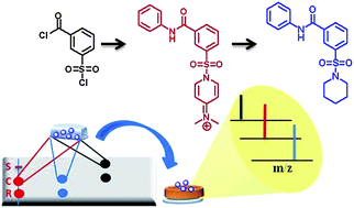
TLC with MALDI-MS to monitor chemical transformations

Direct monitoring of chemical transformations by combining thin layer chromatography with nanoparticle-assisted laser desorption/ionization mass spectrometry
Chun-Chi Chen ,  Yung-Lin Yang ,  Chun-Lin Ou ,  Chih-Hung Chou ,  Chih-Chuang Liaw and Po-Chiao Lin
Analyst, 2013,138, 1379-1385
DOI: 10.1039/C2AN36423A

Mass spectrometry sequencing of transfer ribonucleic acids by the comparative analysis of RNA digests (CARD) approach
Siwei Li and Patrick A. Limbach
Analyst, 2013,138, 1386-1394
DOI: 10.1039/C2AN36515D 

Adsorptive stripping voltammetric determination of imipramine, trimipramine and desipramine employing titanium dioxide nanoparticles and an Amberlite XAD-2 modified glassy carbon paste electrode
Bankim J. Sanghavi and Ashwini K. Srivastava
Analyst, 2013,138, 1395-1404
DOI: 10.1039/C2AN36330E

Electrical, enzymatic graphene biosensing of 5-aminosalicylic acid
Pratima Labroo and Yue Cui
Analyst, 2013,138, 1325-1328
DOI: 10.1039/C3AN36660J

Digg This
Reddit This
Stumble Now!
Share on Facebook
Bookmark this on Delicious
Share on LinkedIn
Bookmark this on Technorati
Post on Twitter
Google Buzz (aka. Google Reader)

Analyst Issue 5 NOW online!

Analyst issue 5 has now gone online! Take a look at these three woderful covers and read all about the new discoveries behind them.

Featured on the outside front cover  is the recent study of Todd Mitchell and his group, from the University of Wollongong, Australia. The team describes a novel approach to identify lipids on the surface of contact lenses by using liquid extraction surface analysis coupled to tandem mass spectrometry, and shows that lipids can be extracted with limited degradation of the contact lenses in only 15 minutes.

Automated surface sampling of lipids from worn contact lenses coupled with tandem mass spectrometry
Simon H. J. Brown ,  Liam H. Huxtable ,  Mark D. P. Willcox ,  Stephen J. Blanksby and Todd W. Mitchell
Analyst, 2013,138, 1316-1320
DOI: 10.1039/C2AN36189B

Our inside front cover showcases work coming from the Republic of Korea. Taesung Kim and researchers at the Ulsan National Institute of Science and Technology introduce a surface-patterned Nafion film to induce ion concentration polarization. This method can be easily used for separation and concentration of proteins extracted from lysed bacterial cells simultaneously and continuously on a chip.

Ion concentration polarization in a single and open microchannel induced by a surface-patterned perm-selective film
Minseok Kim ,  Mingjie Jia and Taesung Kim
Analyst, 2013,138, 1370-1378
DOI: 10.1039/C2AN36346A

Finally, the interesting outisde back cover of this issue shows a novel strategy for efficient ionization of phosphopeptides in mixtures presented by Hao Chen and his group from the Ohio University, USA. Desorption electrospray ionization-mass spectrometry allows direct ionization of analytes without the need of sample separation or enrichment. The new method presented here would provide ionization and examination of analytes in low pH environment.

Highly efficient ionization of phosphopeptides at low pH by desorption electrospray ionization mass spectrometry
Ning Pan ,  Pengyuan Liu ,  Weidong Cui ,  Bo Tang ,  Jingmin Shi and Hao Chen
Analyst, 2013,138, 1321-1324
DOI: 10.1039/C3AN36737A

Digg This
Reddit This
Stumble Now!
Share on Facebook
Bookmark this on Delicious
Share on LinkedIn
Bookmark this on Technorati
Post on Twitter
Google Buzz (aka. Google Reader)

Efficient detection of glutathione and cysteine in human serum

Jennifer A. Dougan is a guest web-writer for Analyst. She is currently a Post-Doctoral Research Associate at Imperial College London, UK.

Ruqin Yu and colleagues from the University of Hunan, China, have developed a label-free fluorescent detection system for glutathione (GSH) and cysteine (Cys). 

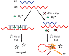
Hg2+ mediated fluorescent sensing strategy for detection of GSH and Cys

Three key interactions are necessary for the development of this assay: two single stranded DNA with thymine-thymine (T•T) mismatches stabilised in the presence of Hg2+, GSH and Cys interaction with Hg2+ with much higher binding affinity than T•T mismatches, and interaction of N-methyl porphyrin IX (MMP) with G-quadruplex structures, which leads to increased fluorescence.
The researchers  judiciously designed two sequences of DNA such that inter-molecular T•T mismatches were stabilised in the presence of Hg2+, inhibiting one of the stands from forming a G-quadruplex.  When the Hg2+ interacts with GSH or Cys, an intramolecular G-quadruplex is formed.  The G-quadruplex interacts with the NMM producing a marked increase in fluorescence.  Contrarily, in the absence of GSH and Cys, the Hg2+ is available to stabilise the mismatched duplex, the G-quadruplex is not formed, no NMM binding occurs and no fluorescent increase is observed.

This system was successfully used to detect GSH or Cys from protein extracted from human serum samples. To read more about this work, please access the full article below. It will be free to read until February 28th.

A Hg2+-mediated label-free fluorescent sensing strategy based on G-quadruplex formation for selective detection of glutathione and cysteine
Jingjin Zhao ,  Chunfei Chen ,  Liangliang Zhang ,  Jianhui Jiang ,  Guoli Shen and Ruqin Yu
Analyst, 2013, Advance Article
DOI: 10.1039/C3AN36657J

Digg This
Reddit This
Stumble Now!
Share on Facebook
Bookmark this on Delicious
Share on LinkedIn
Bookmark this on Technorati
Post on Twitter
Google Buzz (aka. Google Reader)