Archive for the ‘Hot Articles’ Category

New method for studying microalgal growth

The device

The study and optimisation of microalgal growth is a hot topic at the moment due to the use of microalgae in many industrial processes, as well as its potential use as biofuel. Previously, I have written about a Lab on a Chip article from the Sinton lab on optimising microalgal growth by varying irradiance conditions.

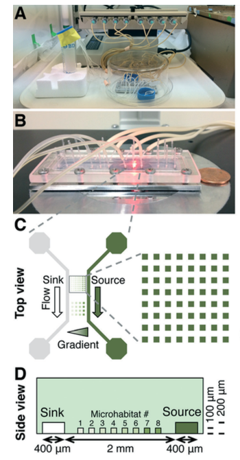
Now Mingming Wu’s group, from Cornell University, have published an article focused on the effect of nitrogen concentration on cell growth rates. Wu has developed a platform based on agarose gel, as shown in the diagram. The nutrient media can flow through this gel while the cells can’t, maintaining separate microhabitats.

The authors decided to study the effect of nitrogen concentration gradients on the microalgae (C. reinhardtii), using ammonium as the nitrogen source. Nitrogen is essential for microalgae, as it is required for protein and nucleic acid synthesis, and ammonium is the preferred source for this particular strain.

An ammonium gradient was obtained by flowing ammonium-containing media through the source channel, and ammonium-free media through the sink channel (diagram C). As expected, increasing the concentration (within the micromolar range) increased the microalgal growth rates. Fluorescence imaging allowed the authors to quantify the growth kinetics using the Monod equation (similar to the Michaelis-Menten equation for enzyme kinetics). This is the first time this has been achieved for this particular microalgal strain with nitrogen concentration as the variable.

Another interesting find was that when the microalgae were subjected to millimolar ammonium concentrations, growth inhibition was seen. The standard medium for microalgae contains 7.5 mM ammonium, so these results suggest that these concentrations need to be reduced by several orders of magnitude in order to maximise growth rates!

Wu and co-workers have nicely demonstrated the capablilty of their agarose-based platform in quantifying growth kinetics and they highlight that it is 50-fold faster, and more cost effective, than the standard chemostat system. They also observed cell heterogeneity during their experiments and plan to use their system to study this further, along with other aspects of cellular behaviour such as quorum sensing.

To download the full article for free* click the link below:

An array microhabitat system for high throughput studies of microalgal growth under controlled nutrient gradients
Beum Jun Kim, Lubna V. Richter, Nicholas Hatter, Chih-kuan Tung, Beth A. Ahner and Mingming Wu
Lab Chip, 2015,15, 3687-3694
DOI: 10.1039/ C5LC00727E

—————-

About the webwriter

Claire Weston is a PhD student in the Fuchter Group, at Imperial College London. Her work is focused on developing novel photoswitches and photoswitchable inhibitors.

—————-

*Access is free until 19/10/2015 through a registered RSC account – click here to register

Digg This
Reddit This
Stumble Now!
Share on Facebook
Bookmark this on Delicious
Share on LinkedIn
Bookmark this on Technorati
Post on Twitter
Google Buzz (aka. Google Reader)

A wireless chemical sensor for long-term monitoring

For effective treatment of many illnesses, in particular cancer, long-term monitoring of certain biomarkers is required. A needle probe with a chemically sensitive tip can be used, however this is invasive as it has to be inserted every time a new measurement is taken. There are also issues with tissue heterogenicity, as it is possible that changes in the measurements are solely due to a different local environment within the tissue.

Size of sensor

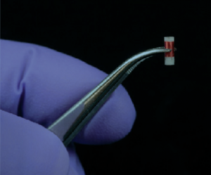
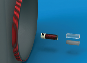
Previously the Cima lab at MIT reported an improved alternative for long-term in vivo monitoring. A capsule containing an NMR contrast agent was inserted in vivo and measurements were recorded using an MRI scanner. They have now done one better and eliminated the need of very costly MRI equipment by developing a small NMR sensor that simply requires a small external reader coil.

Both the sensor and reader contain a circuit with a coil and when both are in range magnetic inductance occurs, causing field amplification inside the chamber of the sensor.  This effectively means that a reading is taken only from tissue within the sensor, rather than surrounding tissue, and this is responsible for the high selectivity seen.

Different components of the system

In order to test their wireless sensor, Cima and coworkers separately measured pH and oxygen tension, both in vivo and in vitro. For the pH experiments, a polymer gel was used as the NMR contrast agent that had an exchangeable H atom with an appropriate pKa value. Using a tumor mouse model, pH readings were found to be lower when the sensor was nearer the tumor, as expected from the acidic nature of tumors. For the oxygen experiments, silicone was used as the contrast agent. The paramagnetic nature of molecular oxygen alters the relaxation time and this can therefore be used to determine the concentration of oxygen in the sensor.

From the success of their experiments, the authors conclude that they have demonstrated the flexibility of the sensor with these two measurements, and indeed there is huge potential for this NMR probe to greatly simplify in vivo monitoring.


To download the full article for free* click the link below:

Miniaturized, biopsy-implantable chemical sensor with wireless, magnetic resonance readout
C. C. Vassiliou, V. H. Liu and M. J. Cima
Lab Chip, 2015, 15, 3465-3472
DOI: 10.1039/C5LC00546A

—————-

About the webwriter

Claire Weston is a PhD student in the Fuchter Group, at Imperial College London. Her work is focused on developing novel photoswitches and photoswitchable inhibitors.

—————-

*Access is free until 01/10/2015  through a registered RSC account.

Digg This
Reddit This
Stumble Now!
Share on Facebook
Bookmark this on Delicious
Share on LinkedIn
Bookmark this on Technorati
Post on Twitter
Google Buzz (aka. Google Reader)

A closer look at erythrocytes in motion

Blood analysis is usually the first step involved in the diagnosis of various diseases, such as typhoid and malaria. The biochemical and cellular equilibrium of blood is very sensitive to physiological variations occurring in the body at various disease stages. Thus, a fast and accurate examination of blood properties is essential. The morphological and biochemical changes in erythrocytes are used as  the pathological signatures of various diseases.

Flow cytometry is used  to examine blood cells, which requires hydrodynamic sheath flow alignment and fluorescence antibody labelling, making it time-consuming and expensive. Advanced light scattering techniques (such as digital holography) are often seen as suitable alternatives, as they provide fast and label-free measurements.

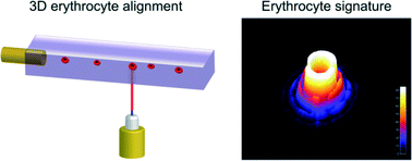
In a recent Lab On A Chip articleNetti et al. from the Italian Institute of Technology, in collaboration with scientists from Germany and Russia, presented a camera-based light scattering approach, coupled with a viscoelasticity -induced cell migration technique. This new system is used to characterise the morphological properties of erythrocytes in microfluidic flows.

They obtained light scattering profiles (LSPs) of individual living cells in microfluidic flows over a wide angular range and matched them with scattering simulations to characterise their morphological properties. A healthy erythrocyte diameter lies between 6 and 9 µm. The diameter values obtained from the experiment lie between 7 and 8.3 µm, which is in good agreement with the existing literature.

‘The results demonstrate the ability of a rapid and cost effective way to measure the average dimensions of an erythrocyte population which can be easily related to the health of a patient,’ concludes Netti.


To gain deeper insight into LSP acquisition and simulation, you can read the full article for free* by following the link below.
Optical signature of erythrocytes by light scattering in microfluidic flows
D. Dannhauser, D. Rossi, F. Causa, P. Memmolo, A. Finizio, T. Wriedt, J. Hellmers, Y. Eremin, P. Ferraro and   P. A. Netti
Lab Chip, 2015,15, 3278-3285
DOI: 10.1039/C5LC00525F

*Access is free until 27/09/2015 through a registered RSC account.
Digg This
Reddit This
Stumble Now!
Share on Facebook
Bookmark this on Delicious
Share on LinkedIn
Bookmark this on Technorati
Post on Twitter
Google Buzz (aka. Google Reader)

Cell pinballs in microfluidic channels

Researchers from the Kaneko Higashimori Lab at Osaka University and the Arai Lab at Nagoya University have observed an interesting phenomenon when studying red blood cells in microfluidic channels. Instead of flowing along the channel in a smooth motion as expected, some cells bounce back and forth between the channel walls in a pinball-like motion at much slower speed. In addition to these ‘cell pinballs’, there are also cells that move at a similar reduced speed, but don’t hit the channel walls.

This altered behaviour could have detrimental effects on microfluidic devices, caused by non-uniform movement of the cells in the channels. In order to prevent these potential problems, the authors have investigated the cause of this behaviour. They noted that cell pinballs only occur when the saline medium is hypotonic, as this causes the cells to inflate due to intake of water. By attaching microbeads to the cells and using a high speed camera, the motion of the cells were studied in more detail. The pinball cells rotated clockwise as they moved to the left of the channel and anticlockwise as they moved to the right of the channel (relative to the direction of the flow).

This observation, combined with the knowledge that the cells were inflated in the hypotonic solution, led the authors to believe that the pinball-motion was occurring due to both the shape of the red blood cell and contact with the channel walls. 3D images obtained using confocal microscopy showed that the upper and lower surfaces of the cells were flattened, confirming that the cells were in contact with the walls.

By studying the different possible deformations of the inflated red blood cells when subjected to flow, the authors found that the contact line (between the cell and wall) and the centre line of the cell were not the same. This explains both types of unexpected cell motion – if the contact line is downstream of the centre line, the cell is unstable to rotational motion and this causes it to move at an angle to the flow, leading to the pinball cells, whereas if the contact line is upstream of the centre line the cell is stable to rotational motion and no displacement occurs, leading to the slow moving non-pinball cells.

From these studies, the authors were able to propose mechanisms that successfully explained the two types of altered red blood cell behaviour in hypotonic solutions, and hopefully in the future this should allow microfluidic systems to be used which will avoid this pinball-motion occurring.


To download the full article for free* click the link below:
Cell pinball: phenomenon and mechanism of inertia-like cell motion in a microfluidic channel
Ryo Murakami, Chia-Hung Dylan Tsai, Makoto Kaneko, Shinya Sakuma and Fumihito Arai
Lab Chip, 2015, 15, 3307-3313
DOI: 10.1039/ c5lc00535c

—————-

About the webwriter

Claire Weston is a PhD student in the Fuchter Group, at Imperial College London. Her work is focused on developing novel photoswitches and photoswitchable inhibitors.

—————-

*Access is free until 06/09/2015  through a registered RSC account.

Digg This
Reddit This
Stumble Now!
Share on Facebook
Bookmark this on Delicious
Share on LinkedIn
Bookmark this on Technorati
Post on Twitter
Google Buzz (aka. Google Reader)

3D Bio-etching is here!

3D technology has revolutionised the entertainment industry by offering viewers the experience of being part of the action, going on in a movie rather than simply watching it. Thanks to 3D technology, we can sky walk hand in hand with George Clooney in ‘Gravity’.

Microfluidics setup for 3D bioetching

The history of 3D technology can be drawn all the way back to the invention of the stereoscope by David Brewster in 1844. Last two decades have seen 3D technology replacing 2D in all walks of life, ranging from entertainment, physics, microelectronics, tissue engineering and regenerative medicine. For e.g., microelectromechanosensors (MEMS) are 3D devices produced by using soft lithography techniques. MEMS installed in air-bags in the cars have saved thousands of lives by sensing pressure levels during accidents.

Can we use 3D technology to have a better look at the complex events happening at cellular level? One of the major challenges in tissue engineering is that the conventional approaches are mainly limited to 2D monolayers systems and do not allow manipulation of complex multilayer tissue. Cells grown on 2D substrates may respond and differentiate distinctly than those in more physiologically relevant 3D environments. The emergence of 3D technology has enabled scientists to mimic the exact cellular environments and helped to provide better insights into the cell signalling, migration and differentiation in cells.

One of the ways of mimicking the cellular architectures is bio-etching which involves subtractive manufacturing. Bioetching of monolayers of cells in response to laser cuts or scratch assays is achieved by using 2D cell culture studies. But the actual biological systems such as tissues and organs are much more complex and cannot be mimicked using simple monolayers. For long time, scientists have been working on developing better technologies to address this problem. One of the ways to achieve this is 3D bio-etching.

William C. Messner et al. from Tufts University in a recent article in Lab on a Chip explain the utility of 3D bioetching technique to create and shape 3D composite tissues using a microfluidics based approach. The ability to shape the 3D form of multicellular tissues and to control 3D stimulation will have a high impact on tissue engineering and regeneration applications in bioengineering and medicine as well as provide significant improvements of highly complex 3D integrated multicellular biosystems.


Can 3D bio-etching help us to design tissue architecture of our choice mimicking different biological events? Find out by reading the full paper for free* using the link below:

3D bio-etching of a complex composite-like embryonic tissue
Melis Hazar, Yong Tae Kim, Jiho Song, Philip R. LeDuc, Lance A. Davidson and William C. Messner
Lab Chip, 2015, Advance Article
DOI: 10.1039/C5LC00530B


*Access is free through a registered RSC account.

Digg This
Reddit This
Stumble Now!
Share on Facebook
Bookmark this on Delicious
Share on LinkedIn
Bookmark this on Technorati
Post on Twitter
Google Buzz (aka. Google Reader)

Recognition of danger in single cells

For more than a century, researchers have been studying how the body detects and defends itself against foreign invaders.1 This process relies on a series of cell-to-cell signaling, starting with the detection of common pathogen components by receptors of immune cells. For example, macrophages, a surveillance immune cell, recognize bacteria using the Toll-Like receptor 4 (TLR4), which binds to lipopolysaccharide (LPS) found in the membrane of gram-negative bacteria.

Upon recognition of danger signals, immune cells engulf and degrade the invaders and activate NF-κB, an intracellular protein that, when activated, migrates into the nucleus and turns on inflammatory response genes. For immune cells, this initiates the secretion of signaling mediators such as tumour necrosis factor (TNF), which helps remove invaders by dilating local blood vessels and recruiting other immune cells.

Although our understanding of the immune system has skyrocketed in the past two decades,2 researchers are still lacking the tools needed to study the signaling of inflammation in a highly controlled manner. Most approaches rely on bulk population measurements, in which important spatial and temporal patterns of signaling are obscured by biological noise from neighbouring cells. To address this challenge, Tino Frank and Prof. Savas Tay from ETH Zurich developed a microfluidic system that can propagate inflammatory signals from a single immune cell (sender cell) and to a population of fibroblast cells (receiver cells).3

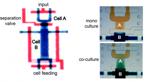
The microfluidic device, formed from polydimethylsiloxane (PDMS), comprises a deflectable membrane sandwiched between a flow layer and a control layer. The flow layer houses supply channels and cell culture chambers, while the control layer regulates input, output, and segregation of these chambers using solenoid-actuated valves.

As shown in picture 1, the chambers for the sender cell (chamber A) and receiver cells (chamber B) can be segregated for mono culture or connected for co-culture, depending on the state of the reversible separation valve. In mono culture mode, the sender cell can be stimulated independent of the receiver cells, whereas in co-culture mode, the signaling mediators secreted by the sender cell can be broadcasted to the receiver cells via diffusion.

Valve PDMS microfluidic device for mono and co-culture

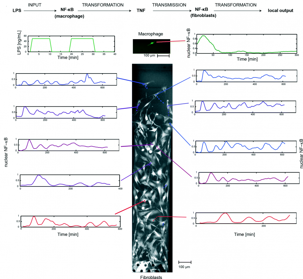
Using this system, the researchers implemented a single-cell model of inflammation arising from bacterial infection (picture 2). This model begins in mono culture mode, with a single macrophage seeded in chamber A and a population fibroblasts seeded in chamber B. Inflammation was initiated by stimulating the macrophage with short pulses of bacterial LPS, leading to the activation of NF-κB and TNF secretion. Subsequently, the device was switched to co-culture mode, allowing the secreted TNF to diffuse across the fibroblasts and activate their NF-κB signaling.

One-way communication chain mimicking an inflammatory process, with macrophage as sender and fibroblasts as receiver.

By tracking the movement of NF-κB in the nucleus of individual cells, the researchers, for the first time, mapped the spatiotemporal distribution of immune response to bacterial infection. They demonstrate that a single macrophage can activate and control over 100 fibroblasts up to 1 mm away, and that fibroblasts located farther away exhibited a time-delayed activation profile and fewer NF-κB oscillations (between the nucleus and cytoplasm). Furthermore, they observed that the NF-κB signaling in some fibroblasts can linger for up to 10 hours, demonstrating that a brief exposure to pathogenic signal can induce long-term inflammatory response in nearby cells.

In summary, Tino Frank and Prof. Savas Tay developed a microfluidic platform to study spatiotemporal dynamics of immune response in single cells. The continued investigation of these dynamics will be important for understanding of the inflammatory process and modeling of relevant signaling pathways.4


To download the full article for free* click the link below:
Automated co-culture system for spatiotemporal analysis of cell-to-cell communication
Tino Franka and Savaş Taya
Lab Chip, 2015, 15, 2192-2200
DOI: 10.1039/C5LC00182J


—————-
References:

1. K. M. Murphy, Janeway’s immunobiology, Garland Science, 2011.
2. L. A. J. O’Neill, D. Golenbock and A. G. Bowie, Nat Rev Immunol, 2013, 13, 453-460.
3. T. Frank and S. Tay, Lab on a Chip, 2015, 15, 2192-2200.
4. M. Junkin and S. Tay, Lab on a Chip, 2014, 14, 1246-1260.

—————-

About the webwriter

Alphonsus Ng is a postdoctoral fellow under the supervision of Dr. Aaron Wheeler in the Department of Chemistry and the Institute of Biomaterials and Biomedical Engineering at the University of Toronto. His research focuses on the development of microfluidic methods for heterogeneous immunoassays, cell-based assays, enzymatic catalysis, sample preparation for proteomics, and chemical synthesis.

—————-

*Access is free until 24/08/2015  through a registered RSC account.

Digg This
Reddit This
Stumble Now!
Share on Facebook
Bookmark this on Delicious
Share on LinkedIn
Bookmark this on Technorati
Post on Twitter
Google Buzz (aka. Google Reader)

Free access to June’s HOT articles

These HOT articles published in June 2015 were recommended by our referees and are free* to access for 4 weeks


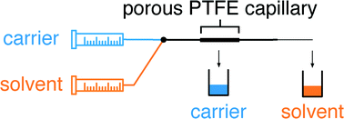
Microscale extraction and phase separation using a porous capillary
Thomas W. Phillips, James H. Bannock and John C. deMello  
Lab Chip, 2015,15, 2960-2967
DOI: 10.1039/C5LC00430F, Paper

Graphical abstract: Microscale extraction and phase separation using a porous capillary


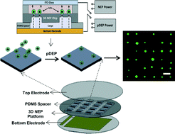
Dielectrophoresis-assisted 3D nanoelectroporation for non-viral cell transfection in adoptive immunotherapy
Lingqian Chang, Daniel Gallego-Perez, Xi Zhao, Paul Bertani, Zhaogang Yang, Chi-Ling Chiang, Veysi Malkoc, Junfeng Shi, Chandan K. Sen, Lynn Odonnell, Jianhua Yu, Wu Lu and L. James Lee  
Lab Chip, 2015,15, 3147-3153
DOI: 10.1039/C5LC00553A, Paper

Graphical abstract: Dielectrophoresis-assisted 3D nanoelectroporation for non-viral cell transfection in adoptive immunotherapy


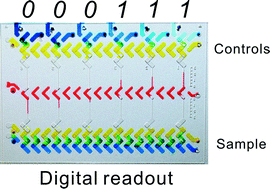
A microfluidic platform with digital readout and ultra-low detection limit for quantitative point-of-care diagnostics
Ying Li, Jie Xuan, Yujun Song, Ping Wang and Lidong Qin  
Lab Chip, 2015, Advance Article
DOI: 10.1039/C5LC00529A, Paper

Graphical abstract: A microfluidic platform with digital readout and ultra-low detection limit for quantitative point-of-care diagnostics


 *Access is free until 17.08.15 through a publishing personal account. It’s quick, easy and free to register
Digg This
Reddit This
Stumble Now!
Share on Facebook
Bookmark this on Delicious
Share on LinkedIn
Bookmark this on Technorati
Post on Twitter
Google Buzz (aka. Google Reader)

A new microfluidic LCD assay for optimising microalgae growth

Research into biofuels as a replacement for fossil fuels is a hugely important area of research. One particular focus is on cultivating photosynthetic organisms, such as microalgae, as these organisms have a high oil content that can be extracted and converted into biofuels. They are grown in photobioreactors in order to control the growth conditions, and there are a large amount of variable factors that need to be taken into account to find the optimal conditions.

For each new microalgal strain used the conditions need to be optimised, and irradiance screening is of particular importance. Currently, the screening process requires multiple culture flasks, each with their own light source. Recent developments have switched to using microwells, again with individual light sources.

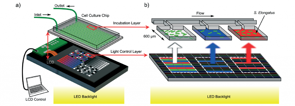
David Sinton and co-workers at the University of Toronto have developed a microfluidic irradiance assay using liquid crystal display (LCD) technology that allows them to rapidly screen irradiance conditions and identify the conditions for optimum growth. Using this technology, they were able to control all irradiance variables (light intensity, time variance, and spectral composition) in over two hundred parallel microreactors.

The diagram below shows the design of their irradiance platform – the LCD screen is lined up so that each pixel is directly below one microreactor, with every pixel individually controlled in order to produce the correct irradiance output. The bacterial growth in each microreactor was characterised by measuring the total fluorescence, emitted by a fluorescent pigment inside the organism.

Design of the irradiance platform

Demonstration of spatial control of microalgal growth

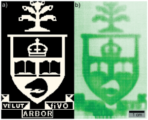
Initially, to demonstrate that their pixel-based method worked, the authors displayed the Toronto University crest on the LCD screen by using high and low irradiance intensities, and you can see from the image that this was successful!

By studying the three major irradiance variables mentioned previously they were able to quantify several important properties, such as the saturation intensity, the threshold frequency for growth and the combined effect of spectral composition and irradiance intensity on growth.

This new method drastically reduces the time needed to screen conditions for bacterial growth and hopefully should have a significant impact on the development of microalgal biofuels.


To download the full article for free* click the link below:
Microalgae on display: a microfluidic pixel-based irradiance assay for photosynthetic growth
Percival J. Graham, Jason Riordon and David Sinton.
Lab Chip, 2015, 15, 3116-3124
DOI: 10.1039/C5LC00527B

—————-

About the webwriter

Claire Weston is a PhD student in the Fuchter Group, at the Imperial College London. Her work is focused on developing novel photoswitches and photoswitchable inhibitors.

—————-

*Access is free until 24/08/2015  through a registered RSC account.

Digg This
Reddit This
Stumble Now!
Share on Facebook
Bookmark this on Delicious
Share on LinkedIn
Bookmark this on Technorati
Post on Twitter
Google Buzz (aka. Google Reader)

Free access to May’s HOT articles

These HOT articles published in May 2015 were recommended by our referees and are free* to access for 4 weeks


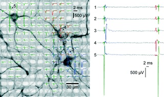
High-resolution CMOS MEA platform to study neurons at subcellular, cellular, and network levels

Jan Müller, Marco Ballini, Paolo Livi, Yihui Chen, Milos Radivojevic, Amir Shadmani, Vijay Viswam, Ian L. Jones, Michele Fiscella, Roland Diggelmann, Alexander Stettler, Urs Frey, Douglas J. Bakkum and Andreas Hierlemann
Lab Chip, 2015,15, 2767-2780
DOI: 10.1039/C5LC00133A


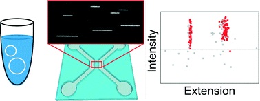
Fast size-determination of intact bacterial plasmids using nanofluidic channels

K. Frykholm, L. K. Nyberg, E. Lagerstedt, C. Noble, J. Fritzsche, N. Karami, T. Ambjörnsson, L. Sandegren and F. Westerlund
Lab Chip, 2015,15, 2739-2743
DOI: 10.1039/C5LC00378D


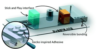
Gecko gaskets for self-sealing and high-strength reversible bonding of microfluidics

A. Wasay and D. Sameoto
Lab Chip, 2015,15, 2749-2753
DOI: 10.1039/C5LC00342C

Take a look at our Lab on a Chip Recent HOT Articles Collection!

*Access is free until 24.07.15 through a publishing personal account. It’s quick, easy and free to register
Digg This
Reddit This
Stumble Now!
Share on Facebook
Bookmark this on Delicious
Share on LinkedIn
Bookmark this on Technorati
Post on Twitter
Google Buzz (aka. Google Reader)

New speedy method for the production of monodisperse droplets!

A group of scientists at IMTEK, University of Freiburg have developed a new method for the production of monodisperse droplets. Previous methods, such as T-junctions and flow focusing require several channels, containing either the disperse phase (which will form the droplet) or the continuous phase (which will surround the droplet), with the droplets forming at a constriction point in the tubing. Extremely precise control of flow rate is therefore required in order to achieve consistent droplet diameters. These methods also have substantial dead volumes due to sample material remaining in the tubing at the end of the process.

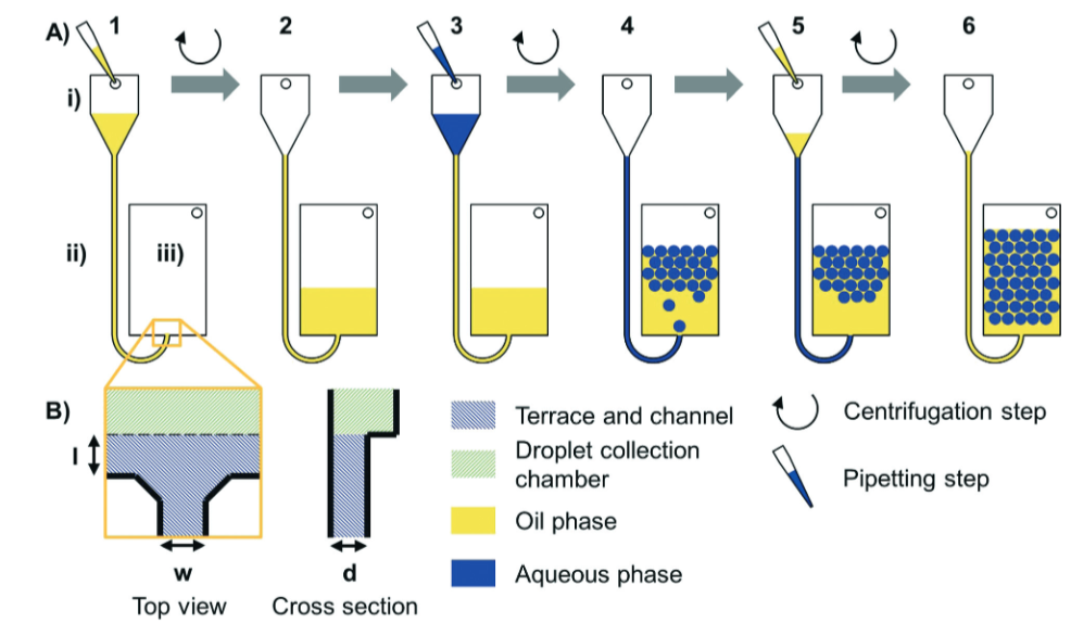
An alternative method is step emulsification (as highlighted recently in a Lab on a Chip HOT article). This only requires one channel, containing both phases, and the droplet formation is caused by a change in capillary pressure. The droplet size depends on the nozzle, rather than on pressure and flow rate, so this method is less sensitive to fluctuations than the methods mentioned previously. The main limitation of step emulsification is the relatively low throughput due to droplet accumulation at the nozzle. This publication reports the use of centrifugal force in order to solve this problem.

Workflow of centrifual step emulsification without dead volume

By spinning the whole system, the disperse phase (water) is overpressured relative to the continuous phase (oil), resulting in the droplet being forced away from the nozzle by the centrifugal gravitational field (step 3 to 4 above). In order to avoid sample material being wasted as dead volume, an additional aliquot of oil is added in order to push the last few droplets of water out of the nozzle (step 5 to 6 above).

Medium throughput monodisperse droplet formation

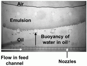
The authors found that droplet diameter is controlled by the nozzle geometry, while rate of formation is controlled by spinning frequency. In light of these findings, they were able to increase droplet production rate from less than 1 droplet per second, to greater than 500 droplets per second, while maintaining monodispersity. They were also able to set up multiple nozzles in parallel (as seen in the microscopic image), all feed by a larger channel, to further increase throughput.

In order to demonstrate the potential applications of this new method, the authors performed digital droplet recombinase polymerase amplification (ddRPA) of L. monocytogenes (a potential contaminant during food production). They found that the number of copies measured with ddRPA was consistent with those measured with digital droplet PCR, and the overall processing was 30 minutes, compared with 2 hourlis for ddPCR.

ddRPA is just one small example of how this new technique can be used – there are a huge range of potential applications where formation of monodisperse particles are a requirement and hopefully we will see this new method being adopted!

To download the full article for free click the link below:

Centrifugal step emulsification applied for absolute quantification of nucleic acids by digital droplet RPA
Friedrich Schuler, Frank Schwemmer, Martin Trotter, Simon Wadle, Roland Zengerle, Felix von Stetten and Nils Paust
DOI: 10.1039/C5LC00291E

Digg This
Reddit This
Stumble Now!
Share on Facebook
Bookmark this on Delicious
Share on LinkedIn
Bookmark this on Technorati
Post on Twitter
Google Buzz (aka. Google Reader)