Archive for the ‘Hot Articles’ Category

Free access to April’s HOT articles

These HOT articles published in April 2015 were recommended by our referees and are free* to access for 4 weeks


Active pneumatic control of centrifugal microfluidic flows for lab-on-a-chip applications
Liviu Clime, Daniel Brassard, Matthias Geissler and Teodor Veres
Lab Chip, 2015,15, 2400-2411
DOI: 10.1039/C4LC01490A

Graphical Abstract


Exosome isolation: A microfluidic road-map
A. Liga, A. D. B. Vliegenthart, W. Oosthuyzen, J. W. Dear and M. Kersaudy-Kerhoas
Lab Chip, 2015,15, 2388-2394
DOI: 10.1039/C5LC00240K

Graphical Abstract


Micromilling: A method for ultra-rapid prototyping of plastic microfluidic devices
David J. Guckenberger, Theodorus E. de Groot, Alwin M. D. Wan, David J. Beebe and Edmond W. K. Young
Lab Chip, 2015,15, 2364-2378
DOI: 10.1039/C5LC00234F

Graphical Abtrsct

Take a look at our Lab on a Chip 2015 HOT Articles Collection!

*Access is free until 29.06.15 through a publishing personal account. It’s quick, easy and free to register

Digg This
Reddit This
Stumble Now!
Share on Facebook
Bookmark this on Delicious
Share on LinkedIn
Bookmark this on Technorati
Post on Twitter
Google Buzz (aka. Google Reader)

Direct measurement of the tensile strength of water using an optofluidic chip

As children, you may remember being fascinated by pond skaters and their ability to walk on water. This is due to water’s high surface tension and there are numerous other ways in which this property is vital to many biological functions. It is also an important factor to take into account when it comes to engineering, and therefore it is essential that there is an accurate and straight forward method for measuring surface tension.

To measure this property, the pressure has to be reduced to such an extent that it causes the water to rupture and form vapour cavities. These vapour cavities must be a result of homogeneous nucleation alone and not heterogeneous nucleation (so should occur spontaneously and randomly, rather than due to nucleation sites). The pressure at which cavitation occurs in termed the tensile strength.

Previous methods have shown a large discrepancy in results, due to the requirement of large volumes of water leading to heterogeneous nucleation. More recently, the mineral inclusion method has overcome this, however has other limitations, such as the requirement of an autoclave. Alternatively, microfluidics allows the use small volumes of water to ensure homogenous nucleation, but this method is limited to low-viscosity liquids.

Graphical Abstract

Dr. Liu Ai Lin and co-workers, at NTU, Singapore, have reported the direct measurement of water’s tensile strength using an optofluidic chip. Their method relies on an infrared laser that is focussed into a microchannel partially filled with water. The laser pulse results in the formation and recombination of plasma, which in turn produces a bubble, causing a spherical shock wave. The reflection of the shock wave on the air-water interface generates a negative pressure and, if this is larger than the tensile strength, the water ruptures, causing nucleation of vapour bubbles near the interface. The pressure value can be attained by both measuring the spreading of the shock wave over time and the displacement of the water-air interface.

By imaging the microchannel at increasing standoff distances (defined in the diagram above), the distance and pressure at which water no longer ruptures can be found. This can be directly converted to a value for the tensile strength.

Graphical Abstract

This work provides a simple, low-cost method for calculating tensile strength that can easily allow rapid testing of a wide range of samples. In order to demonstrate this, the authors also measured the tensile strength of glycerol, a highly viscous fluid.

To download the full article for free* click the link below:

Water’s tensile strength measured using an optofluidic chip
Z. G. Li,  S. Xiong,  L. K. Chin,  K. Ando,  J. B. Zhang and  A. Q. Liu
DOI: 10.1039/ C5LC00048C

*Access is free through a registered RSC personal publishing account

Digg This
Reddit This
Stumble Now!
Share on Facebook
Bookmark this on Delicious
Share on LinkedIn
Bookmark this on Technorati
Post on Twitter
Google Buzz (aka. Google Reader)

Free access to March’s HOT Articles

These HOT articles published in March 2015 were recommended by our referees and are free* to access for 4 weeks


Electromechanical cell lysis using a portable audio device: enabling challenging sample preparation at the point-of-care

J. R. Buser, A. Wollen, E. K. Heiniger, S. A. Byrnes, P. C. Kauffman, P. D. Ladd and P. Yager
Lab Chip, 2015,15, 1994-1997
DOI: 10.1039/C5LC00080G, Technical Innovation


Implementation of in situ SAXS/WAXS characterization into silicon/glass microreactors

Thomas Beuvier, Elvia Anabela Chavez Panduro, Paweł Kwaśniewski, Samuel Marre, Carole Lecoutre, Yves Garrabos, Cyril Aymonier, Brice Calvignac and Alain Gibaud

Lab Chip, 2015,15, 2002-2008
DOI: 10.1039/C5LC00115C, Paper

Reconfigurable microfluidic systems with reversible seals compatible with 2D and 3D surfaces of arbitrary chemical composition

Abhiteja Konda, Jay M. Taylor, Michael A. Stoller and Stephen A. Morin
Lab Chip, 2015, 15, 2009-2017
DOI: 10.1039/C5LC00026B, Paper

Take a look at our Lab on a Chip 2015 HOT Articles Collection!

*Access is free until 31.05.15 through a publishing personal account. It’s quick, easy and free to register!


Digg This
Reddit This
Stumble Now!
Share on Facebook
Bookmark this on Delicious
Share on LinkedIn
Bookmark this on Technorati
Post on Twitter
Google Buzz (aka. Google Reader)

Textile Weaving: a new cheaper alternative to electrochemical sensors

Electrochemical sensors are widely used as analytical tools. They are disposable, cheap to make, and small – making them ideal for many applications.

The current method for commercial electrochemical sensors uses screen printing onto plastic or ceramic surfaces to generate the circuit elements, which requires specialised fabrication equipment. This screen printing method also leads to wastage of electrode inks and reagents. One way of avoiding these issues is using paper microfluidics – Professor George Whitesides is a well-known name in this field.

Dendukuri and coworkers, from Achira Labs, Bangalore, have come at this problem from a different angle. They have developed an alternative approach using textile weaving. Instead of screen printing, they coat their yarn with the required reagents in a way that results in no wastage, as shown in the photo. They use silk as their material which is biodegradable, unlike the plastics usually used. It is also easily processed – initially silk is hydrophobic but it can be made hydrophobic by degumming. This can be achieved by simply boiling the yarn.

To make the sensors, electrode yarns are prepared by coating in conductive inks and reagents, and then woven into the fabric. Large numbers of sensors can be woven as patches on the fabric, which are then stuck onto an adhesive backing and laminated, leaving a window for application of the sample and for contact with a reader.To demonstrate this effectiveness of this new method, the authors developed glucose and haemoglobin sensors. The glucose sensors were found to have a clinically acceptable performance, according to FDA criteria, while the haemoglobin sensors were able to detect physiologically relevant concentrations. Multiplexed sensors capable of detecting more than one analyte were easily prepared by adding an additional electrode.

One of the most pleasing aspects of this new method, is its potential in the developing world, where weaving is still widely used. In addition, the cost of manufacture was calculated as less than 20 USD per 1000 sensors and this could even lower on scale up.

To download the full article for free* click the link below:

Woven electrochemical fabric-based test sensors (WEFTS): a new class of multiplexed electrochemical sensors
Tripurari Choudhary, G. P. Rajamanickam and Dhananjaya Dendukuri
DOI: 10.1039/C5LC00041F

*Access is free until 30.04.2015 through a registered RSC Personal Publishing Account

Digg This
Reddit This
Stumble Now!
Share on Facebook
Bookmark this on Delicious
Share on LinkedIn
Bookmark this on Technorati
Post on Twitter
Google Buzz (aka. Google Reader)

When microfluidics meets inkjet printing

Since its inception over 50 years ago, inkjet printing has become the most widely adopted method for the reproduction of images and text on paper. Inherently a microfluidic technology, inkjet printing is a process in which individual ink droplets, between 10 to 100 µm diameters, are placed at software-configurable locations on a substrate. The resulting dots can combine to create photo-quality images with resolutions of up to several thousand dots per inch. Such exquisite precision and flexibility make inkjet printing particularly attractive for the scalable fabrication of fine features. Indeed, in addition to coloured inks, this technology has also been adapted to deposit a wide range of materials for manufacturing, including metals, ceramics, polymers, and even biological cells.1

Not surprisingly, the lab on a chip community is also beginning to leverage printing technologies for the fabrication of microfluidic components.2 However, this is not straightforward because the quality of the printed features often depends on the interaction of the ink droplets with the substrate material. Since microfluidic devices are predominantly prototyped using polydimethylsiloxane (PDMS),3 recent research has focused on the development of PDMS-compatible methods for inkjet printing.

In one example, Profs Dachao Li at the Tianjin University, China and Robert C. Roberts at the University of Hong Kong used inkjet printing to form robust silver microelectrodes on PDMS-based microfluidics for glucose sensing.4 In forming these electrodes, the PDMS material posed two challenges: 1) poor adhesion of silver to PDMS inhibited robust electrode formation and, 2) the inherent hydrophobicity of PDMS promoted coalescence of neighboring ink droplets, which impaired the precision of printed features.

To overcome these challenges, the researchers modified the substrate using (3-Mercaptopropyl)trimethoxysilane, a coupling reagent that presents thiol groups on the PDMS surface. The thiol groups not only served as covalent anchoring sites for the inkjet-printed silver but also increased the wettability of the ink droplet, which improved the precision of the printed features.

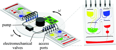
Remarkably, the printed electrodes formed on these substrates tolerated very harsh stress tests, including immersion in water, exposure to high pressure air stream, and even ultrasonication. This new fabrication strategy enabled the team to create an all-in-one PDMS system with fluid handling and a three-electrode electrochemical cell for transdermal detection of glucose (Picture 1).

In another example, Profs Yanlin Song and Fengyu Li at the Chinese Academy of Sciences, China developed an elegant inkjet-printed method 5 for the fabrication of enclosed PDMS microchannels. This process conventionally requires at least 4 steps: 1) create a solid template via photolithography, 2) cast PDMS prepolymer mixture in the template, 3) cure and separate the PDMS from the template, and 4) bond the molded PDMS to a substrate.

To simplify this procedure, the researchers implemented a liquid template formed by inkjet-printing (Picture 2). Here, an immiscible liquid pattern is printed directly on a pool of PDMS prepolymer solution. Within seconds, the liquid spontaneously submerges below the prepolymer solution and acts as a template. Upon heating, the polymer solidifies while the liquid template evaporates, leaving behind an enclosed PDMS microchannel device.

In the development of this method, one major challenge was maintaining the stability of the liquid template in the prepolymer solution. Due to surface tension, printed liquid templates tend to break up and relax into discontinuous spherical droplets.  Although this instability can be inhibited by increasing the liquid viscosity, high viscosities are not compatible with inkjet printing. To address this challenge, the researchers used an ink whose viscosity is tunable by temperature. At room temperature, the ink has relatively low viscosity (10 centipoise), which is compatible with inkjet printing. When the ink is printed on a cooled (3-4oC) pool of prepolymer solution, the viscosity of the liquid increases dramatically (>40 centipoise), which enabled the formation of a stable liquid template with no breakages.

This inkjet-printed approach enabled the formation of remarkably versatile PDMS microchannels. The diameters of the fabricated channels can range from 100 to 900 µm just by changing the template design. In addition, the interior surface of the channel can be modified by including vinyl-terminated functional molecules in the ink composition, which covalently incorporates in the PDMS matrix during thermal curing. Applying this technique with vinyl-terminated poly(ethylene glycol)methacrylate molecules, the researchers effortlessly imbue their devices with protein fouling resistance.

In summary, the lab on a chip community is beginning to leverage the benefits of inkjet printing in the fabrication of microfluidic components. By engineering the interaction of the ink droplets with the substrate material, researchers are devising innovative ways to fabricate robust electrodes and versatile microchannels without the need for cleanroom facilities and complicated procedures.

1.    I. M. Hutchings and G. D. Martin, Inkjet technology for digital fabrication, John Wiley & Sons, 2012.
2.    P. Tseng, C. Murray, D. Kim and D. Di Carlo, Lab on a Chip, 2014, 14, 1491-1495.
3.    E. Berthier, E. W. K. Young and D. Beebe, Lab on a Chip, 2012, 12, 1224-1237.
4.    J. Wu, R. Wang, H. Yu, G. Li, K. Xu, N. C. Tien, R. C. Roberts and D. Li, Lab on a Chip, 2015, 15, 690-695.
5.    Y. Guo, L. Li, F. Li, H. Zhou and Y. Song, Lab on a Chip, 2015, 15, 1759-1764.

Digg This
Reddit This
Stumble Now!
Share on Facebook
Bookmark this on Delicious
Share on LinkedIn
Bookmark this on Technorati
Post on Twitter
Google Buzz (aka. Google Reader)

Free access to February’s HOT Articles

These HOT articles published in February 2015 were recommended by our referees and are free* to access for 4 weeks


On-chip surface acoustic wave lysis and ion-exchange nanomembrane detection of exosomal RNA for pancreatic cancer study and diagnosis
Daniel Taller, Katherine Richards, Zdenek Slouka, Satyajyoti Senapati, Reginald Hill, David B. Go and Hsueh-Chia Chang
Lab Chip, 2015,15, 1656-1666
DOI: 10.1039/C5LC00036J, Paper


Three-dimensional heterogeneous assembly of coded microgels using an untethered mobile microgripper
Su Eun Chung, Xiaoguang Dong and Metin Sitti
Lab Chip, 2015,15, 1667-1676
DOI: 10.1039/C5LC00009B, Paper

A flexible lab-on-a-chip for the synthesis and magnetic separation of magnetite decorated with gold nanoparticles
Flávio C. Cabrera, Antonio F. A. A. Melo, João C. P. de Souza, Aldo E. Job and Frank N. Crespilho
Lab Chip, 2015, Advance Article
DOI: 10.1039/C4LC01483A, Paper

Take a look at our Lab on a Chip 2015 HOT Articles Collection!

*Access is free until 30.04.15 through a publishing personal account. It’s quick, easy and free to register!


Digg This
Reddit This
Stumble Now!
Share on Facebook
Bookmark this on Delicious
Share on LinkedIn
Bookmark this on Technorati
Post on Twitter
Google Buzz (aka. Google Reader)

Scientists can quickly detect waterborne pathogens using a smartphone!

More than half a billion people have to survive with unimproved water, as providing safe drinking water is still a problem in many parts of the developing world. Of the waterborne pathogens, Giardia lambia (G. lambia) is one of the most common intestinal parasites that are difficult to remove using traditional water purification methods. Current methods for their detection take up to two days and require analysis laboratories with trained specialists and expensive equipment. Because of this there is an ongoing effort to design low-cost and field-portable methods that can rapidly analyse large volumes of water.

Ozcan and co-workers at UCLA have developed a method for the detection of G. lambia cysts in water using a light weight attachment to a smartphone. The attachment consists of a fluorescence microscope, aligned to the smartphone camera, and a disposable water sample cassette that can hold 20 mL of water. The whole test can be carried out in just 1 hour, from taking the sample from the source, to receiving the total number of cysts detected in the sample.

The process is relatively simple, with the test sample first being fluorescently labelled and then filtered through a membrane that traps the G. lambia cysts. A fluorescence image is taken and wirelessly transmitted to servers using an app designed by the group. Digital analysis is carried out using a machine learning algorithm that can specifically recognise the cysts over other fluorescent micro-objects. The results of this analysis are then transmitted back to the phone and displayed on the app.

The group were able to achieve an impressive limit of detection of 12 cysts per 10 mL of sample, citing several factors that led to this limit. They have put forward a number of suggestions for how they hope to further improve their system, so it will be interesting to hear more from this group.

To download the full article for free* click the link below:

Rapid imaging, detection and quantification of Giardia lamblia cysts using mobile-phone based fluorescent microscopy and machine learning
Hatice Ceylan Koydemir, Zoltan Gorocs, Derek Tseng, Bingen Cortazar, Steve Feng, Raymond Yan Lok Chan, Jordi Burbano, Euan McLeod and Aydogan Ozcan
DOI: 10.1039/C4LC01358A

*Access is free until 31st March 2015 through a publishing personal account. It’s quick, easy and free to register.

About the web writer

Claire Weston is currently studying for a PhD at Imperial College London, focussing on developing novel photoswitches and photoswitchable inhibitors.

Digg This
Reddit This
Stumble Now!
Share on Facebook
Bookmark this on Delicious
Share on LinkedIn
Bookmark this on Technorati
Post on Twitter
Google Buzz (aka. Google Reader)

Hooked on a Feeling: measuring cell-substrate adhesion with ISFET devices

By developing an ion-sensitive field-effect transistor with small gate dimensions, scientists at the University of Applied Sciences Kaiserslautern in Germany were able to measure cell-substrate adhesion on the single cell scale.

To survive, most mammalian cells attach to other cells and the extracellular environment in order to regulate their growth, proliferation, and migration. Electrical impedance spectroscopy is one way to quantitatively monitor cell-substrate interactions. The strength of cellular adhesion to a substrate with integrated electrodes can be measured by comparing the ratio of the readout voltage to the applied alternating current. Yet this method is limited groups of many cells as the size of the microelectrode must be larger than 100 μm in diameter. Smaller features are subject to greater interface impedance between the electrode and liquid media and this background impedance overwhelms the desired cell-substrate measurements. Suslorapova and colleagues thus used an ion-sensitive field-effect transistor (ISFET) with small gate dimensions to overcome this limitation. The group was able measure the effects of enzymatic digestion with trypsin and an apoptosis-inducing drug on single cell detachment using the ISFET devices with a 16 by 2 square micron gate.

The authors create an equivalent circuit model to interpret recorded impedance spectra from their single cell and small cell groups grown in contact with the field-effect transistor devices. The seal resistance and membrane capacitance parameters which can be extracted from the measured transistor transfer function (TTF) provide measures of cell shape and adhesion to the substrate. Changes in TTF correspond to adhesion of individual cells on top of the ISFET gates. This platform and the model developed to interpret TTF signal opens exciting avenues to monitoring cell adhesion in high throughput yet still at single cell resolution.

Download the full research paper paper for free* for a limited time only!

Electrical cell-substrate impedance sensing with field-effect transistors is able to unravel cellular adhesion and detachment processes on a single cell level
A. Susloparova , D. Koppenhöfer , J. K. Y. Law , X. T. Vu and S. Ingebrandt. Lab Chip, 2015, 15, 668-679. DOI: 10.1039/C4LC00593G

*Access is free until 27.03.15 through a publishing personal account. It’s quick, easy and free to register!

Digg This
Reddit This
Stumble Now!
Share on Facebook
Bookmark this on Delicious
Share on LinkedIn
Bookmark this on Technorati
Post on Twitter
Google Buzz (aka. Google Reader)

January’s Free HOT Articles

These HOT articles, published in January 2015 were recommended by our referees and are free* to access for 4 weeks

Microfluidic single sperm entrapment and analysis
B. de Wagenaar, J. T. W. Berendsen, J. G. Bomer, W. Olthuis, A. van den Berg and L. I. Segerink
Lab Chip, 2015, Advance Article
DOI: 10.1039/C4LC01425A, Paper

Graphical abstract: Microfluidic single sperm entrapment and analysis

Optofluidic ultrahigh-throughput detection of fluorescent drops
Minkyu Kim, Ming Pan, Ya Gai, Shuo Pang, Chao Han, Changhuei Yang and Sindy K. Y. Tang
Lab Chip, 2015, Advance Article
DOI: 10.1039/C4LC01465K, Paper

Graphical abstract: Optofluidic ultrahigh-throughput detection of fluorescent drops

Hydrogel-droplet microfluidic platform for high-resolution imaging and sorting of early larval Caenorhabditis elegans
Guillaume Aubry, Mei Zhan and Hang Lu
Lab Chip, 2015, Advance Article
DOI: 10.1039/C4LC01384K, Paper

Graphical abstract: Hydrogel-droplet microfluidic platform for high-resolution imaging and sorting of early larval Caenorhabditis elegans

Take a look at our Lab on a Chip 2015 HOT Articles Collection!

*Access is free until 11.03.15 through a publishing personal account. It’s quick, easy and free to register!

Digg This
Reddit This
Stumble Now!
Share on Facebook
Bookmark this on Delicious
Share on LinkedIn
Bookmark this on Technorati
Post on Twitter
Google Buzz (aka. Google Reader)

Culturing Cells on Druggable Bubbles

When testing the effects of drugs on cells and tissues, laboratory scientists generally use a pretty crude approach. They simply mix the desired concentration of the drug with the culture medium and then add it to separate plastic wells in which cells or tissues have been cultured. Of course, this approach has its limitations: when testing many different concentrations and mixtures of drugs, the amount of wells needed for an experiment grows very quickly to overwhelming numbers. Moreover, all of the drug is generally added at once, not taking into account the gradual pharmacokinetic profiles that you would normally find in the human body.

In a paper in Lab on a Chip, scientists from Corning, Inc. and Massachusetts General Hospital demonstrate a new, microengineered approach for treating cultured cells with drugs. Instead of growing the cells on flat surfaces, cells are grown on micro-modified wells plates that contain regular patterns of tiny holes. The microholes can be filled with dried-up drug and then sealed off with a semi-permeable layer of collagen. When cells are grown on the collagen, culture medium starts seeping into the air-filled microholes. The dried-up drug then dissolves and diffuses through the collagen layer, exposing the microscopic patch of cells around the microhole to the drug (see also the figure below).

A schematic overview of how to will microwells with drug and culture hepatocytes on them

The authors only show the results of a simple proof-of-principle experiment with Nefazodone-filled holes leading to local toxicity for cultured liver cells. The technique looks promising however, and it will be interesting to see further development. How easy will it be to load holes with different concentrations and mixtures of drugs? Can the process of drugs diffusing into the medium be controlled? Can we control time-release profiles by changing the shape of the holes, by changing the surface properties of the holes, or maybe by changing the gas content of the medium?

It will also be interesting to see whether the technique can be combined with high-throughput imaging, like fluorescence microscopy or scanning electrochemical microscopy as was recently shown for cells in microscopic wells by Sridhar, et al.

Make sure to check out the paper by Goral, et al. in which they outline their technique while it is still free* to access.

*Access is free until 9.03.15 through a publishing personal account. It’s quick, easy and free to register!

Digg This
Reddit This
Stumble Now!
Share on Facebook
Bookmark this on Delicious
Share on LinkedIn
Bookmark this on Technorati
Post on Twitter
Google Buzz (aka. Google Reader)