Archive for the ‘Web writers’ Category

Magnetic nanoparticles as contrast agents for MRI-based stem cell tracking and labeling.

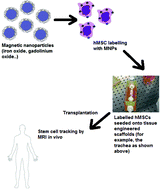
Tracking the distribution of stem cells (SCs) in tissue engineered organ scaffolds is of great importance in order to understand the role, movement and distribution of SCs after they have been implanted.   A recent minireview, by Hachani et al. at University College London, highlights the recent application of nanotechnology in SC labeling and magnetic resonance imaging (MRI).

The authors provide an overview of magnetic nanoparticles (MNPs), such as iron oxide and gadolinium nanoparticles, for use as contrast agents in MRI-based cell tracking and labeling.  The minireview introduces SC research and how conventional MRI is used as a tool for tracking and labeling.  However, the review’s main focus is discussing the recent developments in magnetic nanoparticle research, particularly detailing the efforts made by researchers in controlling the physical properties of the MNPs, such as size, shape, surface chemistry, etc, in order to produce more biocompatible and stable MNP contrast agents.

For researchers wishing to gain an insight into the application of nanotechnology in MRI and stem cell research, this review provides a valuable starting point for becoming familiar with recent developments in this area.

By Lee Barrett

You can read this Minireview in Nanoscale by clicking the link below:

Tracking stem cells in tissue-engineered organs using magnetic nanoparticles
Roxanne Hachani, Mark Lowdell, Martin Birchall and Nguyễn Thi Kim Thanh
DOI: 10.1039/C3NR03861K, Minireview

Digg This
Reddit This
Stumble Now!
Share on Facebook
Bookmark this on Delicious
Share on LinkedIn
Bookmark this on Technorati
Post on Twitter
Google Buzz (aka. Google Reader)

Measuring the environmental toxicity of engineered nanomaterials

Advances in nanomaterial production for device implementation, biotechnology and biomedical areas require methods to measure the possible health and safety risks associated with nanomaterials exposed to a particular biological environment.  In this manuscript, researchers from the Health Research Institute, Japan, investigate the environmental toxicity of engineered CdSe quantum dots (QDs) and ZnO nanopowder (NP) in human lung cells.

Table of contents imageTo evaluate the toxicity, the researchers induce the breakdown of the nanomaterials by exposing them to simulated solar UV light.  This releases Cd2+ and Zn2+ ions for the QDs and NPs, respectively, which were then detected by measuring the quenching of the fluorescence signal in the presence of tetrakis (4-carboxyphenyl) porphyrin (TCPP) or by measuring the enhancement of the fluorescence signal in the presence of the commercial Measure iT Pd/Cd sensor.

The researchers conclude that Cd2+ and Zn2+ ions are not cytotoxic at low concentrations and for short UV exposure times but become toxic at longer exposure times, i.e. 72 hours or longer.  In addition, cells exposed in the long term to Cd2+ ions result in extensive DNA damage as measured by comet assays and gel electrophoresis.  This manuscript details the importance of measuring the health and safety aspects of engineered nanomaterials, in particular the environmental impact of released meal ions, exposed to UV light.

by Dr Lee Barrett

Read the full Nanoscale communication here:

Impairments of cells and genomic DNA by environmentally transformed engineered nanomaterials
Philip Jones, Sakiko Sugino, Shohei Yamamura, Fred Lacyb and Vasudevanpillai Biju*
Nanoscale, 2013, Advance Article
DOI: 10.1039/C3NR03118G

Digg This
Reddit This
Stumble Now!
Share on Facebook
Bookmark this on Delicious
Share on LinkedIn
Bookmark this on Technorati
Post on Twitter
Google Buzz (aka. Google Reader)

Rainbow coloured Ag nanostructrues for nanosensing applications

In this article, researchers from Southwest University, China, investigate the size-dependent nature of the localized surface plasmon resonance (LSPR) of silver nanoparticles (AgNPs) using a combination of darkfield microscopy (DFM), scanning electron microscopy (SEM) and UV-Vis spectroscopy.

The AgNPs were synthesized with different shapes via a sulfide-mediated reduction of AgNO3 with ethylene glycol in the presence of PVP and Na2S.  By combining SEM and DFM analysis, the authors were able to confirm the presence of nanospheres, nanocubes, triangular nanobipyramids and nanorods, which emit blue,

cyan, yellow, and red light, respectively.  In addition, the researchers adsorbed small alkanethiol molecules on the surface of the various nanostructures in order to test their viability as nanosensors.  By measuring the change in the λmax before and after the addition of the small molecule, the researchers concluded that the nanorods offer the greatest degree of sensitivity.

The authors have reported a simple method whereby AgNPs of different shapes and colours can be synthesized and utilized as multiple labels, in a similar fashion to quantum dots, in bioassays and imaging.

By Dr Lee Barrett

Read more about this interesting research:

Screening sensitive nanosensors via the investigation of shape-dependent localized surface plasmon resonance of single Ag nanoparticles
Yue Liu and Cheng Zhi Huang
DOI: 10.1039/C3NR01952G

Digg This
Reddit This
Stumble Now!
Share on Facebook
Bookmark this on Delicious
Share on LinkedIn
Bookmark this on Technorati
Post on Twitter
Google Buzz (aka. Google Reader)

A model platform for the optical sensing of proteases

Researchers from the Chinese Academy of Science have developed a simple and sensitive strategy for the detection of trypsin using surface enhanced Raman scattering (SERS).

Ultrasensitive surface-enhanced Raman scattering detection of trypsin based on anti-aggregation of 4-mercaptopyridine-functionalized silver nanoparticles: an optical sensing platform toward proteasesThis method is based upon the anti-aggregation of silver nanoparticles (AgNPs), resulting in a reduced SERS signal in the presence of trypsin.  The authors functionalized AgNPs with protamine, a low molecular weight protein, which adsorbs to the negatively charged AgNP surface via electrostatic interaction of the polycationic arginine residues abundant in the protein.  Adsorption of protamine resulted in AgNP aggregation due to the neutralization of the negative charge on the NP surface.  This resulted in an increase in the SERS signal of a Raman reporter molecule, 4-mercaptopyridine (4-MPY), adsorbed on the NP surface.

The authors observed that upon addition of trypsin, a protease enzyme responsible for the digestion of proteins, AgNP aggregation was reversed.  This was due to hydrolysis of the protamine causing it to break down into smaller protein fragments, thereby eliminating its neutralizing effect on the NP surface.  Consequently, this resulted in a reduction in the observed SERS signal since the AgNPs were no longer in close enough  proximity to one another to facilitate the strong SERS enhancements in NP “hot spots”.  The authors cited a detection limit of 0.1 ng ml-1 and compared their method to other analytical methods for the determination of trypsin, concluding that SERS-based assays provide a simple, rapid, sensitive, selective and reproducible strategy for protease detection.

by Dr Lee Barrett

For more detail, read the full article here:

Ultrasensitive surface-enhanced Raman scattering detection of trypsin based on anti-aggregation of 4-mercaptopyridine-functionalized silver nanoparticles: an optical sensing platform toward proteases
Lingxin Chen, Xiuli Fu and Jinhua Li
DOI: 10.1039/C3NR00637A

Digg This
Reddit This
Stumble Now!
Share on Facebook
Bookmark this on Delicious
Share on LinkedIn
Bookmark this on Technorati
Post on Twitter
Google Buzz (aka. Google Reader)

SERS – a potential tool for sensitive metal pollutant detection

Researchers from Spain have investigated surface enhanced Raman scattering (SERS) as a potential tool for the simultaneous detection of two metal pollutants, Co(II) and Cu(II), at ultratrace levels.

The authors functionalized silver nanoparticles with a metal ion receptor molecule, terpyridine (TPY), which is known to bind to first-row transition metal ions with high affinity.  Dithiocarbamate (DTC) is introduced to the TPY structure in order to facilitate adsorption onto the nanoparticle surface. Upon addition of metal ions, such as Co(II) and Cu(II), a conformational change takes place, which can be detected as a peak shift in the Raman spectra. This shift is unique to the ion that is conjugated to the TPY-DTC ligand, allowing the simultaneous detection of both Co(II) and Cu(II) ions, which are known to cause teratogenic or carcinogenic effects when bioaccumulated to high concentrations.

The authors demonstrate a limit of detection of 6.5 ppb and 60 ppt for Cu(II) and Co(II), respectively.  This sensitivity is significantly higher when compared to analogous techniques, such as AAS or AES, demonstrating the applicability of SERS as tool for the sensitive detection of metal ions.

by Dr Lee Barrett

Full details can be found in the Nanoscale article:

Simultaneous SERS detection of copper and cobalt at ultratrace levels

Dionysia Tsoutsi, Luca Guerrini, Jose Manuel Hermida-Ramon, Vincenzo Giannini, Luis M. Liz-Marzán, Alex Wei and Ramon A Alvarez-Puebla
DOI: 10.1039/C3NR01518A

Digg This
Reddit This
Stumble Now!
Share on Facebook
Bookmark this on Delicious
Share on LinkedIn
Bookmark this on Technorati
Post on Twitter
Google Buzz (aka. Google Reader)

Origami electronics for foldable devices

Researchers from Osaka University, Japan, have developed foldable nanopaper antennas by fabricating a cellulose nanofiber substrate and imprinting silver nanowires on the surface.

The authors compiled nanopaper sheets from nanofibrillated pulp fibers, without conventional high pressure processing, to produce sheets with diameters ranging from 15 to 60 nm and a high smoothness of 0.16 μm.  The nanopaper surfaces were found to be 15-80 times smoother than pulp papers – a quality required for effective electrical properties in devices.  The silver nanowires were synthesized by reducing silver nitrate in the presence of PVP in ethylene glycol.  This produced silver nanowires 100 nm in diameter with lengths of between 5-10 μm.  The nanowires were mixed with ethylene glycol to produce pastes, which were subsequently mask-printed onto the nanopaper substrate.

To test the durability of the nanopaper-silver nanowire devices, the authors folded the paper into origami cranes, which was used to power an LED light.  This demonstrated that, even with multiple folding, the nanopaper devices retain their electrical properties more consistently than folded pulp papers.  This research has demonstrated, for the first time, that durable folded nanopaper devices with printed silver nanowire antennas can be easily and reproducibly fabricated.  The authors envisage these antennas could herald a new wave of foldable electronic devices, such as those used in smart phones and laptop computers.

by Dr Lee Barrett

Read this HOT Nanoscale article in full:

Foldable nanopaper antennas for origami electronics
Masaya Nogi, Natsuki Komoda, Kanji Otsuka and Katsuaki Suganuma
DOI: 10.1039/C3NR00231D

Table of contents image

Digg This
Reddit This
Stumble Now!
Share on Facebook
Bookmark this on Delicious
Share on LinkedIn
Bookmark this on Technorati
Post on Twitter
Google Buzz (aka. Google Reader)

Nanoparticle system for simultaneous drug delivery and biomedical imaging

Huanxin Cai and Ping Yao, from Fudan University, have developed a facile and green approach for the synthesis of gold nanoparticle conjugates prepared from a lys-dex nangel, comprising a lysozyme core with a dextran shell. The Au@lys-dex nangels loaded with doxorubicin show the same antitumour activity as free doxorubicin, showing the potential of the nanogels for drug delivery applications.

Table of contents imageThe lys-dex conjugates were spherical in shape with a hydrodynamic radius of 200 nm.  Due to the stability of the lys-dex nanogels against changes in pH and ionic strength, in addition to the net positive charge of the lys core produced at pH < 10.7, the nanogels are a suitable substrate for the synthesis of gold nanoparticles.

By mixing the lys-dex nanogel and chloroauric acid at pH 4, gold nanoparticles can be synthesized by inducing the reduction of Au3+ using UV photo-irradiation.  The synthesis process was monitored by UV-Vis spectroscopy indicating that 2 hours of UV-irradiation is sufficient to produce gold nanoparticles with a surface plasmon band centered at 536 nm.   The authors report that the gold nanoparticle morphology can be controlled by altering the pH of the reaction, thereby leading to nanoparticles with sizes of 11, 8 and 4 nm at pH 2, 4 and 6, respectively.

Due to the plasmonic properties of the nanoparticles, the Au@lys-dex nanogels can also be used as contrast agents for optical microscopy imaging.  The authors have therefore devised a nanoparticle system for simultaneous drug delivery and biomedical imaging applications.

by Dr Lee Barrett

Read the full details of this HOT Nanoscale paper today:

In situ preparation of gold nanoparticle-loaded lysozyme–dextran nanogels and applications for cell imaging and drug delivery
Huanxin Cai and Ping Yao
DOI: 10.1039/C3NR00178D

Digg This
Reddit This
Stumble Now!
Share on Facebook
Bookmark this on Delicious
Share on LinkedIn
Bookmark this on Technorati
Post on Twitter
Google Buzz (aka. Google Reader)

An immunoassay based on fully autonomous, self-propelling microengines

Table of contents imageIn this recent publication, Joseph Wang et al. report the first example of a self-propelling antibody functionalized catalytic microengine for targeting and delivering target proteins in a microfluidic device.

The microengines, 8 µm long, were prepared by a template-based electrodeposition of multilayer polymer/ Pt/ Ni microtubes.  The outermost polymer layer (PEDOT: PEDOT-COOH) facilitates the immobilization of the antibodies for biomolecular recognition, the innermost Pt layer catalyses the breakdown of the hydrogen peroxide fuel, thereby releasing oxygen bubbles that propel the microengines through the microfluidic chip.  Finally, the intermediate Ni layer is used to guide the microengines through the chip via magnetic guidance.

The authors demonstrated that the microengines could be used for the direct, real-time visualisation of antibody-protein binding events on the surface of Staphylococcus aureus bacterial cells.  The autonomous transport of antibody-functionalized microengines in a lab-on-a-chip (LOC) format allows the user to avoid laborious and time-consuming washing steps associated with conventional immunoassay protocols.  The authors envisage that the LOC self-propelling microengines could be potential tools for medical diagnostics and food safety analysis.

By Dr Lee Barrett

Read this HOT Nanoscale article today:

Micromotor-based lab-on-chip immunoassays

Miguel García, Jahir Orozco, Maria Guix, Wei Gao, Sirilak Sattayasamitsathit, Alberto Escarpa, Arben Merkoçi and Joseph Wang
DOI: 10.1039/C2NR32400H

This article is part of the Nanoscale themed collection on Self-propelled nano and microsystems Guest Edited by Martin Pumera and Samuel Sanchez. Check out the rest of the articles in this exciting collection.

Digg This
Reddit This
Stumble Now!
Share on Facebook
Bookmark this on Delicious
Share on LinkedIn
Bookmark this on Technorati
Post on Twitter
Google Buzz (aka. Google Reader)

A rapid multiplexing assay based on the colorimetric properties of gold and silver nanoparticles

Researchers from Cornell University have developed an assay for the multiplexed detection of Kaposi’s sarcoma-associated herpesvirus (KSHV, a precursor to an infectious type of cancer found mainly in developing countries and difficult to diagnose) and bacillary angiomatosis (BA) based on the colorimetric properties of thiol-modified oligonucleotide-gold and silver nanoparticle conjugates.

Table of contents imageThe BA-conjugated silver and KSHV-conjugated gold nanoparticles were combined in a single tube and aggregation of the nanoparticles was induced by addition of complementary DNA.  Addition of BA complementary DNA resulted in a red-coloured solution associated with unaggregated gold nanoparticles, while the addition of KSHV complementary DNA resulted in a yellow-orange solution associated with unaggregated silver nanoparticles.  The sensitivity of the assay was 2 nM and 1 nM complementary DNA for gold and silver nanoparticle conjugates, respectively.

The authors propose that this simple strategy for DNA detection could be integrated into a microfluidic device for rapid point-of-care detection of KSHV.

By Dr Lee Barrett

Read the full details of this exciting research published in Nanoscale:

Multiplexed colorimetric detection of Kaposi’s sarcoma associated herpesvirus and Bartonella DNA using gold and silver nanoparticles
Matthew Mancuso, Li Jiang, Ethel Cesarman and David Erickson
DOI: 10.1039/C3NR33492A

Digg This
Reddit This
Stumble Now!
Share on Facebook
Bookmark this on Delicious
Share on LinkedIn
Bookmark this on Technorati
Post on Twitter
Google Buzz (aka. Google Reader)

Magnetic nanoparticles show potential use as gene delivery vehicles

Researchers from the Chinese Academy of Sciences have developed a potential magnetic resonance imaging (MRI) gene delivery vehicle by functionalising superparamagnetic iron oxide (SPIO) nanoparticles with an amphiphilic stearic acid-modified gene transfection agent, polyethylenimine (PEI).

The stearic acid-PEI conjugates were used to form clusters of SPIO nanoparticles via self-assembly.  The stearic acid-PEI-SPIO nanocomplexes were employed as MRI contrast agents and displayed an imaging sensitivity limit of 1.5 µg ml-1 Fe, which is essential for effective molecular imaging applications.  The authors demonstrated that the nanoparticle conjugates could be used to effectively shield minicircle (mc) DNA from enzymatic degradation, with transefected MCF-7 cells revealing increased luciferase expression with minimal cytotoxicity.  The magnetic nanoparticle conjugates show potential use in nanomedicine for non-invasive MRI gene delivery.

By Dr Lee Barrett

Read this Nanoscale article in full today:

Self-assembled magnetic theranostic nanoparticles for highly sensitive MRI of minicircle DNA delivery
Qian Wan, Lisi Xie, Lin Gao, Zhiyong Wang, Xiang Nan, Hulong Lei, Xiaojing Long, Zhi-Ying Chen, Cheng-Yi He, Gang Liu, Xin Liu and Bensheng Qiu
DOI: 10.1039/C2NR32438E

Scheme showing: cell with nanoparticle + minicircle DNA and Gene delivery goes to MR imaging and Gene expression

Digg This
Reddit This
Stumble Now!
Share on Facebook
Bookmark this on Delicious
Share on LinkedIn
Bookmark this on Technorati
Post on Twitter
Google Buzz (aka. Google Reader)