Magnetic nanoparticles as contrast agents for MRI-based stem cell tracking and labeling.

Lee Barrett is a guest web-writer for Nanoscale. He is currently a post-doctoral researcher at the University of Strathclyde, UK

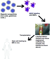
Tracking the distribution of stem cells (SCs) in tissue engineered organ scaffolds is of great importance in order to understand the role, movement and distribution of SCs after they have been implanted.   A recent minireview, by Hachani et al. at University College London, highlights the recent application of nanotechnology in SC labeling and magnetic resonance imaging (MRI).

The authors provide an overview of magnetic nanoparticles (MNPs), such as iron oxide and gadolinium nanoparticles, for use as contrast agents in MRI-based cell tracking and labeling.  The minireview introduces SC research and how conventional MRI is used as a tool for tracking and labeling.  However, the review’s main focus is discussing the recent developments in magnetic nanoparticle research, particularly detailing the efforts made by researchers in controlling the physical properties of the MNPs, such as size, shape, surface chemistry, etc, in order to produce more biocompatible and stable MNP contrast agents.

For researchers wishing to gain an insight into the application of nanotechnology in MRI and stem cell research, this review provides a valuable starting point for becoming familiar with recent developments in this area.

By Lee Barrett

You can read this Minireview in Nanoscale by clicking the link below:

Tracking stem cells in tissue-engineered organs using magnetic nanoparticles
Roxanne Hachani, Mark Lowdell, Martin Birchall and Nguyễn Thi Kim Thanh
DOI: 10.1039/C3NR03861K, Minireview

Digg This
Reddit This
Stumble Now!
Share on Facebook
Bookmark this on Delicious
Share on LinkedIn
Bookmark this on Technorati
Post on Twitter
Google Buzz (aka. Google Reader)